 Movie
Movie Controller

[English] 日本語
 Yorodumi
- EMDB-1883: Three-dimensional structure of the RNA polymerase II-Iwr1 complex -
+ Open data

- Basic information
| Entry | Database: EMDB / ID: EMD-1883 | |||||||||
|---|---|---|---|---|---|---|---|---|---|---|
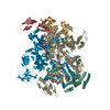
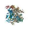
| Title | Three-dimensional structure of the RNA polymerase II-Iwr1 complex | |||||||||
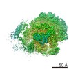
 Map data Map data | This is a map of the RNA Polymerase II-Iwr1 complex. | |||||||||
 Sample Sample |
| |||||||||
 Keywords Keywords | transcription / Pol II / nuclear import | |||||||||
| Biological species |  | |||||||||
| Method | single particle reconstruction / cryo EM / Resolution: 20.9 Å | |||||||||
 Authors Authors | Czeko E / Seizl M / Augsberger C / Mielke T / Cramer P | |||||||||
 Citation Citation | Journal: Mol Cell / Year: 2011 Title: Iwr1 directs RNA polymerase II nuclear import. Authors: Elmar Czeko / Martin Seizl / Christian Augsberger / Thorsten Mielke / Patrick Cramer /  Abstract: RNA polymerase (Pol) II transcribes protein-coding genes in the nucleus of eukaryotic cells and consists of 12 polypeptide subunits. It is unknown how Pol II is imported into the nucleus. Here we ...RNA polymerase (Pol) II transcribes protein-coding genes in the nucleus of eukaryotic cells and consists of 12 polypeptide subunits. It is unknown how Pol II is imported into the nucleus. Here we show that Pol II nuclear import requires the protein Iwr1 and provide evidence for cyclic Iwr1 function. Iwr1 binds Pol II in the active center cleft between the two largest subunits, maybe facilitating or sensing complete Pol II assembly in the cytoplasm. Iwr1 then uses an N-terminal bipartite nuclear localization signal that is recognized by karyopherin α to direct Pol II nuclear import. In the nucleus, Iwr1 is displaced from Pol II by transcription initiation factors and nucleic acids, enabling its export and recycling. Iwr1 function is Pol II specific, transcription independent, and apparently conserved from yeast to human. | |||||||||
| History |
|
- Structure visualization
| Movie |
 Movie viewer Movie viewer |
|---|---|
| Structure viewer | EM map: SurfViewMolmilJmol/JSmol |
| Supplemental images |

- Downloads & links
-EMDB archive
| Map data | emd_1883.map.gz | 1.2 MB | EMDB map data format | |
|---|---|---|---|---|
| Header (meta data) | emd-1883-v30.xmlemd-1883.xml | 10 KB 10 KB | Display Display | EMDB header |
| Images | 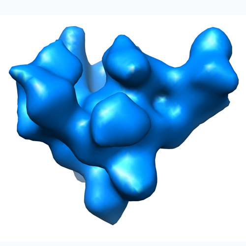 EMD-1883.jpg EMD-1883.jpg | 65.4 KB | ||
| Archive directory |  http://ftp.pdbj.org/pub/emdb/structures/EMD-1883ftp://ftp.pdbj.org/pub/emdb/structures/EMD-1883 http://ftp.pdbj.org/pub/emdb/structures/EMD-1883ftp://ftp.pdbj.org/pub/emdb/structures/EMD-1883 | HTTPS FTP |
-Related structure data
| Similar structure data |
|---|


-Links
| EMDB pages | EMDB (EBI/PDBe) / EMDataResource |
|---|
-Map
| File | Download / File: emd_1883.map.gz / Format: CCP4 / Size: 1.3 MB / Type: IMAGE STORED AS FLOATING POINT NUMBER (4 BYTES) | ||||||||||||||||||||||||||||||||||||||||||||||||||||||||||||||||||||
|---|---|---|---|---|---|---|---|---|---|---|---|---|---|---|---|---|---|---|---|---|---|---|---|---|---|---|---|---|---|---|---|---|---|---|---|---|---|---|---|---|---|---|---|---|---|---|---|---|---|---|---|---|---|---|---|---|---|---|---|---|---|---|---|---|---|---|---|---|---|
| Annotation | This is a map of the RNA Polymerase II-Iwr1 complex. | ||||||||||||||||||||||||||||||||||||||||||||||||||||||||||||||||||||
| Projections & slices | Image control
Images are generated by Spider. | ||||||||||||||||||||||||||||||||||||||||||||||||||||||||||||||||||||
| Voxel size | X=Y=Z: 3.71 Å | ||||||||||||||||||||||||||||||||||||||||||||||||||||||||||||||||||||
| Density |
| ||||||||||||||||||||||||||||||||||||||||||||||||||||||||||||||||||||
| Symmetry | Space group: 1 | ||||||||||||||||||||||||||||||||||||||||||||||||||||||||||||||||||||
| Details | EMDB XML:
CCP4 map header:
| ||||||||||||||||||||||||||||||||||||||||||||||||||||||||||||||||||||
 Z (Sec.)
 Y (Row.)
 X (Col.)


-Supplemental data
- Sample components
-Entire : RNA polymerase II-Iwr1
| Entire | Name: RNA polymerase II-Iwr1 |
|---|---|
| Components |
|
-Supramolecule #1000: RNA polymerase II-Iwr1
| Supramolecule | Name: RNA polymerase II-Iwr1 / type: sample / ID: 1000 / Oligomeric state: Monomeric / Number unique components: 2 |
|---|---|
| Molecular weight | Theoretical: 550 KDa |
-Macromolecule #1: DNA-directed RNA polymerase
| Macromolecule | Name: DNA-directed RNA polymerase / type: protein_or_peptide / ID: 1 / Name.synonym: Pol II / Number of copies: 1 / Recombinant expression: No |
|---|---|
| Source (natural) | Organism: |
| Molecular weight | Theoretical: 510 KDa |
-Macromolecule #2: Import adaptor
| Macromolecule | Name: Import adaptor / type: protein_or_peptide / ID: 2 / Name.synonym: Iwr1 / Number of copies: 1 / Oligomeric state: Monomer / Recombinant expression: Yes |
|---|---|
| Source (natural) | Organism: |
| Molecular weight | Theoretical: 40 KDa |
| Recombinant expression | Organism:  |
-Experimental details
-Structure determination
| Method | cryo EM |
|---|---|
 Processing Processing | single particle reconstruction |
| Aggregation state | particle |
-Sample preparation
| Concentration | 0.1 mg/mL |
|---|---|
| Buffer | pH: 7.25 Details: 5 mM HEPES, 40 mM Ammonium sulfate, 10 microM Zinc chloride, 10 mM DTT |
| Vitrification | Cryogen name: ETHANE / Chamber humidity: 95 % / Chamber temperature: 80 K / Instrument: OTHER / Details: Vitrification instrument: FEI Vitrobot / Method: Blot for 10 s before plunging |
- Electron microscopy
| Microscope | FEI POLARA 300 |
|---|---|
| Temperature | Average: 80 K |
| Image recording | Category: FILM / Film or detector model: KODAK SO-163 FILM / Digitization - Scanner: OTHER / Number real images: 35 / Average electron dose: 25 e/Å2 |
| Electron beam | Acceleration voltage: 300 kV / Electron source:  FIELD EMISSION GUN FIELD EMISSION GUN |
| Electron optics | Illumination mode: SPOT SCAN / Imaging mode: BRIGHT FIELD / Cs: 2.0 mm / Nominal defocus max: 3.5 µm / Nominal defocus min: 1.5 µm / Nominal magnification: 67000 |
| Sample stage | Specimen holder: Multispecimen holder / Specimen holder model: OTHER |
| Experimental equipment |  Model: Tecnai Polara / Image courtesy: FEI Company |
-Image processing
| Details | Particles were selected using Signature and thereafter checked manually |
|---|---|
| CTF correction | Details: Defocus groups |
| Final reconstruction | Applied symmetry - Point group: C1 (asymmetric) / Algorithm: OTHER / Resolution.type: BY AUTHOR / Resolution: 20.9 Å / Resolution method: FSC 0.5 CUT-OFF / Software - Name: SPIDER / Number images used: 25206 |
| Final angle assignment | Details: SPIDER |
-Atomic model buiding 1
| Initial model | PDB ID: |
|---|---|
| Software | Name: Chimera |
| Details | Protocol: Rigid body |
| Refinement | Space: REAL / Protocol: RIGID BODY FIT / Target criteria: Least-square |

