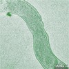
Journal: Appl Environ Microbiol / Year: 2017 Title: Three-Dimensional Structure of the Ultraoligotrophic Marine Bacterium "Candidatus Pelagibacter ubique". Authors: Xiaowei Zhao / Cindi L Schwartz / Jason Pierson / Stephen J Giovannoni / J Richard McIntosh / Daniela Nicastro / Abstract: SAR11 bacteria are small, heterotrophic, marine alphaproteobacteria found throughout the oceans. They thrive at the low nutrient concentrations typical of open ocean conditions, although the ...SAR11 bacteria are small, heterotrophic, marine alphaproteobacteria found throughout the oceans. They thrive at the low nutrient concentrations typical of open ocean conditions, although the adaptations required for life under those conditions are not well understood. To illuminate this issue, we used cryo-electron tomography to study "Candidatus Pelagibacter ubique" strain HTCC1062, a member of the SAR11 clade. Our results revealed its cellular dimensions and details of its intracellular organization. Frozen-hydrated cells, which were preserved in a life-like state, had an average cell volume (enclosed by the outer membrane) of 0.037 ± 0.011 μm Strikingly, the periplasmic space occupied ∼20% to 50% of the total cell volume in log-phase cells and ∼50% to 70% in stationary-phase cells. The nucleoid occupied the convex side of the crescent-shaped cells and the ribosomes predominantly occupied the concave side, at a relatively high concentration of 10,000 to 12,000 ribosomes/μm Outer membrane pore complexes, likely composed of PilQ, were frequently observed in both log-phase and stationary-phase cells. Long filaments, most likely type IV pili, were found on dividing cells. The physical dimensions, intracellular organization, and morphological changes throughout the life cycle of "Ca. Pelagibacter ubique" provide structural insights into the functional adaptions of these oligotrophic ultramicrobacteria to their habitat. IMPORTANCE: Bacterioplankton of the SAR11 clade (Pelagibacterales) are of interest because of their global biogeochemical significance and because they appear to have been molded by unusual ...IMPORTANCE: Bacterioplankton of the SAR11 clade (Pelagibacterales) are of interest because of their global biogeochemical significance and because they appear to have been molded by unusual evolutionary circumstances that favor simplicity and efficiency. They have adapted to an ecosystem in which nutrient concentrations are near the extreme limits at which transport systems can function adequately, and they have evolved streamlined genomes to execute only functions essential for life. However, little is known about the actual size limitations and cellular features of living oligotrophic ultramicrobacteria. In this study, we have used cryo-electron tomography to obtain accurate physical information about the cellular architecture of "Candidatus Pelagibacter ubique," the first cultivated member of the SAR11 clade. These results provide foundational information for answering questions about the cell architecture and functions of these ultrasmall oligotrophic bacteria.
History
Deposition
Aug 12, 2016
-
Header (metadata) release
Oct 5, 2016
-
Map release
Feb 21, 2018
-
Update
Apr 24, 2019
-
Current status
Apr 24, 2019
Processing site: RCSB / Status: Released
-
Structure visualization
Movie
Surface view with section colored by density value
In the structure databanks used in Yorodumi, some data are registered as the other names, "COVID-19 virus" and "2019-nCoV". Here are the details of the virus and the list of structure data.
Jan 31, 2019. EMDB accession codes are about to change! (news from PDBe EMDB page)
EMDB accession codes are about to change! (news from PDBe EMDB page)
The allocation of 4 digits for EMDB accession codes will soon come to an end. Whilst these codes will remain in use, new EMDB accession codes will include an additional digit and will expand incrementally as the available range of codes is exhausted. The current 4-digit format prefixed with “EMD-” (i.e. EMD-XXXX) will advance to a 5-digit format (i.e. EMD-XXXXX), and so on. It is currently estimated that the 4-digit codes will be depleted around Spring 2019, at which point the 5-digit format will come into force.
The EM Navigator/Yorodumi systems omit the EMD- prefix.
Related info.:Q: What is EMD? / ID/Accession-code notation in Yorodumi/EM Navigator
Yorodumi is a browser for structure data from EMDB, PDB, SASBDB, etc.
This page is also the successor to EM Navigator detail page, and also detail information page/front-end page for Omokage search.
The word "yorodu" (or yorozu) is an old Japanese word meaning "ten thousand". "mi" (miru) is to see.
Related info.:EMDB / PDB / SASBDB / Comparison of 3 databanks / Yorodumi Search / Aug 31, 2016. New EM Navigator & Yorodumi / Yorodumi Papers / Jmol/JSmol / Function and homology information / Changes in new EM Navigator and Yorodumi
 Movie
Movie Controller


 Open data
 Basic information
 Map data
 Sample
 Candidatus pelagibacter (bacteria)
 Authors
 Citation
 Structure visualization
 Movie viewer
 Downloads & links
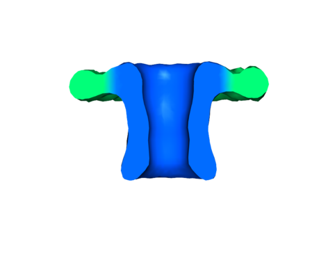 emd_8330.png
 http://ftp.pdbj.org/pub/emdb/structures/EMD-8330


 Z (Sec.)
 Y (Row.)
 X (Col.)


 Sample components
 Processing
 Electron microscopy
 FIELD EMISSION GUN
