 ムービー
ムービー コントローラー


+ データを開く

- 基本情報
基本情報
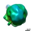
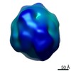
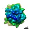
| 登録情報 | データベース: EMDB / ID: EMD-8042 | |||||||||
|---|---|---|---|---|---|---|---|---|---|---|
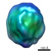
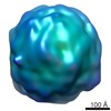
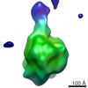
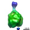
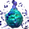
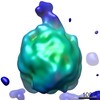
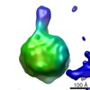
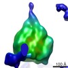
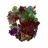
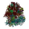
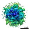
| タイトル | Electron tomographic structure of an individual human plasma VLDL particle (No.001) | |||||||||
 マップデータ マップデータ | human plasma VLDL particle | |||||||||
 試料 試料 |
| |||||||||
| 生物種 |  Homo sapiens (ヒト) Homo sapiens (ヒト) | |||||||||
| 手法 | 電子線トモグラフィー法 / クライオ電子顕微鏡法 / 解像度: 50.0 Å | |||||||||
 データ登録者 データ登録者 | Yu Y / Kuang Y / Lei D / Zhai X / Krauss R / Ren G | |||||||||
| 資金援助 |  米国, 2件 米国, 2件
| |||||||||
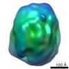
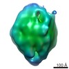
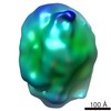
 引用 引用 | ジャーナル: J Lipid Res / 年: 2016 タイトル: Polyhedral 3D structure of human plasma very low density lipoproteins by individual particle cryo-electron tomography1. 著者: Yadong Yu / Yu-Lin Kuang / Dongsheng Lei / Xiaobo Zhai / Meng Zhang / Ronald M Krauss / Gang Ren / 要旨: Human VLDLs assembled in the liver and secreted into the circulation supply energy to peripheral tissues. VLDL lipolysis yields atherogenic LDLs and VLDL remnants that strongly correlate with CVD. ...Human VLDLs assembled in the liver and secreted into the circulation supply energy to peripheral tissues. VLDL lipolysis yields atherogenic LDLs and VLDL remnants that strongly correlate with CVD. Although the composition of VLDL particles has been well-characterized, their 3D structure is elusive because of their variations in size, heterogeneity in composition, structural flexibility, and mobility in solution. Here, we employed cryo-electron microscopy and individual-particle electron tomography to study the 3D structure of individual VLDL particles (without averaging) at both below and above their lipid phase transition temperatures. The 3D reconstructions of VLDL and VLDL bound to antibodies revealed an unexpected polyhedral shape, in contrast to the generally accepted model of a spherical emulsion-like particle. The smaller curvature of surface lipids compared with HDL may also reduce surface hydrophobicity, resulting in lower binding affinity to the hydrophobic distal end of the N-terminal β-barrel domain of cholesteryl ester transfer protein (CETP) compared with HDL. The directional binding of CETP to HDL and VLDL may explain the function of CETP in transferring TGs and cholesteryl esters between these particles. This first visualization of the 3D structure of VLDL could improve our understanding of the role of VLDL in atherogenesis. | |||||||||
| 履歴 |
|
- 構造の表示
構造の表示
| ムービー |
 ムービービューア ムービービューア |
|---|---|
| 構造ビューア | EMマップ: SurfViewMolmilJmol/JSmol |
| 添付画像 |
 UCSF Chimera
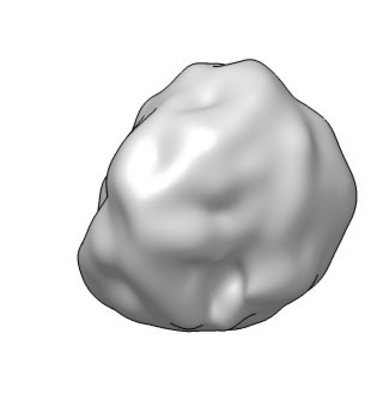
- ダウンロードとリンク
-EMDBアーカイブ
| マップデータ | emd_8042.map.gz | 28.1 MB | EMDBマップデータ形式 | |
|---|---|---|---|---|
| ヘッダ (付随情報) | emd-8042-v30.xmlemd-8042.xml | 15.8 KB 15.8 KB | 表示 表示 | EMDBヘッダ |
| 画像 |  emd_8042.png emd_8042.png | 36.1 KB | ||
| アーカイブディレクトリ |  http://ftp.pdbj.org/pub/emdb/structures/EMD-8042ftp://ftp.pdbj.org/pub/emdb/structures/EMD-8042 http://ftp.pdbj.org/pub/emdb/structures/EMD-8042ftp://ftp.pdbj.org/pub/emdb/structures/EMD-8042 | HTTPS FTP |
-関連構造データ

-リンク
| EMDBのページ | EMDB (EBI/PDBe) / EMDataResource |
|---|
-マップ
| ファイル | ダウンロード / ファイル: emd_8042.map.gz / 形式: CCP4 / 大きさ: 30.5 MB / タイプ: IMAGE STORED AS FLOATING POINT NUMBER (4 BYTES) | ||||||||||||||||||||||||||||||||||||||||||||||||||||||||||||||||||||
|---|---|---|---|---|---|---|---|---|---|---|---|---|---|---|---|---|---|---|---|---|---|---|---|---|---|---|---|---|---|---|---|---|---|---|---|---|---|---|---|---|---|---|---|---|---|---|---|---|---|---|---|---|---|---|---|---|---|---|---|---|---|---|---|---|---|---|---|---|---|
| 注釈 | human plasma VLDL particle | ||||||||||||||||||||||||||||||||||||||||||||||||||||||||||||||||||||
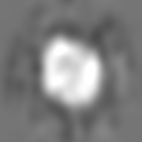
| 投影像・断面図 | 画像のコントロール
画像は Spider により作成 | ||||||||||||||||||||||||||||||||||||||||||||||||||||||||||||||||||||
| ボクセルのサイズ | X=Y=Z: 2.4 Å | ||||||||||||||||||||||||||||||||||||||||||||||||||||||||||||||||||||
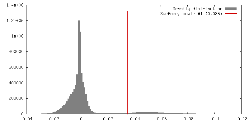
| 密度 |
| ||||||||||||||||||||||||||||||||||||||||||||||||||||||||||||||||||||
| 対称性 | 空間群: 1 | ||||||||||||||||||||||||||||||||||||||||||||||||||||||||||||||||||||
| 詳細 | EMDB XML:
CCP4マップ ヘッダ情報:
| ||||||||||||||||||||||||||||||||||||||||||||||||||||||||||||||||||||
 Z (Sec.)
 Y (Row.)
 X (Col.)


-添付データ
- 試料の構成要素
-全体 : Human Plasma Very-Low-Density Lipoprotein
| 全体 | 名称: Human Plasma Very-Low-Density Lipoprotein |
|---|---|
| 要素 |
|
-超分子 #1: Human Plasma Very-Low-Density Lipoprotein
| 超分子 | 名称: Human Plasma Very-Low-Density Lipoprotein / タイプ: complex / ID: 1 / 親要素: 0 詳細: VLDL isolated from human plasma by density gradient centrifugation. |
|---|---|
| 由来(天然) | 生物種: Homo sapiens (ヒト) / 組織: blood |
| 分子量 | 実験値: 4.0 MDa |
-実験情報
-構造解析
| 手法 | クライオ電子顕微鏡法 |
|---|---|
 解析 解析 | 電子線トモグラフィー法 |
| 試料の集合状態 | particle |
-試料調製
| 濃度 | 0.4 mg/mL | |||||||||||||||
|---|---|---|---|---|---|---|---|---|---|---|---|---|---|---|---|---|
| 緩衝液 | pH: 7.3 構成要素:
詳細: DPBS | |||||||||||||||
| グリッド | モデル: EMS Lacey carbon / 材質: COPPER / メッシュ: 200 / 支持フィルム - 材質: CARBON / 支持フィルム - トポロジー: LACEY / 支持フィルム - Film thickness: 50.0 nm / 前処理 - タイプ: GLOW DISCHARGE / 前処理 - 雰囲気: AIR / 前処理 - 気圧: 0.039 kPa | |||||||||||||||
| 凍結 | 凍結剤: ETHANE / チャンバー内湿度: 90 % / チャンバー内温度: 277 K / 装置: LEICA EM GP 詳細: 3 microliter of specimen blotted for 3 seconds before plunging. | |||||||||||||||
| 詳細 | This sample was monodisperse. | |||||||||||||||
| 切片作成 | その他: NO SECTIONING |
- 電子顕微鏡法
| 顕微鏡 | ZEISS LIBRA120PLUS |
|---|---|
| 温度 | 最低: 90.0 K / 最高: 93.0 K |
| 特殊光学系 | エネルギーフィルター - 名称: Zeiss in-column Omega エネルギーフィルター - エネルギー下限: 0 eV エネルギーフィルター - エネルギー上限: 20 eV |
| 撮影 | フィルム・検出器のモデル: GATAN ULTRASCAN 4000 (4k x 4k) デジタル化 - サイズ - 横: 4096 pixel / デジタル化 - サイズ - 縦: 4096 pixel / デジタル化 - サンプリング間隔: 15.0 µm / 撮影したグリッド数: 1 / 実像数: 85 / 平均露光時間: 0.5 sec. / 平均電子線量: 1.8 e/Å2 詳細: Images were collected by using the Gatan Tomography module of Digital Micrograph. XY tracking and focusing were done manually. |
| 電子線 | 加速電圧: 120 kV / 電子線源: LAB6 |
| 電子光学系 | C2レンズ絞り径: 75.0 µm / 最大 デフォーカス(補正後): 2.5 µm / 最小 デフォーカス(補正後): 1.5 µm / 倍率(補正後): 50000 / 照射モード: FLOOD BEAM / 撮影モード: BRIGHT FIELD / Cs: 2.2 mm / 最大 デフォーカス(公称値): 2.5 µm / 最小 デフォーカス(公称値): 1.5 µm / 倍率(公称値): 50000 |
| 試料ステージ | 試料ホルダーモデル: GATAN 626 SINGLE TILT LIQUID NITROGEN CRYO TRANSFER HOLDER ホルダー冷却材: NITROGEN |
-画像解析
| 詳細 | Image defects such as bad pixels and X-ray were removed prior to alignment and 3D reconstruction. |
|---|---|
| 最終 再構成 | アルゴリズム: BACK PROJECTION / 解像度のタイプ: BY AUTHOR / 解像度: 50.0 Å / 解像度の算出法: FSC 0.5 CUT-OFF / ソフトウェア - 名称: IPET 詳細: The 3D reconstruction was conducted by using Individual Particle Electron Tomography (IPET) 使用した粒子像数: 85 |
| CTF補正 | ソフトウェア - 名称: TOMOCTF (ver. V. October 2012) |
