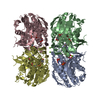
Movie
Movie Controller

[English] 日本語
 Yorodumi
- EMDB-80332: Peptidoglycan and lipopolysaccharide biosynthesis enzymes with in... -
+ Open data

- Basic information
| Entry |  | |||||||||
|---|---|---|---|---|---|---|---|---|---|---|
| Title | Peptidoglycan and lipopolysaccharide biosynthesis enzymes with inhibitor | |||||||||
 Map data Map data | ||||||||||
 Sample Sample |
| |||||||||
 Keywords Keywords | biogenesis / peptidoglycan synthesis enzyme / lipopolysaccharide synthesis enzyme / bacterial cell wall / bacterial outer membrane / BIOSYNTHETIC PROTEIN | |||||||||
| Function / homology |  Function and homology information Function and homology informationUDP-N-acetylglucosamine 1-carboxyvinyltransferase activity / UDP-N-acetylgalactosamine biosynthetic process / UDP-N-acetylglucosamine 1-carboxyvinyltransferase / UDP-3-O-acyl-N-acetylglucosamine deacetylase / UDP-3-O-acyl-N-acetylglucosamine deacetylase activity / lipid A biosynthetic process / peptidoglycan biosynthetic process / cell wall organization / regulation of cell shape / cell division / cytoplasm Similarity search - Function | |||||||||
| Biological species |  Pseudomonas aeruginosa PAO1 (bacteria) Pseudomonas aeruginosa PAO1 (bacteria) | |||||||||
| Method | single particle reconstruction / cryo EM / Resolution: 2.35 Å | |||||||||
 Authors Authors | Yeo JY / Yan XF / Gao YG | |||||||||
| Funding support |  Singapore, 1 items Singapore, 1 items
| |||||||||
 Citation Citation | Journal: J Struct Biol / Year: 2026 Title: Structure of the MurA-LpxC enzyme complex in modulating peptidoglycan and lipopolysaccharide biosynthesis. Authors: Joshua Yi Yeo / Xin-Fu Yan / Zhu Qiao / Yan Yu Liew / Phong Hoa Do / Yuguang Mu / Yong-Gui Gao / Abstract: Coordination of peptidoglycan and lipopolysaccharide biosynthesis is essential for maintaining Gram-negative cell envelope homeostasis. Two enzymes, MurA and LpxC, catalyze the first committed steps ...Coordination of peptidoglycan and lipopolysaccharide biosynthesis is essential for maintaining Gram-negative cell envelope homeostasis. Two enzymes, MurA and LpxC, catalyze the first committed steps in peptidoglycan and lipopolysaccharide biosynthesis, respectively. Here, we determined cryo-electron microscopy (cryo-EM) structures of the Pseudomonas aeruginosa MurA-LpxC complex in the absence and presence of the LpxC inhibitor CHIR-090, providing molecular insights into complex formation. Structure-guided mutagenesis of MurA, together with in vitro pull-down assays, identified residues crucial for complex formation. We show that MurA G58 favors, but is not sufficient for complex formation, as substitution of this residue to mimic Escherichia coli MurA (G58S) weakens the interaction. Together, our study advances our structural understanding of how two biosynthesis pathways for peptidoglycan and lipopolysaccharide are coordinated to maintain a synergistic and balanced cell envelope. | |||||||||
| History |
|
- Structure visualization
| Supplemental images |
|---|

- Downloads & links
-EMDB archive
| Map data | emd_80332.map.gz | 32.2 MB | EMDB map data format | |
|---|---|---|---|---|
| Header (meta data) | emd-80332-v30.xmlemd-80332.xml | 16.5 KB 16.5 KB | Display Display | EMDB header |
| FSC (resolution estimation) | emd_80332_fsc.xml | 8.4 KB | Display | FSC data file |
| Images |  emd_80332.png emd_80332.png | 73.2 KB | ||
| Filedesc metadata | emd-80332.cif.gz | 6.1 KB | ||
| Others | emd_80332_half_map_1.map.gzemd_80332_half_map_2.map.gz | 59.5 MB 59.5 MB | ||
| Archive directory |  https://data.pdbj.org/pub/emdb/structures/EMD-80332ftp://data.pdbj.org/pub/emdb/structures/EMD-80332 https://data.pdbj.org/pub/emdb/structures/EMD-80332ftp://data.pdbj.org/pub/emdb/structures/EMD-80332 | HTTPS FTP |
-Related structure data
| Related structure data |  25rwMC  25rvC M: atomic model generated by this map C: citing same article ( |
|---|---|
| Similar structure data |

-Links
| EMDB pages | EMDB (EBI/PDBe) / EMDataResource |
|---|---|
| Related items in Molecule of the Month |
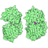
-Map
| File | Download / File: emd_80332.map.gz / Format: CCP4 / Size: 64 MB / Type: IMAGE STORED AS FLOATING POINT NUMBER (4 BYTES) | ||||||||||||||||||||||||||||||||||||
|---|---|---|---|---|---|---|---|---|---|---|---|---|---|---|---|---|---|---|---|---|---|---|---|---|---|---|---|---|---|---|---|---|---|---|---|---|---|
| Projections & slices | Image control
Images are generated by Spider. | ||||||||||||||||||||||||||||||||||||
| Voxel size | X=Y=Z: 0.76 Å | ||||||||||||||||||||||||||||||||||||
| Density |
| ||||||||||||||||||||||||||||||||||||
| Symmetry | Space group: 1 | ||||||||||||||||||||||||||||||||||||
| Details | EMDB XML:
|
 Z (Sec.)
 Y (Row.)
 X (Col.)


-Supplemental data
-Half map: #2
| File | emd_80332_half_map_1.map | ||||||||||||
|---|---|---|---|---|---|---|---|---|---|---|---|---|---|
| Projections & Slices |
| ||||||||||||
| Density Histograms |


-Half map: #1
| File | emd_80332_half_map_2.map | ||||||||||||
|---|---|---|---|---|---|---|---|---|---|---|---|---|---|
| Projections & Slices |
| ||||||||||||
| Density Histograms |


- Sample components
-Entire : PaMurA-PaLpxC complex in the presence of CHIR-090
| Entire | Name: PaMurA-PaLpxC complex in the presence of CHIR-090 |
|---|---|
| Components |
|
-Supramolecule #1: PaMurA-PaLpxC complex in the presence of CHIR-090
| Supramolecule | Name: PaMurA-PaLpxC complex in the presence of CHIR-090 / type: complex / ID: 1 / Parent: 0 / Macromolecule list: #1 |
|---|---|
| Source (natural) | Organism: Pseudomonas aeruginosa PAO1 (bacteria) |
| Molecular weight | Theoretical: 78.06 kDa/nm |
-Macromolecule #1: UDP-N-acetylglucosamine 1-carboxyvinyltransferase,UDP-3-O-acyl-N-...
| Macromolecule | Name: UDP-N-acetylglucosamine 1-carboxyvinyltransferase,UDP-3-O-acyl-N-acetylglucosamine deacetylase type: protein_or_peptide / ID: 1 / Number of copies: 2 / Enantiomer: LEVO EC number: UDP-N-acetylglucosamine 1-carboxyvinyltransferase |
|---|---|
| Source (natural) | Organism: Pseudomonas aeruginosa PAO1 (bacteria) |
| Molecular weight | Theoretical: 78.149594 KDa |
| Recombinant expression | Organism:  Escherichia coli BL21(DE3) (bacteria) Escherichia coli BL21(DE3) (bacteria) |
| Sequence | String: MDKLIITGGN RLDGEIRISG AKNSALPILA ATLLADTPVT VCNLPHLHDI TTMIELFGRM GVQPIIDEKL NVEVDASSIK TLVAPYELV KTMRASILVL GPMLARFGEA EVALPGGCAI GSRPVDLHIR GLEAMGAQIE VEGGYIKAKA PAGGLRGGHF F FDTVSVTG ...String: MDKLIITGGN RLDGEIRISG AKNSALPILA ATLLADTPVT VCNLPHLHDI TTMIELFGRM GVQPIIDEKL NVEVDASSIK TLVAPYELV KTMRASILVL GPMLARFGEA EVALPGGCAI GSRPVDLHIR GLEAMGAQIE VEGGYIKAKA PAGGLRGGHF F FDTVSVTG TENLMMAAAL ANGRTVLQNA AREPEVVDLA NCLNAMGANV QGAGSDTIVI EGVKRLGGAR YDVLPDRIET GT YLVAAAA TGGRVKLKDT DPTILEAVLQ KLEEAGAHIS TGSNWIELDM KGNRPKAVNV RTAPYPAFPT DMQAQFISMN AVA EGTGAV IETVFENRFM HVYEMNRMGA QILVEGNTAI VTGVPKLKGA PVMATDLRAS ASLVIAGLVA EGDTLIDRIY HIDR GYECI EEKLQLLGAK IRRVPGMIKQ RTLKNIIRAT GVGLHSGEKV YLTLKPAPVD TGIVFCRTDL DPVVEIPARA ENVGE TTMS TTLVKGDVKV DTVEHLLSAM AGLGIDNAYV ELSASEVPIM DGSAGPFVFL IQSAGLQEQE AAKKFIRIKR EVSVEE GDK RAVFVPFDGF KVSFEIDFDH PVFRGRTQQA SVDFSSTSFV KEVSRARTFG FMRDIEYLRS QNLALGGSVE NAIVVDE NR VLNEDGLRYE DEFVKHKILD AIGDLYLLGN SLIGEFRGFK SGHALNNQLL RTLIADKDAW EVVTFEDART APISYMRP A AAV UniProtKB: UDP-N-acetylglucosamine 1-carboxyvinyltransferase, UDP-3-O-acyl-N-acetylglucosamine deacetylase |
-Macromolecule #2: N-{(1S,2R)-2-hydroxy-1-[(hydroxyamino)carbonyl]propyl}-4-{[4-(mor...
| Macromolecule | Name: N-{(1S,2R)-2-hydroxy-1-[(hydroxyamino)carbonyl]propyl}-4-{[4-(morpholin-4-ylmethyl)phenyl]ethynyl}benzamide type: ligand / ID: 2 / Number of copies: 1 / Formula: C90 |
|---|---|
| Molecular weight | Theoretical: 437.488 Da |
| Chemical component information |  ChemComp-C90: |
-Experimental details
-Structure determination
| Method | cryo EM |
|---|---|
 Processing Processing | single particle reconstruction |
| Aggregation state | particle |
-Sample preparation
| Buffer | pH: 7.5 Component:
| ||||||
|---|---|---|---|---|---|---|---|
| Grid | Model: Quantifoil R1.2/1.3 / Material: COPPER / Mesh: 300 / Support film - Material: GRAPHENE | ||||||
| Vitrification | Cryogen name: ETHANE |
- Electron microscopy
| Microscope | TFS KRIOS |
|---|---|
| Image recording | Film or detector model: GATAN K3 (6k x 4k) / Average electron dose: 50.0 e/Å2 |
| Electron beam | Acceleration voltage: 300 kV / Electron source:  FIELD EMISSION GUN FIELD EMISSION GUN |
| Electron optics | Illumination mode: OTHER / Imaging mode: BRIGHT FIELD / Nominal defocus max: 1.2 µm / Nominal defocus min: 0.6 µm |
| Experimental equipment |  Model: Titan Krios / Image courtesy: FEI Company |