 Movie
Movie Controller

[English] 日本語
 Yorodumi
- PDB-25rw: Peptidoglycan and lipopolysaccharide biosynthesis enzymes with in... -
+ Open data

- Basic information
| Entry | Database: PDB / ID: 25rw | |||||||||
|---|---|---|---|---|---|---|---|---|---|---|
| Title | Peptidoglycan and lipopolysaccharide biosynthesis enzymes with inhibitor | |||||||||
 Components Components | UDP-N-acetylglucosamine 1-carboxyvinyltransferase,UDP-3-O-acyl-N-acetylglucosamine deacetylase | |||||||||
 Keywords Keywords | BIOSYNTHETIC PROTEIN / biogenesis / peptidoglycan synthesis enzyme / lipopolysaccharide synthesis enzyme / bacterial cell wall / bacterial outer membrane | |||||||||
| Function / homology |  Function and homology information Function and homology informationUDP-N-acetylglucosamine 1-carboxyvinyltransferase activity / UDP-N-acetylgalactosamine biosynthetic process / UDP-N-acetylglucosamine 1-carboxyvinyltransferase / UDP-3-O-acyl-N-acetylglucosamine deacetylase / UDP-3-O-acyl-N-acetylglucosamine deacetylase activity / lipid A biosynthetic process / peptidoglycan biosynthetic process / cell wall organization / regulation of cell shape / cell division / cytoplasm Similarity search - Function | |||||||||
| Biological species |  Pseudomonas aeruginosa PAO1 (bacteria) Pseudomonas aeruginosa PAO1 (bacteria) | |||||||||
| Method | ELECTRON MICROSCOPY / single particle reconstruction / cryo EM / Resolution: 2.35 Å | |||||||||
 Authors Authors | Yeo, J.Y. / Yan, X.F. / Gao, Y.G. | |||||||||
| Funding support |  Singapore, 1items Singapore, 1items
| |||||||||
 Citation Citation | Journal: J Struct Biol / Year: 2026 Title: Structure of the MurA-LpxC enzyme complex in modulating peptidoglycan and lipopolysaccharide biosynthesis. Authors: Joshua Yi Yeo / Xin-Fu Yan / Zhu Qiao / Yan Yu Liew / Phong Hoa Do / Yuguang Mu / Yong-Gui Gao / Abstract: Coordination of peptidoglycan and lipopolysaccharide biosynthesis is essential for maintaining Gram-negative cell envelope homeostasis. Two enzymes, MurA and LpxC, catalyze the first committed steps ...Coordination of peptidoglycan and lipopolysaccharide biosynthesis is essential for maintaining Gram-negative cell envelope homeostasis. Two enzymes, MurA and LpxC, catalyze the first committed steps in peptidoglycan and lipopolysaccharide biosynthesis, respectively. Here, we determined cryo-electron microscopy (cryo-EM) structures of the Pseudomonas aeruginosa MurA-LpxC complex in the absence and presence of the LpxC inhibitor CHIR-090, providing molecular insights into complex formation. Structure-guided mutagenesis of MurA, together with in vitro pull-down assays, identified residues crucial for complex formation. We show that MurA G58 favors, but is not sufficient for complex formation, as substitution of this residue to mimic Escherichia coli MurA (G58S) weakens the interaction. Together, our study advances our structural understanding of how two biosynthesis pathways for peptidoglycan and lipopolysaccharide are coordinated to maintain a synergistic and balanced cell envelope. | |||||||||
| History |
|
- Structure visualization
| Structure viewer | Molecule: MolmilJmol/JSmol |
|---|
- Downloads & links
-Download
| PDBx/mmCIF format | 25rw.cif.gz | 145.4 KB | Display | PDBx/mmCIF format |
|---|---|---|---|---|
| PDB format | pdb25rw.ent.gz | 102.2 KB | Display | PDB format |
| PDBx/mmJSON format | 25rw.json.gz | Tree view | PDBx/mmJSON format | |
| Others |  Other downloads Other downloads |
-Validation report
| Arichive directory | https://data.pdbj.org/pub/pdb/validation_reports/5r/25rwftp://data.pdbj.org/pub/pdb/validation_reports/5r/25rw | HTTPS FTP |
|---|
-Related structure data
| Related structure data |  80332MC  25rvC M: map data used to model this data C: citing same article ( |
|---|---|
| Similar structure data |

-Links
 PDBj
PDBj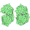
- Assembly
| Deposited unit | 
|
|---|---|
| 1 |
|
-Components
| #1: Protein | Mass: 78149.594 Da / Num. of mol.: 2 Source method: isolated from a genetically manipulated source Source: (gene. exp.) Pseudomonas aeruginosa PAO1 (bacteria) / Gene: murA, PA4450, lpxC, envA, PA4406 / Production host:  Escherichia coli BL21(DE3) (bacteria) Escherichia coli BL21(DE3) (bacteria)References: UniProt: Q9HVW7, UniProt: P47205, UDP-N-acetylglucosamine 1-carboxyvinyltransferase, UDP-3-O-acyl-N-acetylglucosamine deacetylase #2: Chemical | ChemComp-C90 / |  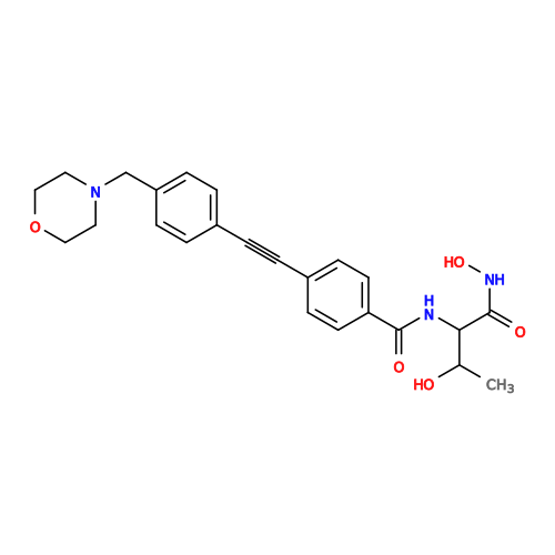 Mass: 437.488 Da / Num. of mol.: 1 / Source method: obtained synthetically / Formula: C24H27N3O5 / Feature type: SUBJECT OF INVESTIGATION Mass: 437.488 Da / Num. of mol.: 1 / Source method: obtained synthetically / Formula: C24H27N3O5 / Feature type: SUBJECT OF INVESTIGATIONHas ligand of interest | Y | Has protein modification | N | |
|---|
-Experimental details
-Experiment
| Experiment | Method: ELECTRON MICROSCOPY |
|---|---|
| EM experiment | Aggregation state: PARTICLE / 3D reconstruction method: single particle reconstruction |
- Sample preparation
| Component | Name: PaMurA-PaLpxC complex in the presence of CHIR-090 / Type: COMPLEX / Entity ID: #1 / Source: RECOMBINANT | ||||||||||||
|---|---|---|---|---|---|---|---|---|---|---|---|---|---|
| Molecular weight | Value: 78.06 kDa/nm / Experimental value: NO | ||||||||||||
| Source (natural) | Organism: Pseudomonas aeruginosa PAO1 (bacteria) | ||||||||||||
| Source (recombinant) | Organism: Escherichia coli BL21(DE3) (bacteria) | ||||||||||||
| Buffer solution | pH: 7.5 | ||||||||||||
| Buffer component |
| ||||||||||||
| Specimen | Embedding applied: NO / Shadowing applied: NO / Staining applied: NO / Vitrification applied: YES | ||||||||||||
| Specimen support | Grid material: COPPER / Grid mesh size: 300 divisions/in. / Grid type: Quantifoil R1.2/1.3 | ||||||||||||
| Vitrification | Cryogen name: ETHANE |
- Electron microscopy imaging
| Experimental equipment |  Model: Titan Krios / Image courtesy: FEI Company |
|---|---|
| Microscopy | Model: TFS KRIOS |
| Electron gun | Electron source:  FIELD EMISSION GUN / Accelerating voltage: 300 kV / Illumination mode: OTHER FIELD EMISSION GUN / Accelerating voltage: 300 kV / Illumination mode: OTHER |
| Electron lens | Mode: BRIGHT FIELD / Nominal defocus max: 1200 nm / Nominal defocus min: 600 nm |
| Image recording | Electron dose: 50 e/Å2 / Film or detector model: GATAN K3 (6k x 4k) |
- Processing
| EM software |
| ||||||||||||||||
|---|---|---|---|---|---|---|---|---|---|---|---|---|---|---|---|---|---|
| CTF correction | Type: NONE | ||||||||||||||||
| 3D reconstruction | Resolution: 2.35 Å / Resolution method: FSC 0.143 CUT-OFF / Num. of particles: 275131 / Symmetry type: POINT | ||||||||||||||||
| Refinement | Cross valid method: NONE |
