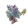
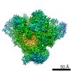
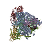
Journal: Nat Struct Mol Biol / Year: 2018 Title: Architecture of Pol II(G) and molecular mechanism of transcription regulation by Gdown1. Authors: Miki Jishage / Xiaodi Yu / Yi Shi / Sai J Ganesan / Wei-Yi Chen / Andrej Sali / Brian T Chait / Francisco J Asturias / Robert G Roeder / Abstract: Tight binding of Gdown1 represses RNA polymerase II (Pol II) function in a manner that is reversed by Mediator, but the structural basis of these processes is unclear. Although Gdown1 is ...Tight binding of Gdown1 represses RNA polymerase II (Pol II) function in a manner that is reversed by Mediator, but the structural basis of these processes is unclear. Although Gdown1 is intrinsically disordered, its Pol II interacting domains were localized and shown to occlude transcription factor IIF (TFIIF) and transcription factor IIB (TFIIB) binding by perfect positioning on their Pol II interaction sites. Robust binding of Gdown1 to Pol II is established by cooperative interactions of a strong Pol II binding region and two weaker binding modulatory regions, thus providing a mechanism both for tight Pol II binding and transcription inhibition and for its reversal. In support of a physiological function for Gdown1 in transcription repression, Gdown1 co-localizes with Pol II in transcriptionally silent nuclei of early Drosophila embryos but re-localizes to the cytoplasm during zygotic genome activation. Our study reveals a self-inactivation through Gdown1 binding as a unique mode of repression in Pol II function.
History
Deposition
Jun 11, 2018
-
Header (metadata) release
Jul 25, 2018
-
Map release
Jun 12, 2019
-
Update
Dec 23, 2020
-
Current status
Dec 23, 2020
Processing site: RCSB / Status: Released
-
Structure visualization
Movie
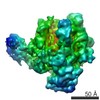
Surface view with section colored by density value
In the structure databanks used in Yorodumi, some data are registered as the other names, "COVID-19 virus" and "2019-nCoV". Here are the details of the virus and the list of structure data.
Jan 31, 2019. EMDB accession codes are about to change! (news from PDBe EMDB page)
EMDB accession codes are about to change! (news from PDBe EMDB page)
The allocation of 4 digits for EMDB accession codes will soon come to an end. Whilst these codes will remain in use, new EMDB accession codes will include an additional digit and will expand incrementally as the available range of codes is exhausted. The current 4-digit format prefixed with “EMD-” (i.e. EMD-XXXX) will advance to a 5-digit format (i.e. EMD-XXXXX), and so on. It is currently estimated that the 4-digit codes will be depleted around Spring 2019, at which point the 5-digit format will come into force.
The EM Navigator/Yorodumi systems omit the EMD- prefix.
Related info.:Q: What is EMD? / ID/Accession-code notation in Yorodumi/EM Navigator
Yorodumi is a browser for structure data from EMDB, PDB, SASBDB, etc.
This page is also the successor to EM Navigator detail page, and also detail information page/front-end page for Omokage search.
The word "yorodu" (or yorozu) is an old Japanese word meaning "ten thousand". "mi" (miru) is to see.
Related info.:EMDB / PDB / SASBDB / Comparison of 3 databanks / Yorodumi Search / Aug 31, 2016. New EM Navigator & Yorodumi / Yorodumi Papers / Jmol/JSmol / Function and homology information / Changes in new EM Navigator and Yorodumi
 Movie
Movie Controller


 Open data
 Basic information
 Map data
 Sample
 Homo sapiens (human)
 Authors
 Citation

 Structure visualization
 Movie viewer
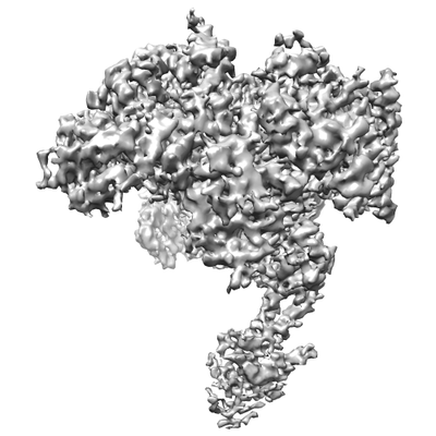
 Downloads & links
 emd_7998.png
 http://ftp.pdbj.org/pub/emdb/structures/EMD-7998

 Z (Sec.)
 Y (Row.)
 X (Col.)


 Sample components
 Processing
 Electron microscopy
 FIELD EMISSION GUN