+ Open data
- Basic information
| Entry | Database: EMDB / ID: EMD-7963 | |||||||||
|---|---|---|---|---|---|---|---|---|---|---|
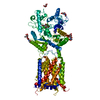
| Title | Cryo-EM structure of human Ptch1 | |||||||||
 Map data Map data | ||||||||||
 Sample Sample |
| |||||||||
 Keywords Keywords | Receptor / RND family / PROTEIN BINDING | |||||||||
| Function / homology |  Function and homology information Function and homology informationneural plate axis specification / response to chlorate / cell differentiation involved in kidney development / hedgehog receptor activity / neural tube patterning / cell proliferation involved in metanephros development / smoothened binding / hedgehog family protein binding / Ligand-receptor interactions / hindlimb morphogenesis ...neural plate axis specification / response to chlorate / cell differentiation involved in kidney development / hedgehog receptor activity / neural tube patterning / cell proliferation involved in metanephros development / smoothened binding / hedgehog family protein binding / Ligand-receptor interactions / hindlimb morphogenesis / epidermal cell fate specification / spinal cord motor neuron differentiation / prostate gland development / patched binding / negative regulation of cell division / somite development / limb morphogenesis / Activation of SMO / smooth muscle tissue development / dorsal/ventral neural tube patterning / pharyngeal system development / mammary gland duct morphogenesis / mammary gland epithelial cell differentiation / cellular response to cholesterol / commissural neuron axon guidance / cell fate determination / metanephric collecting duct development / regulation of smoothened signaling pathway / dorsal/ventral pattern formation / Class B/2 (Secretin family receptors) / embryonic limb morphogenesis / negative regulation of multicellular organism growth / branching involved in ureteric bud morphogenesis / ciliary membrane / cholesterol binding / positive regulation of epidermal cell differentiation / dendritic growth cone / keratinocyte proliferation / spermatid development / positive regulation of cholesterol efflux / embryonic organ development / negative regulation of keratinocyte proliferation / response to retinoic acid / negative regulation of osteoblast differentiation / response to mechanical stimulus / axonal growth cone / heart morphogenesis / negative regulation of stem cell proliferation / Hedgehog 'off' state / liver regeneration / regulation of mitotic cell cycle / cyclin binding / animal organ morphogenesis / stem cell proliferation / protein localization to plasma membrane / negative regulation of smoothened signaling pathway / neural tube closure / Hedgehog 'on' state / protein processing / brain development / caveola / apical part of cell / endocytic vesicle membrane / response to estradiol / glucose homeostasis / heparin binding / regulation of protein localization / midbody / in utero embryonic development / postsynaptic membrane / response to xenobiotic stimulus / intracellular membrane-bounded organelle / positive regulation of DNA-templated transcription / protein-containing complex binding / perinuclear region of cytoplasm / negative regulation of transcription by RNA polymerase II / signal transduction / plasma membrane Similarity search - Function | |||||||||
| Biological species |  Homo sapiens (human) Homo sapiens (human) | |||||||||
| Method | single particle reconstruction / cryo EM / Resolution: 3.9 Å | |||||||||
 Authors Authors | Yan N / Gong X | |||||||||
| Funding support |  China, 2 items China, 2 items
| |||||||||
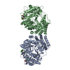
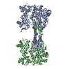
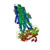
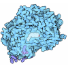
 Citation Citation |  Journal: Science / Year: 2018 Journal: Science / Year: 2018Title: Structural basis for the recognition of Sonic Hedgehog by human Patched1. Authors: Xin Gong / Hongwu Qian / Pingping Cao / Xin Zhao / Qiang Zhou / Jianlin Lei / Nieng Yan /  Abstract: The Hedgehog (Hh) pathway involved in development and regeneration is activated by the extracellular binding of Hh to the membrane receptor Patched (Ptch). We report the structures of human Ptch1 ...The Hedgehog (Hh) pathway involved in development and regeneration is activated by the extracellular binding of Hh to the membrane receptor Patched (Ptch). We report the structures of human Ptch1 alone and in complex with the N-terminal domain of human Sonic hedgehog (ShhN) at resolutions of 3.9 and 3.6 angstroms, respectively, as determined by cryo-electron microscopy. Ptch1 comprises two interacting extracellular domains, ECD1 and ECD2, and 12 transmembrane segments (TMs), with TMs 2 to 6 constituting the sterol-sensing domain (SSD). Two steroid-shaped densities are resolved in both structures, one enclosed by ECD1/2 and the other in the membrane-facing cavity of the SSD. Structure-guided mutational analysis shows that interaction between ShhN and Ptch1 is steroid-dependent. The structure of a steroid binding-deficient Ptch1 mutant displays pronounced conformational rearrangements. | |||||||||
| History |
|
- Structure visualization
| Movie |
 Movie viewer Movie viewer |
|---|---|
| Structure viewer | EM map:  SurfView SurfView Molmil Molmil Jmol/JSmol Jmol/JSmol |
| Supplemental images |
- Downloads & links
-EMDB archive
| Map data |  emd_7963.map.gz emd_7963.map.gz | 40.1 MB |  EMDB map data format EMDB map data format | |
|---|---|---|---|---|
| Header (meta data) |  emd-7963-v30.xml emd-7963-v30.xml emd-7963.xml emd-7963.xml | 18.5 KB 18.5 KB | Display Display |  EMDB header EMDB header |
| Images |  emd_7963.png emd_7963.png | 50.3 KB | ||
| Filedesc metadata |  emd-7963.cif.gz emd-7963.cif.gz | 7 KB | ||
| Archive directory |  http://ftp.pdbj.org/pub/emdb/structures/EMD-7963 http://ftp.pdbj.org/pub/emdb/structures/EMD-7963 ftp://ftp.pdbj.org/pub/emdb/structures/EMD-7963 ftp://ftp.pdbj.org/pub/emdb/structures/EMD-7963 | HTTPS FTP |
-Validation report
| Summary document |  emd_7963_validation.pdf.gz emd_7963_validation.pdf.gz | 547.8 KB | Display |  EMDB validaton report EMDB validaton report |
|---|---|---|---|---|
| Full document |  emd_7963_full_validation.pdf.gz emd_7963_full_validation.pdf.gz | 547.4 KB | Display | |
| Data in XML |  emd_7963_validation.xml.gz emd_7963_validation.xml.gz | 5.7 KB | Display | |
| Data in CIF |  emd_7963_validation.cif.gz emd_7963_validation.cif.gz | 6.6 KB | Display | |
| Arichive directory |  https://ftp.pdbj.org/pub/emdb/validation_reports/EMD-7963 https://ftp.pdbj.org/pub/emdb/validation_reports/EMD-7963 ftp://ftp.pdbj.org/pub/emdb/validation_reports/EMD-7963 ftp://ftp.pdbj.org/pub/emdb/validation_reports/EMD-7963 | HTTPS FTP |
-Related structure data
| Related structure data |  6dmbMC  7964C  7968C  6dmoC  6dmyC M: atomic model generated by this map C: citing same article ( |
|---|---|
| Similar structure data |
- Links
Links
| EMDB pages |  EMDB (EBI/PDBe) / EMDB (EBI/PDBe) /  EMDataResource EMDataResource |
|---|---|
| Related items in Molecule of the Month |
- Map
Map
| File |  Download / File: emd_7963.map.gz / Format: CCP4 / Size: 42.9 MB / Type: IMAGE STORED AS FLOATING POINT NUMBER (4 BYTES) Download / File: emd_7963.map.gz / Format: CCP4 / Size: 42.9 MB / Type: IMAGE STORED AS FLOATING POINT NUMBER (4 BYTES) | ||||||||||||||||||||||||||||||||||||||||||||||||||||||||||||
|---|---|---|---|---|---|---|---|---|---|---|---|---|---|---|---|---|---|---|---|---|---|---|---|---|---|---|---|---|---|---|---|---|---|---|---|---|---|---|---|---|---|---|---|---|---|---|---|---|---|---|---|---|---|---|---|---|---|---|---|---|---|
| Projections & slices | Image control
Images are generated by Spider. | ||||||||||||||||||||||||||||||||||||||||||||||||||||||||||||
| Voxel size | X=Y=Z: 1.091 Å | ||||||||||||||||||||||||||||||||||||||||||||||||||||||||||||
| Density |
| ||||||||||||||||||||||||||||||||||||||||||||||||||||||||||||
| Symmetry | Space group: 1 | ||||||||||||||||||||||||||||||||||||||||||||||||||||||||||||
| Details | EMDB XML:
CCP4 map header:
| ||||||||||||||||||||||||||||||||||||||||||||||||||||||||||||
-Supplemental data
- Sample components
-Entire : Patch1
| Entire | Name: Patch1 |
|---|---|
| Components |
|
-Supramolecule #1: Patch1
| Supramolecule | Name: Patch1 / type: organelle_or_cellular_component / ID: 1 / Parent: 0 / Macromolecule list: #1 |
|---|---|
| Source (natural) | Organism:  Homo sapiens (human) Homo sapiens (human) |
-Macromolecule #1: Protein patched homolog 1
| Macromolecule | Name: Protein patched homolog 1 / type: protein_or_peptide / ID: 1 / Number of copies: 1 / Enantiomer: LEVO |
|---|---|
| Source (natural) | Organism:  Homo sapiens (human) Homo sapiens (human) |
| Molecular weight | Theoretical: 150.189578 KDa |
| Recombinant expression | Organism:  Homo sapiens (human) Homo sapiens (human) |
| Sequence | String: MADYKDDDDK SGPDEVDASG RMASAGNAAE PQDRGGGGSG CIGAPGRPAG GGRRRRTGGL RRAAAPDRDY LHRPSYCDAA FALEQISKG KATGRKAPLW LRAKFQRLLF KLGCYIQKNC GKFLVVGLLI FGAFAVGLKA ANLETNVEEL WVEVGGRVSR E LNYTRQKI ...String: MADYKDDDDK SGPDEVDASG RMASAGNAAE PQDRGGGGSG CIGAPGRPAG GGRRRRTGGL RRAAAPDRDY LHRPSYCDAA FALEQISKG KATGRKAPLW LRAKFQRLLF KLGCYIQKNC GKFLVVGLLI FGAFAVGLKA ANLETNVEEL WVEVGGRVSR E LNYTRQKI GEEAMFNPQL MIQTPKEEGA NVLTTEALLQ HLDSALQASR VHVYMYNRQW KLEHLCYKSG ELITETGYMD QI IEYLYPC LIITPLDCFW EGAKLQSGTA YLLGKPPLRW TNFDPLEFLE ELKKINYQVD SWEEMLNKAE VGHGYMDRPC LNP ADPDCP ATAPNKNSTK PLDMALVLNG GCHGLSRKYM HWQEELIVGG TVKNSTGKLV SAHALQTMFQ LMTPKQMYEH FKGY EYVSH INWNEDKAAA ILEAWQRTYV EVVHQSVAQN STQKVLSFTT TTLDDILKSF SDVSVIRVAS GYLLMLAYAC LTMLR WDCS KSQGAVGLAG VLLVALSVAA GLGLCSLIGI SFNAATTQVL PFLALGVGVD DVFLLAHAFS ETGQNKRIPF EDRTGE CLK RTGASVALTS ISNVTAFFMA ALIPIPALRA FSLQAAVVVV FNFAMVLLIF PAILSMDLYR REDRRLDIFC CFTSPCV SR VIQVEPQAYT DTHDNTRYSP PPPYSSHSFA HETQITMQST VQLRTEYDPH THVYYTTAEP RSEISVQPVT VTQDTLSC Q SPESTSSTRD LLSQFSDSSL HCLEPPCTKW TLSSFAEKHY APFLLKPKAK VVVIFLFLGL LGVSLYGTTR VRDGLDLTD IVPRETREYD FIAAQFKYFS FYNMYIVTQK ADYPNIQHLL YDLHRSFSNV KYVMLEENKQ LPKMWLHYFR DWLQGLQDAF DSDWETGKI MPNNYKNGSD DGVLAYKLLV QTGSRDKPID ISQLTKQRLV DADGIINPSA FYIYLTAWVS NDPVAYAASQ A NIRPHRPE WVHDKADYMP ETRLRIPAAE PIEYAQFPFY LNGLRDTSDF VEAIEKVRTI CSNYTSLGLS SYPNGYPFLF WE QYIGLRH WLLLFISVVL ACTFLVCAVF LLNPWTAGII VMVLALMTVE LFGMMGLIGI KLSAVPVVIL IASVGIGVEF TVH VALAFL TAIGDKNRRA VLALEHMFAP VLDGAVSTLL GVLMLAGSEF DFIVRYFFAV LAILTILGVL NGLVLLPVLL SFFG PYPEV SPANGLNRLP TPSPEPPPSV VRFAMPPGHT HSGSDSSDSE YSSQTTVSGL SEELRHYEAQ QGAGGPAHQV IVEAT ENPV FAHSTVVHPE SRHHPPSNPR QQPHLDSGSL PPGRQGQQPR RDLEGSDEVD AVEGSHHHHH HHHHH UniProtKB: Protein patched homolog 1 |
-Macromolecule #3: 2-acetamido-2-deoxy-beta-D-glucopyranose
| Macromolecule | Name: 2-acetamido-2-deoxy-beta-D-glucopyranose / type: ligand / ID: 3 / Number of copies: 5 / Formula: NAG |
|---|---|
| Molecular weight | Theoretical: 221.208 Da |
| Chemical component information |  ChemComp-NAG: |
-Macromolecule #4: CHOLESTEROL HEMISUCCINATE
| Macromolecule | Name: CHOLESTEROL HEMISUCCINATE / type: ligand / ID: 4 / Number of copies: 2 / Formula: Y01 |
|---|---|
| Molecular weight | Theoretical: 486.726 Da |
| Chemical component information |  ChemComp-Y01: |
-Experimental details
-Structure determination
| Method | cryo EM |
|---|---|
 Processing Processing | single particle reconstruction |
| Aggregation state | particle |
- Sample preparation
| Concentration | 15 mg/mL |
|---|---|
| Buffer | pH: 8 |
| Vitrification | Cryogen name: ETHANE |
- Electron microscopy
| Microscope | FEI TITAN KRIOS |
|---|---|
| Image recording | Film or detector model: GATAN K2 SUMMIT (4k x 4k) / Detector mode: SUPER-RESOLUTION / Average electron dose: 50.0 e/Å2 |
| Electron beam | Acceleration voltage: 300 kV / Electron source:  FIELD EMISSION GUN FIELD EMISSION GUN |
| Electron optics | Illumination mode: FLOOD BEAM / Imaging mode: BRIGHT FIELD |
| Experimental equipment |  Model: Titan Krios / Image courtesy: FEI Company |
 Movie
Movie Controller






 Z (Sec.)
 Y (Row.)
 X (Col.)


