Movie
Movie Controller

[English] 日本語
 Yorodumi
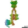
- EMDB-78696: Local Refinement of G protein heterotrimer bound to scFv14 when i... -
+ Open data

- Basic information
| Entry |  | ||||||||||||
|---|---|---|---|---|---|---|---|---|---|---|---|---|---|
| Title | Local Refinement of G protein heterotrimer bound to scFv14 when in complex with active mGluR8 | ||||||||||||
 Map data Map data | Local refinement of G protein heterotrimer bound to scFv14 when in complex with mGluR8 | ||||||||||||
 Sample Sample |
| ||||||||||||
 Keywords Keywords | GPCR / metabotropic glutamate receptor / membrane protein / synaptic protein / signaling protein | ||||||||||||
| Biological species |  Homo sapiens (human) / Homo sapiens (human) /   Mus musculus (house mouse) Mus musculus (house mouse) | ||||||||||||
| Method | single particle reconstruction / cryo EM / Resolution: 5.9 Å | ||||||||||||
 Authors Authors | Marx DC / Levitz JT | ||||||||||||
| Funding support |  United States, 3 items United States, 3 items
| ||||||||||||
 Citation Citation | Journal: Nat Commun / Year: 2026 Title: Structural basis of active state coupling of metabotropic glutamate receptor 8 to beta-arrestins. Authors: Dagan C Marx / Kevin Huynh / Alberto J Gonzalez-Hernandez / Alexa Strauss / Carlos Rico / Pamela N Gallo / Sheida Sharghi Moshtaghin / Anisul Arefin / Johannes Broichhagen / David Eliezer / ...Authors: Dagan C Marx / Kevin Huynh / Alberto J Gonzalez-Hernandez / Alexa Strauss / Carlos Rico / Pamela N Gallo / Sheida Sharghi Moshtaghin / Anisul Arefin / Johannes Broichhagen / David Eliezer / George Khelashvili / Joshua Levitz /  Abstract: Metabotropic glutamate receptors (mGluRs) are prototypical, dimeric family C G protein-coupled receptors (GPCR) that perform crucial modulatory roles throughout the nervous system. While mGluR ...Metabotropic glutamate receptors (mGluRs) are prototypical, dimeric family C G protein-coupled receptors (GPCR) that perform crucial modulatory roles throughout the nervous system. While mGluR activation and signaling through G proteins has been studied extensively, how these receptors interact with and are desensitized by β-arrestins (β-arrs) is not well understood. Here, we use an integrative biophysical and structural approach to probe the coupling of mGluR8 and β-arrs. Using negative stain electron microscopy (EM), we identify tail- and core-bound orientations and stoichiometries of mGluR8/β-arr complexes. Cryo-EM structures of mGluR8 alone or bound to either G proteins or β-arr1 reveal mGluR8 active states with transducer-specific differences. The mGluR8/β-arr structure shows a distinct complex orientation compared to other GPCR/β-arr structures which supports a steric mechanism of mGluR desensitization involving interactions with both subunits and the lipid bilayer. Coupling of mGluR8 to β-arr1 in an active-like conformation is verified by live-cell and single molecule FRET analysis. Finally, molecular dynamics simulations further define the positioning and dynamics of mGluR8-bound β-arr1 and the importance of critical mGluR8 residues for stabilizing β-arr1 complexes. Together, our data provide a framework for agonist-driven family C GPCR/β-arr coupling. | ||||||||||||
| History |
|
- Structure visualization
| Supplemental images |
|---|

- Downloads & links
-EMDB archive
| Map data | emd_78696.map.gz | 255.6 MB |  EMDB map data format EMDB map data format | |
|---|---|---|---|---|
| Header (meta data) | emd-78696-v30.xmlemd-78696.xml | 22.3 KB 22.3 KB | Display Display | EMDB header |
| FSC (resolution estimation) | emd_78696_fsc.xml | 17 KB | Display | FSC data file |
| Images |  emd_78696.png emd_78696.png | 53.7 KB | ||
| Filedesc metadata | emd-78696.cif.gz | 6.8 KB | ||
| Others | emd_78696_half_map_1.map.gzemd_78696_half_map_2.map.gz | 474.8 MB 474.8 MB | ||
| Archive directory |  https://data.pdbj.org/pub/emdb/structures/EMD-78696ftp://data.pdbj.org/pub/emdb/structures/EMD-78696 https://data.pdbj.org/pub/emdb/structures/EMD-78696ftp://data.pdbj.org/pub/emdb/structures/EMD-78696 | HTTPS FTP |
-Related structure data


-Links
| EMDB pages | EMDB (EBI/PDBe) / EMDataResource |
|---|
-Map
| File | Download / File: emd_78696.map.gz / Format: CCP4 / Size: 512 MB / Type: IMAGE STORED AS FLOATING POINT NUMBER (4 BYTES) | ||||||||||||||||||||||||||||||||||||
|---|---|---|---|---|---|---|---|---|---|---|---|---|---|---|---|---|---|---|---|---|---|---|---|---|---|---|---|---|---|---|---|---|---|---|---|---|---|
| Annotation | Local refinement of G protein heterotrimer bound to scFv14 when in complex with mGluR8 | ||||||||||||||||||||||||||||||||||||
| Projections & slices | Image control
Images are generated by Spider. | ||||||||||||||||||||||||||||||||||||
| Voxel size | X=Y=Z: 1.16 Å | ||||||||||||||||||||||||||||||||||||
| Density |
| ||||||||||||||||||||||||||||||||||||
| Symmetry | Space group: 1 | ||||||||||||||||||||||||||||||||||||
| Details | EMDB XML:
|
 Z (Sec.)
 Y (Row.)
 X (Col.)


-Supplemental data
-Half map: #1
| File | emd_78696_half_map_1.map | ||||||||||||
|---|---|---|---|---|---|---|---|---|---|---|---|---|---|
| Projections & Slices |
| ||||||||||||
| Density Histograms |


-Half map: #2
| File | emd_78696_half_map_2.map | ||||||||||||
|---|---|---|---|---|---|---|---|---|---|---|---|---|---|
| Projections & Slices |
| ||||||||||||
| Density Histograms |


- Sample components
-Entire : Metabotropic Glutamate receptor bound to G proteins alpha i-1, be...
| Entire | Name: Metabotropic Glutamate receptor bound to G proteins alpha i-1, beta-1, and gamma-2 in the presence of agonist and PAM and scFv14 |
|---|---|
| Components |
|
-Supramolecule #1: Metabotropic Glutamate receptor bound to G proteins alpha i-1, be...
| Supramolecule | Name: Metabotropic Glutamate receptor bound to G proteins alpha i-1, beta-1, and gamma-2 in the presence of agonist and PAM and scFv14 type: complex / ID: 1 / Parent: 0 / Macromolecule list: all |
|---|---|
| Source (natural) | Organism: Homo sapiens (human) |
| Molecular weight | Theoretical: 188 KDa |
-Supramolecule #2: G protein heterotrimer bound to scFv14
| Supramolecule | Name: G protein heterotrimer bound to scFv14 / type: complex / ID: 2 / Parent: 1 / Macromolecule list: #2-#5 |
|---|---|
| Source (natural) | Organism: Homo sapiens (human) |
-Supramolecule #3: mGluR8 homodimer bound to agonist and PAM
| Supramolecule | Name: mGluR8 homodimer bound to agonist and PAM / type: complex / ID: 3 / Parent: 1 / Macromolecule list: #1 |
|---|---|
| Source (natural) | Organism: Homo sapiens (human) |
-Macromolecule #1: Metabotropic glutamate receptor 8
| Macromolecule | Name: Metabotropic glutamate receptor 8 / type: protein_or_peptide / ID: 1 / Number of copies: 2 / Enantiomer: LEVO |
|---|---|
| Source (natural) | Organism: Homo sapiens (human) |
| Molecular weight | Theoretical: 94.195492 KDa |
| Recombinant expression | Organism: Homo sapiens (human) |
| Sequence | String: MKTIIALSYI FCLVFADYKD DDDAAAQEYA HSIRVDGDII LGGLFPVHAK GERGVPCGEL KKEKGIHRLE AMLYAIDQIN KDPDLLSNI TLGVRILDTC SRDTYALEQS LTFVQALIEK DASDVKCANG DPPIFTKPDK ISGVIGAAAS SVSIMVANIL R LFKIPQIS ...String: MKTIIALSYI FCLVFADYKD DDDAAAQEYA HSIRVDGDII LGGLFPVHAK GERGVPCGEL KKEKGIHRLE AMLYAIDQIN KDPDLLSNI TLGVRILDTC SRDTYALEQS LTFVQALIEK DASDVKCANG DPPIFTKPDK ISGVIGAAAS SVSIMVANIL R LFKIPQIS YASTAPELSD NTRYDFFSRV VPPDSYQAQA MVDIVTALGW NYVSTLASEG NYGESGVEAF TQISREIGGV CI AQSQKIP REPRPGEFEK IIKRLLETPN ARAVIMFANE DDIRRILEAA KKLNQSGHFL WIGSDSWGSK IAPVYQQEEI AEG AVTILP KRASIDGFDR YFRSRTLANN RRNVWFAEFW EENFGCKLGS HGKRNSHIKK CTGLERIARD SSYEQEGKVQ FVID AVYSM AYALHNMHKD LCPGYIGLCP RMSTIDGKEL LGYIRAVNFN GSAGTPVTFN ENGDAPGRYD IFQYQITNKS TEYKV IGHW TNQLHLKVED MQWAHREHTH PASVCSLPCK PGERKKTVKG VPCCWHCERC EGYNYQVDEL SCELCPLDQR PNMNRT GCQ LIPIIKLEWH SPWAVVPVFV AILGIIATTF VIVTFVRYND TPIVRASGRE LSYVLLTGIF LCYSITFLMI AAPDTII CS FRRVFLGLGM CFSYAALLTK TNRIHRIFEQ GKKSVTAPKF ISPASQLVIT FSLISVQLLG VFVWFVVDPP HIIIDYGE Q RTLDPEKARG VLKCDISDLS LICSLGYSIL LMVTCTVYAI KTRGVPETFN EAKPIGFTMY TTCIIWLAFI PIFFGTAQS AEKMYIQTTT LTVSMSLSAS VSLGMLYMPK VYIIIFHPEQ N |
-Macromolecule #2: Guanine nucleotide-binding protein G(I)/G(S)/G(O) subunit gamma-2
| Macromolecule | Name: Guanine nucleotide-binding protein G(I)/G(S)/G(O) subunit gamma-2 type: protein_or_peptide / ID: 2 / Number of copies: 1 / Enantiomer: LEVO |
|---|---|
| Source (natural) | Organism: Homo sapiens (human) |
| Molecular weight | Theoretical: 7.861143 KDa |
| Recombinant expression | Organism: Homo sapiens (human) |
| Sequence | String: MASNNTASIA QARKLVEQLK MEANIDRIKV SKAAADLMAY CEAHAKEDPL LTPVPASENP FREKKFFCAI L |
-Macromolecule #3: scFv-14
| Macromolecule | Name: scFv-14 / type: protein_or_peptide / ID: 3 / Number of copies: 1 / Enantiomer: LEVO |
|---|---|
| Source (natural) | Organism: Mus musculus (house mouse) |
| Molecular weight | Theoretical: 27.784896 KDa |
| Recombinant expression | Organism: Homo sapiens (human) |
| Sequence | String: DVQLVESGGG LVQPGGSRKL SCSASGFAFS SFGMHWVRQA PEKGLEWVAY ISSGSGTIYY ADTVKGRFTI SRDDPKNTLF LQMTSLRSE DTAMYYCVRS IYYYGSSPFD FWGQGTTLTV SSGGGGSGGG GSGGGGSDIV MTQATSSVPV TPGESVSISC R SSKSLLHS ...String: DVQLVESGGG LVQPGGSRKL SCSASGFAFS SFGMHWVRQA PEKGLEWVAY ISSGSGTIYY ADTVKGRFTI SRDDPKNTLF LQMTSLRSE DTAMYYCVRS IYYYGSSPFD FWGQGTTLTV SSGGGGSGGG GSGGGGSDIV MTQATSSVPV TPGESVSISC R SSKSLLHS NGNTYLYWFL QRPGQSPQLL IYRMSNLASG VPDRFSGSGS GTAFTLTISR LEAEDVGVYY CMQHLEYPLT FG AGTKLEL KAAAHHHHHH HH |
-Macromolecule #4: Guanine nucleotide-binding protein G(i) subunit alpha-1
| Macromolecule | Name: Guanine nucleotide-binding protein G(i) subunit alpha-1 type: protein_or_peptide / ID: 4 / Number of copies: 1 / Enantiomer: LEVO EC number: Hydrolases; Acting on acid anhydrides; Acting on GTP to facilitate cellular and subcellular movement |
|---|---|
| Source (natural) | Organism: Homo sapiens (human) |
| Molecular weight | Theoretical: 40.415031 KDa |
| Recombinant expression | Organism: Homo sapiens (human) |
| Sequence | String: MGCTLSAEDK AAVERSKMID RNLREDGEKA AREVKLLLLG AGESGKSTIV KQMKIIHEAG YSEEECKQYK AVVYSNTIQS IIAIIRAMG RLKIDFGDSA RADDARQLFV LAGAAEEGFM TAELAGVIKR LWKDSGVQAC FNRSREYQLN DSAAYYLNDL D RIAQPNYI ...String: MGCTLSAEDK AAVERSKMID RNLREDGEKA AREVKLLLLG AGESGKSTIV KQMKIIHEAG YSEEECKQYK AVVYSNTIQS IIAIIRAMG RLKIDFGDSA RADDARQLFV LAGAAEEGFM TAELAGVIKR LWKDSGVQAC FNRSREYQLN DSAAYYLNDL D RIAQPNYI PTQQDVLRTR VKTTGIVETH FTFKDLHFKM FDVGGQRSER KKWIHCFEGV TAIIFCVALS DYDLVLAEDE EM NRMHESM KLFDSICNNK WFTDTSIILF LNKKDLFEEK IKKSPLTICY PEYAGSNTYE EAAAYIQCQF EDLNKRKDTK EIY THFTCA TDTKNVQFVF DAVTDVIIKN NLKDCGLF |
-Macromolecule #5: Guanine nucleotide-binding protein G(I)/G(S)/G(T) subunit beta-1
| Macromolecule | Name: Guanine nucleotide-binding protein G(I)/G(S)/G(T) subunit beta-1 type: protein_or_peptide / ID: 5 / Number of copies: 1 / Enantiomer: LEVO |
|---|---|
| Source (natural) | Organism: Homo sapiens (human) |
| Molecular weight | Theoretical: 39.286891 KDa |
| Recombinant expression | Organism: Homo sapiens (human) |
| Sequence | String: HHHHHHLEVL FQGPGSSGSE LDQLRQEAEQ LKNQIRDARK ACADATLSQI TNNIDPVGRI QMRTRRTLRG HLAKIYAMHW GTDSRLLVS ASQDGKLIIW DSYTTNKVHA IPLRSSWVMT CAYAPSGNYV ACGGLDNICS IYNLKTREGN VRVSRELAGH T GYLSCCRF ...String: HHHHHHLEVL FQGPGSSGSE LDQLRQEAEQ LKNQIRDARK ACADATLSQI TNNIDPVGRI QMRTRRTLRG HLAKIYAMHW GTDSRLLVS ASQDGKLIIW DSYTTNKVHA IPLRSSWVMT CAYAPSGNYV ACGGLDNICS IYNLKTREGN VRVSRELAGH T GYLSCCRF LDDNQIVTSS GDTTCALWDI ETGQQTTTFT GHTGDVMSLS LAPDTRLFVS GACDASAKLW DVREGMCRQT FT GHESDIN AICFFPNGNA FATGSDDATC RLFDLRADQE LMTYSHDNII CGITSVSFSK SGRLLLAGYD DFNCNVWDAL KAD RAGVLA GHDNRVSCLG VTDDGMAVAT GSWDSFLKIW N |
-Experimental details
-Structure determination
| Method | cryo EM |
|---|---|
 Processing Processing | single particle reconstruction |
| Aggregation state | particle |
-Sample preparation
| Concentration | 4.5 mg/mL |
|---|---|
| Buffer | pH: 7.5 |
| Vitrification | Cryogen name: ETHANE / Instrument: FEI VITROBOT MARK IV |
- Electron microscopy
| Microscope | TFS GLACIOS |
|---|---|
| Image recording | Film or detector model: FEI FALCON IV (4k x 4k) / Average electron dose: 48.0 e/Å2 |
| Electron beam | Acceleration voltage: 200 kV / Electron source:  FIELD EMISSION GUN FIELD EMISSION GUN |
| Electron optics | Illumination mode: FLOOD BEAM / Imaging mode: BRIGHT FIELD / Nominal defocus max: 2.0 µm / Nominal defocus min: 0.7000000000000001 µm |