National Institutes of Health/National Institute of General Medical Sciences (NIH/NIGMS)
F32GM148001
United States
National Institutes of Health/National Institute of Neurological Disorders and Stroke (NIH/NINDS)
R01NS129904
United States
National Institutes of Health/National Institute of Neurological Disorders and Stroke (NIH/NINDS)
F31NS129320
United States
Citation
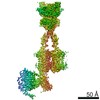
Journal: Nat Commun / Year: 2026 Title: Structural basis of active state coupling of metabotropic glutamate receptor 8 to beta-arrestins. Authors: Dagan C Marx / Kevin Huynh / Alberto J Gonzalez-Hernandez / Alexa Strauss / Carlos Rico / Pamela N Gallo / Sheida Sharghi Moshtaghin / Anisul Arefin / Johannes Broichhagen / David Eliezer / ...Authors: Dagan C Marx / Kevin Huynh / Alberto J Gonzalez-Hernandez / Alexa Strauss / Carlos Rico / Pamela N Gallo / Sheida Sharghi Moshtaghin / Anisul Arefin / Johannes Broichhagen / David Eliezer / George Khelashvili / Joshua Levitz / Abstract: Metabotropic glutamate receptors (mGluRs) are prototypical, dimeric family C G protein-coupled receptors (GPCR) that perform crucial modulatory roles throughout the nervous system. While mGluR ...Metabotropic glutamate receptors (mGluRs) are prototypical, dimeric family C G protein-coupled receptors (GPCR) that perform crucial modulatory roles throughout the nervous system. While mGluR activation and signaling through G proteins has been studied extensively, how these receptors interact with and are desensitized by β-arrestins (β-arrs) is not well understood. Here, we use an integrative biophysical and structural approach to probe the coupling of mGluR8 and β-arrs. Using negative stain electron microscopy (EM), we identify tail- and core-bound orientations and stoichiometries of mGluR8/β-arr complexes. Cryo-EM structures of mGluR8 alone or bound to either G proteins or β-arr1 reveal mGluR8 active states with transducer-specific differences. The mGluR8/β-arr structure shows a distinct complex orientation compared to other GPCR/β-arr structures which supports a steric mechanism of mGluR desensitization involving interactions with both subunits and the lipid bilayer. Coupling of mGluR8 to β-arr1 in an active-like conformation is verified by live-cell and single molecule FRET analysis. Finally, molecular dynamics simulations further define the positioning and dynamics of mGluR8-bound β-arr1 and the importance of critical mGluR8 residues for stabilizing β-arr1 complexes. Together, our data provide a framework for agonist-driven family C GPCR/β-arr coupling.
In the structure databanks used in Yorodumi, some data are registered as the other names, "COVID-19 virus" and "2019-nCoV". Here are the details of the virus and the list of structure data.
Jan 31, 2019. EMDB accession codes are about to change! (news from PDBe EMDB page)
EMDB accession codes are about to change! (news from PDBe EMDB page)
The allocation of 4 digits for EMDB accession codes will soon come to an end. Whilst these codes will remain in use, new EMDB accession codes will include an additional digit and will expand incrementally as the available range of codes is exhausted. The current 4-digit format prefixed with “EMD-” (i.e. EMD-XXXX) will advance to a 5-digit format (i.e. EMD-XXXXX), and so on. It is currently estimated that the 4-digit codes will be depleted around Spring 2019, at which point the 5-digit format will come into force.
The EM Navigator/Yorodumi systems omit the EMD- prefix.
Related info.:Q: What is EMD? / ID/Accession-code notation in Yorodumi/EM Navigator
Yorodumi is a browser for structure data from EMDB, PDB, SASBDB, etc.
This page is also the successor to EM Navigator detail page, and also detail information page/front-end page for Omokage search.
The word "yorodu" (or yorozu) is an old Japanese word meaning "ten thousand". "mi" (miru) is to see.
Related info.:EMDB / PDB / SASBDB / Comparison of 3 databanks / Yorodumi Search / Aug 31, 2016. New EM Navigator & Yorodumi / Yorodumi Papers / Jmol/JSmol / Function and homology information / Changes in new EM Navigator and Yorodumi
 Movie
Movie Controller

 Yorodumi
 Open data
 Basic information
 Map data
 Sample
 Keywords
 Homo sapiens (human)
 Authors
 United States, 3 items
  Citation
 Structure visualization
 Downloads & links
 EMDB map data format
 emd_78692.png
 https://data.pdbj.org/pub/emdb/structures/EMD-78692


 Z (Sec.)
 Y (Row.)
 X (Col.)


 Sample components
 Processing
 Electron microscopy
 FIELD EMISSION GUN