Movie
Movie Controller

[English] 日本語
 Yorodumi
- EMDB-77065: CryoEM structure of AdhE spirosome from Clostridium thermocellum ... -
+ Open data

- Basic information
| Entry |  | |||||||||
|---|---|---|---|---|---|---|---|---|---|---|
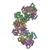
| Title | CryoEM structure of AdhE spirosome from Clostridium thermocellum uncovered by visual proteomics. | |||||||||
 Map data Map data | Sharpened cryoEM map of Ct spirosome obtained by helical refinement in cryoSPARC | |||||||||
 Sample Sample |
| |||||||||
 Keywords Keywords | Dehydrogenase / Spirosome / Filament / OXIDOREDUCTASE | |||||||||
| Function / homology |  Function and homology information Function and homology informationbutanol dehydrogenase (NAD+) activity / acetaldehyde dehydrogenase (acetylating) activity / methylglyoxal reductase (NADPH) (acetol producing) activity / alcohol metabolic process / carbon utilization / alcohol dehydrogenase (NADP+) activity / aldehyde dehydrogenase (NAD+) activity / metal ion binding / cytosol Similarity search - Function | |||||||||
| Biological species |  Acetivibrio thermocellus DSM 1313 (bacteria) Acetivibrio thermocellus DSM 1313 (bacteria) | |||||||||
| Method | helical reconstruction / cryo EM / Resolution: 4.07 Å | |||||||||
 Authors Authors | Agdanowski MP / Rodriguez JA / Moser T / Evans JE | |||||||||
| Funding support |  United States, 2 items United States, 2 items
| |||||||||
 Citation Citation | Journal: bioRxiv / Year: 2026 Title: Visual exoproteomics of during anaerobic biomass-degradation identifies functional spirosomes. Authors: Matthew P Agdanowski / Matthew J Kensil / Trevor H Moser / Ethan Humm / Yaneli I Guandique / Kayleigh Mason-Chalmers / Tracy Al-Set / Rachel R Ogorzalek Loo / James E Evans / Robert P ...Authors: Matthew P Agdanowski / Matthew J Kensil / Trevor H Moser / Ethan Humm / Yaneli I Guandique / Kayleigh Mason-Chalmers / Tracy Al-Set / Rachel R Ogorzalek Loo / James E Evans / Robert P Gunsalus / Joseph A Loo / Jose A Rodriguez Abstract: Visual proteomics enables the study of low-abundance proteins and identification of unknown complexes from heterogeneous samples by complementing high-resolution cryogenic electron microscopy (cryoEM) ...Visual proteomics enables the study of low-abundance proteins and identification of unknown complexes from heterogeneous samples by complementing high-resolution cryogenic electron microscopy (cryoEM) with external inputs on protein identity such as mass spectrometry. Using this approach, we interrogated the exoproteome of the anaerobic cellulose-degrading bacterium as it carried out biomass degradation. Mass spectrometry indicated a broad exoproteome composition, including cellulose degrading machinery CelA and CipA. A focus on large exoproteome assemblies revealed abundant protein filaments and pleomorphic vesicular structures. Analysis of the most abundant protein filaments yielded an ∼4 □ resolution native structure that, aided by mass spectrometry, modeling, and structural searching, was found to be the aldehyde-alcohol dehydrogenase (AdhE) spirosome. AdhE contained both NAD and Fe in their expected binding sites and biochemical and structural analyses of enriched spirosome preparations indicated they were functional. Altered NADH solution concentrations triggered conformational changes in the exoproteomic spirosomes, and the constituent AdhE remained capable of ethanol production. Although the basis for functional extracellular spirosome accumulation in live anaerobic cultures remains unclear, their abundance in crude exoproteomes suggests their presence could influence biomass fueled growth. | |||||||||
| History |
|
- Structure visualization
| Supplemental images |
|---|

- Downloads & links
-EMDB archive
| Map data | emd_77065.map.gz | 483.8 MB | EMDB map data format | |
|---|---|---|---|---|
| Header (meta data) | emd-77065-v30.xmlemd-77065.xml | 25.8 KB 25.8 KB | Display Display | EMDB header |
| FSC (resolution estimation) | emd_77065_fsc.xml | 14.8 KB | Display | FSC data file |
| Images |  emd_77065.png emd_77065.png | 43.3 KB | ||
| Filedesc metadata | emd-77065.cif.gz | 7.9 KB | ||
| Others | emd_77065_additional_1.map.gzemd_77065_half_map_1.map.gzemd_77065_half_map_2.map.gz | 253.2 MB 475 MB 475 MB | ||
| Archive directory |  http://ftp.pdbj.org/pub/emdb/structures/EMD-77065ftp://ftp.pdbj.org/pub/emdb/structures/EMD-77065 http://ftp.pdbj.org/pub/emdb/structures/EMD-77065ftp://ftp.pdbj.org/pub/emdb/structures/EMD-77065 | HTTPS FTP |
-Related structure data
| Related structure data |  13heMC M: atomic model generated by this map C: citing same article ( |
|---|---|
| Similar structure data |
-Links
| EMDB pages | EMDB (EBI/PDBe) / EMDataResource |
|---|---|
| Related items in Molecule of the Month |


-Map
| File | Download / File: emd_77065.map.gz / Format: CCP4 / Size: 512 MB / Type: IMAGE STORED AS FLOATING POINT NUMBER (4 BYTES) | ||||||||||||||||||||||||||||||||||||
|---|---|---|---|---|---|---|---|---|---|---|---|---|---|---|---|---|---|---|---|---|---|---|---|---|---|---|---|---|---|---|---|---|---|---|---|---|---|
| Annotation | Sharpened cryoEM map of Ct spirosome obtained by helical refinement in cryoSPARC | ||||||||||||||||||||||||||||||||||||
| Projections & slices | Image control
Images are generated by Spider. | ||||||||||||||||||||||||||||||||||||
| Voxel size | X=Y=Z: 0.68 Å | ||||||||||||||||||||||||||||||||||||
| Density |
| ||||||||||||||||||||||||||||||||||||
| Symmetry | Space group: 1 | ||||||||||||||||||||||||||||||||||||
| Details | EMDB XML:
|
 Z (Sec.)
 Y (Row.)
 X (Col.)


-Supplemental data
-Additional map: Unsharpened cryoEM map of Ct spirosome obtained by...
| File | emd_77065_additional_1.map | ||||||||||||
|---|---|---|---|---|---|---|---|---|---|---|---|---|---|
| Annotation | Unsharpened cryoEM map of Ct spirosome obtained by helical refinement in cryoSPARC | ||||||||||||
| Projections & Slices |
| ||||||||||||
| Density Histograms |


-Half map: Half Map A of helical Ct Spirosome
| File | emd_77065_half_map_1.map | ||||||||||||
|---|---|---|---|---|---|---|---|---|---|---|---|---|---|
| Annotation | Half Map A of helical Ct Spirosome | ||||||||||||
| Projections & Slices |
| ||||||||||||
| Density Histograms |


-Half map: Half Map B of helical Ct Spirosome
| File | emd_77065_half_map_2.map | ||||||||||||
|---|---|---|---|---|---|---|---|---|---|---|---|---|---|
| Annotation | Half Map B of helical Ct Spirosome | ||||||||||||
| Projections & Slices |
| ||||||||||||
| Density Histograms |


- Sample components
-Entire : AdhE spirosome
| Entire | Name: AdhE spirosome |
|---|---|
| Components |
|
-Supramolecule #1: AdhE spirosome
| Supramolecule | Name: AdhE spirosome / type: complex / ID: 1 / Parent: 0 / Macromolecule list: #1 |
|---|---|
| Source (natural) | Organism: Acetivibrio thermocellus DSM 1313 (bacteria) |
-Macromolecule #1: Aldehyde-alcohol dehydrogenase
| Macromolecule | Name: Aldehyde-alcohol dehydrogenase / type: protein_or_peptide / ID: 1 / Number of copies: 6 / Enantiomer: LEVO |
|---|---|
| Source (natural) | Organism: Acetivibrio thermocellus DSM 1313 (bacteria) |
| Molecular weight | Theoretical: 94.788023 KDa |
| Sequence | String: EVIDNVEKLE KALKRLREAQ SVYATYTQEQ VDKIFFEAAM AANKMRIPLA KMAVEETGMG VVEDKVIKNH YASEYIYNAY KNTKTCGVI EEDPAFGIKK IAEPLGVIAA VIPTTNPTST AIFKTLIALK TRNAIIISPH PRAKNSTIEA AKIVLEAAVK A GAPEGIIG ...String: EVIDNVEKLE KALKRLREAQ SVYATYTQEQ VDKIFFEAAM AANKMRIPLA KMAVEETGMG VVEDKVIKNH YASEYIYNAY KNTKTCGVI EEDPAFGIKK IAEPLGVIAA VIPTTNPTST AIFKTLIALK TRNAIIISPH PRAKNSTIEA AKIVLEAAVK A GAPEGIIG WIDVPSLELT NLVMREADVI LATGGPGLVK AAYSSGKPAI GVGAGNTPAI IDDSADIVLA VNSIIHSKTF DN GMICASE QSVIVLDGVY KEVKKEFEKR GCYFLNEDET EKVRKTIIIN GALNAKIVGQ KAHTIANLAG FEVPETTKIL IGE VTSVDI SEEFAHEKLC PVLAMYRAKD FDDALDKAER LVADGGFGHT SSLYIDTVTQ KEKLQKFSER MKTCRILVNT PSSQ GGIGD LYNFKLAPSL TLGCGSWGGN SVSDNVGVKH LLNIKTVAER RENMLWFRTP EKIYIKRGCL PVALDELKNV MGKKK AFIV TDNFLYNNGY TKPITDKLDE MGIVHKTFFD VSPDPSLASA KAGAAEMLAF QPDTIIAVGG GSAMDAAKIM WVMYEH PEV DFMDMAMRFM DIRKRVYTFP KMGQKAYFIA IPTSAGTGSE VTPFAVITDE KTGIKYPLAD YELLPDMAIV DADMMMN AP KGLTAASGID ALTHALEAYV SMLATDYTDS LALRAIKMIF EYLPRAYENG ASDPVAREKM ANAATIAGMA FANAFLGV C HSMAHKLGAF YHLPHGVANA LMINEVIRFN SSEAPTKMGT FPQYDHPRTL ERYAEIADYI GLKGKNNEEK VENLIKAID ELKEKVGIRK TIKDYDIDEK EFLDRLDEMV EQAFDDQCTG TNPRYPLMNE IRQMYLNAYY G UniProtKB: Aldehyde-alcohol dehydrogenase |
-Macromolecule #2: FE (III) ION
| Macromolecule | Name: FE (III) ION / type: ligand / ID: 2 / Number of copies: 6 / Formula: FE |
|---|---|
| Molecular weight | Theoretical: 55.845 Da |
-Macromolecule #3: NICOTINAMIDE-ADENINE-DINUCLEOTIDE
| Macromolecule | Name: NICOTINAMIDE-ADENINE-DINUCLEOTIDE / type: ligand / ID: 3 / Number of copies: 6 / Formula: NAD |
|---|---|
| Molecular weight | Theoretical: 663.425 Da |
| Chemical component information |  ChemComp-NAD: |
-Experimental details
-Structure determination
| Method | cryo EM |
|---|---|
 Processing Processing | helical reconstruction |
| Aggregation state | filament |
-Sample preparation
| Concentration | 3 mg/mL | ||||||||||||
|---|---|---|---|---|---|---|---|---|---|---|---|---|---|
| Buffer | pH: 8 Component:
Details: 20mM Tris pH 8.0, 150mM NaCl, 2mM CaCl2 | ||||||||||||
| Grid | Model: EMS Formvar Carbon / Material: COPPER / Mesh: 300 / Support film - Material: CARBON / Support film - topology: HOLEY / Support film - Film thickness: 20 / Pretreatment - Type: GLOW DISCHARGE / Pretreatment - Time: 20 sec. Details: CFlat 1.2/1.3 300 mesh holey carbon Cu support grids were negatively glow discharged for 20 seconds on the sample side. | ||||||||||||
| Vitrification | Cryogen name: ETHANE / Chamber humidity: 100 % / Chamber temperature: 285.15 K / Instrument: FEI VITROBOT MARK IV Details: Vitification was performed on CFlat 1.2/1.3 300 mesh holey carbon grids with copper support.. | ||||||||||||
| Details | Sample was obtained from partially-purified extracellular media and contained an array of unidentified species. |
- Electron microscopy
| Microscope | TFS KRIOS |
|---|---|
| Specialist optics | Energy filter - Name: GIF Bioquantum / Energy filter - Slit width: 20 eV |
| Image recording | Film or detector model: GATAN K3 (6k x 4k) / Digitization - Dimensions - Width: 11520 pixel / Digitization - Dimensions - Height: 88184 pixel / Number grids imaged: 1 / Number real images: 4863 / Average exposure time: 1.03 sec. / Average electron dose: 48.0 e/Å2 Details: Images were recorded as movies consisting of 50 frames over an exposure time of 1.03 seconds, with a total accumulated dose of 48 electrons per Angstrom |
| Electron beam | Acceleration voltage: 300 kV / Electron source:  FIELD EMISSION GUN FIELD EMISSION GUN |
| Electron optics | Illumination mode: FLOOD BEAM / Imaging mode: BRIGHT FIELD / Nominal defocus max: 0.5 µm / Nominal defocus min: 0.1 µm / Nominal magnification: 130000 |
| Experimental equipment |  Model: Titan Krios / Image courtesy: FEI Company |
+Image processing

-Atomic model buiding 1
| Initial model | PDB ID: Chain - Chain ID: A / Chain - Residue range: 11-869 / Chain - Source name: PDB / Chain - Initial model type: experimental model Details: published structure was fast relaxed and then used as an initial model for model building and refinement |
|---|---|
| Details | A existing structure's (PDB:8UHW) coordinates were relaxed using the Rosetta Fast Relax algorithm and refined against the EM density from cryoSPARC using Phenix and Coot. Histidine residues coordinate the iron ion; short distances flagged as clashes correspond to metal coordination geometry. |
| Refinement | Protocol: RIGID BODY FIT |
| Output model | PDB-13he: |