 ムービー
ムービー コントローラー


+ データを開く

- 基本情報
基本情報
| 登録情報 | データベース: EMDB / ID: EMD-7627 | |||||||||
|---|---|---|---|---|---|---|---|---|---|---|
| タイトル | Apoferritin 0.5 mM TCEP with spot-to-plunge time of 170ms | |||||||||
 マップデータ マップデータ | Apoferritin 0.5 mM TCEP with spot-to-plunge time of 170ms | |||||||||
 試料 試料 |
| |||||||||
| 生物種 |  Tabanus atratus (昆虫) Tabanus atratus (昆虫) | |||||||||
| 手法 | 電子線トモグラフィー法 / クライオ電子顕微鏡法 | |||||||||
 データ登録者 データ登録者 | Noble AJ / Wei H / Dandey VP / Zhang Z / Potter CS / Carragher B | |||||||||
 引用 引用 | ジャーナル: Nat Methods / 年: 2018 タイトル: Reducing effects of particle adsorption to the air-water interface in cryo-EM. 著者: Alex J Noble / Hui Wei / Venkata P Dandey / Zhening Zhang / Yong Zi Tan / Clinton S Potter / Bridget Carragher /  要旨: Most protein particles prepared in vitreous ice for single-particle cryo-electron microscopy (cryo-EM) are adsorbed to air-water or substrate-water interfaces, which can cause the particles to adopt ...Most protein particles prepared in vitreous ice for single-particle cryo-electron microscopy (cryo-EM) are adsorbed to air-water or substrate-water interfaces, which can cause the particles to adopt preferred orientations. By using a rapid plunge-freezing robot and nanowire grids, we were able to reduce some of the deleterious effects of the air-water interface by decreasing the dwell time of particles in thin liquid films. We demonstrated this by using single-particle cryo-EM and cryo-electron tomography (cryo-ET) to examine hemagglutinin, insulin receptor complex, and apoferritin. | |||||||||
| 履歴 |
|
- 構造の表示
構造の表示
| ムービー |
 ムービービューア ムービービューア |
|---|---|
| 添付画像 |

- ダウンロードとリンク
-EMDBアーカイブ
| マップデータ | emd_7627.map.gz | 1.3 GB | EMDBマップデータ形式 | |
|---|---|---|---|---|
| ヘッダ (付随情報) | emd-7627-v30.xmlemd-7627.xml | 8.1 KB 8.1 KB | 表示 表示 | EMDBヘッダ |
| 画像 | 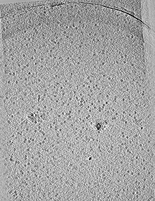 emd_7627.png emd_7627.png | 110.7 KB | ||
| アーカイブディレクトリ |  http://ftp.pdbj.org/pub/emdb/structures/EMD-7627ftp://ftp.pdbj.org/pub/emdb/structures/EMD-7627 http://ftp.pdbj.org/pub/emdb/structures/EMD-7627ftp://ftp.pdbj.org/pub/emdb/structures/EMD-7627 | HTTPS FTP |
-関連構造データ
| 関連構造データ |  7623C  7624C  7625C  7628C  7629C  7630C 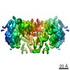 7788C 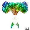 7791C 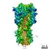 7792C C: 同じ文献を引用 ( |
|---|---|
| 電子顕微鏡画像生データ | EMPIAR-10171 (タイトル: CryoET of apoferritin with 0.5 mM TCEP single particle with spot-to-plunge time of 170ms Data size: 31.6 / Data #1: Tilt-series image stacks [tilt series] Data #2: Manual particle picking in Dynamo [particle picking] Data #3: Appion-Protomo tilt-series alignments [tilt series]) |
-リンク
| EMDBのページ | EMDB (EBI/PDBe) / EMDataResource |
|---|
-マップ
| ファイル | ダウンロード / ファイル: emd_7627.map.gz / 形式: CCP4 / 大きさ: 1.5 GB / タイプ: IMAGE STORED AS FLOATING POINT NUMBER (4 BYTES) | ||||||||||||||||||||||||||||||||||||||||||||||||||||||||||||||||||||
|---|---|---|---|---|---|---|---|---|---|---|---|---|---|---|---|---|---|---|---|---|---|---|---|---|---|---|---|---|---|---|---|---|---|---|---|---|---|---|---|---|---|---|---|---|---|---|---|---|---|---|---|---|---|---|---|---|---|---|---|---|---|---|---|---|---|---|---|---|---|
| 注釈 | Apoferritin 0.5 mM TCEP with spot-to-plunge time of 170ms | ||||||||||||||||||||||||||||||||||||||||||||||||||||||||||||||||||||
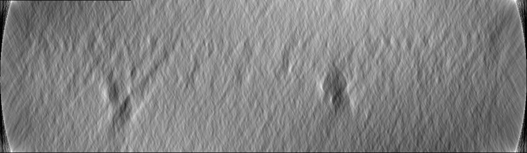
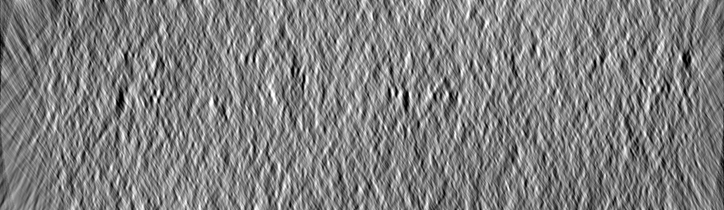
| 投影像・断面図 | 画像のコントロール
画像は Spider により作成 これらの図は立方格子座標系で作成されたものです | ||||||||||||||||||||||||||||||||||||||||||||||||||||||||||||||||||||
| ボクセルのサイズ | X=Y=Z: 2.34 Å | ||||||||||||||||||||||||||||||||||||||||||||||||||||||||||||||||||||
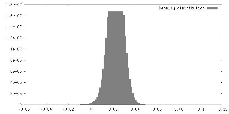
| 密度 |
| ||||||||||||||||||||||||||||||||||||||||||||||||||||||||||||||||||||
| 対称性 | 空間群: 1 | ||||||||||||||||||||||||||||||||||||||||||||||||||||||||||||||||||||
| 詳細 | EMDB XML:
CCP4マップ ヘッダ情報:
| ||||||||||||||||||||||||||||||||||||||||||||||||||||||||||||||||||||
 Z (Sec.)
 Y (Row.)
 X (Col.)


-添付データ
- 試料の構成要素
-全体 : Apoferritin
| 全体 | 名称: Apoferritin |
|---|---|
| 要素 |
|
-超分子 #1: Apoferritin
| 超分子 | 名称: Apoferritin / タイプ: complex / ID: 1 / 親要素: 0 / 詳細: with 0.5 mM TCEP |
|---|---|
| 由来(天然) | 生物種: Tabanus atratus (昆虫) |
| 組換発現 | 生物種: unidentified (未定義) |
-実験情報
-構造解析
| 手法 | クライオ電子顕微鏡法 |
|---|---|
 解析 解析 | 電子線トモグラフィー法 |
| 試料の集合状態 | particle |
-試料調製
| 濃度 | 15 mg/mL |
|---|---|
| 緩衝液 | pH: 0.0001 |
| グリッド | モデル: Homemade |
| 凍結 | 凍結剤: ETHANE / 装置: OTHER 詳細: The value given for _emd_vitrification.instrument is SPOTITON. This is not in a list of allowed values set(['LEICA EM CPC', 'GATAN CRYOPLUNGE 3', 'LEICA PLUNGER', 'FEI VITROBOT MARK II', ...詳細: The value given for _emd_vitrification.instrument is SPOTITON. This is not in a list of allowed values set(['LEICA EM CPC', 'GATAN CRYOPLUNGE 3', 'LEICA PLUNGER', 'FEI VITROBOT MARK II', 'HOMEMADE PLUNGER', 'REICHERT-JUNG PLUNGER', 'FEI VITROBOT MARK I', 'LEICA KF80', 'FEI VITROBOT MARK III', 'LEICA EM GP', 'OTHER', 'FEI VITROBOT MARK IV']) so OTHER is written into the XML file. |
| 切片作成 | その他: NO SECTIONING |
- 電子顕微鏡法
| 顕微鏡 | FEI TECNAI F20 |
|---|---|
| 撮影 | フィルム・検出器のモデル: DIRECT ELECTRON DE-20 (5k x 3k) 検出モード: INTEGRATING / 平均電子線量: 3.0 e/Å2 |
| 電子線 | 加速電圧: 200 kV / 電子線源:  FIELD EMISSION GUN FIELD EMISSION GUN |
| 電子光学系 | 照射モード: FLOOD BEAM / 撮影モード: BRIGHT FIELD / Cs: 2.0 mm |
| 実験機器 |  モデル: Tecnai F20 / 画像提供: FEI Company |
-画像解析
| 最終 再構成 | アルゴリズム: SIMULTANEOUS ITERATIVE (SIRT) / ソフトウェア - 名称: TOMO3D / ソフトウェア - 詳細: SIRT, 30 iterations / 使用した粒子像数: 30 |
|---|
