[English] 日本語
Yorodumi
- EMDB-76188: In situ subtomogram average of the transition zone doublet microt... -

+
Open data


ID or keywords:

Loading...

-
Basic information

Entry
Database: EMDB / ID: EMD-76188
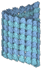
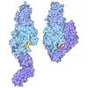
TitleIn situ subtomogram average of the transition zone doublet microtubule in human airway cilia (composite map)
Map dataA composite map of Transition Zone DMT in human airway cilia
Sample
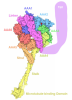
  • Organelle or cellular component: Transition zone DMT with MIPs repeat every 8 nm
    • Protein or peptide: Tubulin alpha-1A chain
    • Protein or peptide: Tubulin beta-4B chain
    • Protein or peptide: Cilia- and flagella-associated protein 20
    • Protein or peptide: Centrosomal protein of 41 kDa
    • Protein or peptide: Calcyphosin-like protein
    • Protein or peptide: Microtubule-associated protein 9
    • Protein or peptide: Enkurin domain-containing protein 1
  • Ligand: GUANOSINE-5'-TRIPHOSPHATE
  • Ligand: GUANOSINE-5'-DIPHOSPHATE
KeywordsCilia / Transition Zone / DMT / MIPs / cryoET / STRUCTURAL PROTEIN
Function / homology
Function and homology information


axonemal B tubule inner sheath / organelle / protein polyglutamylation / positive regulation of feeding behavior / regulation of cilium beat frequency involved in ciliary motility / pyramidal neuron differentiation / mitotic spindle midzone / motile cilium assembly / glial cell differentiation / Post-chaperonin tubulin folding pathway ...axonemal B tubule inner sheath / organelle / protein polyglutamylation / positive regulation of feeding behavior / regulation of cilium beat frequency involved in ciliary motility / pyramidal neuron differentiation / mitotic spindle midzone / motile cilium assembly / glial cell differentiation / Post-chaperonin tubulin folding pathway / cytoskeleton-dependent intracellular transport / Cargo trafficking to the periciliary membrane / dentate gyrus development / Carboxyterminal post-translational modifications of tubulin / Microtubule-dependent trafficking of connexons from Golgi to the plasma membrane / axonemal microtubule / organelle transport along microtubule / forebrain morphogenesis / astral microtubule / 9+0 non-motile cilium / cerebellar cortex morphogenesis / positive regulation of cell motility / Sealing of the nuclear envelope (NE) by ESCRT-III / Intraflagellar transport / neuron projection arborization / Formation of tubulin folding intermediates by CCT/TriC / regulation of mitotic centrosome separation / non-motile cilium assembly / Gap junction assembly / smoothened signaling pathway / homeostasis of number of cells within a tissue / motor behavior / Kinesins / motile cilium / Prefoldin mediated transfer of substrate to CCT/TriC / response to L-glutamate / Assembly and cell surface presentation of NMDA receptors / COPI-independent Golgi-to-ER retrograde traffic / centrosome cycle / natural killer cell mediated cytotoxicity / COPI-dependent Golgi-to-ER retrograde traffic / startle response / 'de novo' protein folding / intercellular bridge / regulation of mitotic cytokinesis / spindle midzone / ciliary base / flagellated sperm motility / regulation of synapse organization / locomotory exploration behavior / Recycling pathway of L1 / microtubule polymerization / mitotic cytokinesis / response to tumor necrosis factor / mitotic spindle assembly / MHC class I protein binding / response to mechanical stimulus / neuron apoptotic process / axoneme / alpha-tubulin binding / sperm flagellum / adult locomotory behavior / RHO GTPases activate IQGAPs / microtubule-based process / cilium assembly / Hedgehog 'off' state / COPI-mediated anterograde transport / cytoplasmic microtubule / condensed chromosome / Activation of AMPK downstream of NMDARs / Loss of Nlp from mitotic centrosomes / Loss of proteins required for interphase microtubule organization from the centrosome / Recruitment of mitotic centrosome proteins and complexes / centriole / MHC class II antigen presentation / cellular response to calcium ion / Recruitment of NuMA to mitotic centrosomes / visual learning / Anchoring of the basal body to the plasma membrane / HSP90 chaperone cycle for steroid hormone receptors (SHR) in the presence of ligand / Mitotic Prometaphase / EML4 and NUDC in mitotic spindle formation / AURKA Activation by TPX2 / Resolution of Sister Chromatid Cohesion / regulation of mitotic spindle organization / Translocation of SLC2A4 (GLUT4) to the plasma membrane / establishment of mitotic spindle orientation / neuromuscular junction / neuron migration / memory / cerebral cortex development / RHO GTPases Activate Formins / intracellular protein transport / microtubule cytoskeleton organization / synapse organization / recycling endosome / PKR-mediated signaling / mitotic spindle / structural constituent of cytoskeleton / cytoplasmic ribonucleoprotein granule
Similarity search - Function
Microtubule-associated protein 9 / : / : / Enkurin domain / : / Calmodulin-binding / Enkurin domain profile. / CFA20 domain / Cilia- and flagella-associated protein 20/CFAP20DC / CFA20 domain ...Microtubule-associated protein 9 / : / : / Enkurin domain / : / Calmodulin-binding / Enkurin domain profile. / CFA20 domain / Cilia- and flagella-associated protein 20/CFAP20DC / CFA20 domain / Rhodanese Homology Domain / Rhodanese-like domain / Rhodanese domain profile. / Rhodanese-like domain superfamily / Rhodanese-like domain / Alpha tubulin / Tubulin-beta mRNA autoregulation signal. / Beta tubulin, autoregulation binding site / Beta tubulin / Tubulin / Tubulin, C-terminal / Tubulin C-terminal domain / Tubulin, conserved site / Tubulin subunits alpha, beta, and gamma signature. / Tubulin/FtsZ family, C-terminal domain / Tubulin/FtsZ-like, C-terminal domain / Tubulin/FtsZ, C-terminal / Tubulin/FtsZ, 2-layer sandwich domain / Tubulin/FtsZ family, GTPase domain / Tubulin/FtsZ family, GTPase domain / Tubulin/FtsZ, GTPase domain / Tubulin/FtsZ, GTPase domain superfamily / EF-hand domain pair / EF-hand, calcium binding motif / EF-Hand 1, calcium-binding site / EF-hand calcium-binding domain. / EF-hand calcium-binding domain profile. / EF-hand domain / EF-hand domain pair
Similarity search - Domain/homology
Tubulin beta-4B chain / Microtubule-associated protein 9 / Tubulin alpha-1A chain / Calcyphosin-like protein / Centrosomal protein of 41 kDa / Enkurin domain-containing protein 1 / Cilia- and flagella-associated protein 20
Similarity search - Component
Biological speciesHomo sapiens (human)
Methodsubtomogram averaging / cryo EM / Resolution: 7.3 Å
AuthorsZhou H / Brown A
Funding support United States, 3 items
OrganizationGrant numberCountry
National Institutes of Health/National Institute of General Medical Sciences (NIH/NIGMS) United States
Richard and Susan Smith Family Foundation United States
The Giovanni Armenise-Harvard Foundation United States
CitationJournal: Science / Year: 2026
Title: In situ structure of the human ciliary transition zone links linker defects to primary ciliary dyskinesia
Authors: Zhou H / Brown A
History
DepositionMar 18, 2026-
Header (metadata) releaseSep 9, 2026-
Map releaseSep 9, 2026-
UpdateSep 9, 2026-
Current statusSep 9, 2026Processing site: RCSB / Status: Released

-
Structure visualization

Supplemental images

Downloads & links

-
Map

FileDownload / File: emd_76188.map.gz / Format: CCP4 / Size: 91.8 MB / Type: IMAGE STORED AS FLOATING POINT NUMBER (4 BYTES)
AnnotationA composite map of Transition Zone DMT in human airway cilia
Projections & slices

Image control

Size
Brightness
Contrast
Others
AxesZ (Sec.)Y (Row.)X (Col.)
1.92 Å/pix.
x 240 pix.
= 461.76 Å
1.92 Å/pix.
x 288 pix.
= 554.112 Å
1.92 Å/pix.
x 348 pix.
= 669.552 Å

Surface

Projections

Slices (1/3)

Slices (1/2)

Slices (2/3)

Images are generated by Spider.

generated in cubic-lattice coordinate

Voxel sizeX=Y=Z: 1.924 Å
Density
Contour LevelBy AUTHOR: 0.001
Minimum - Maximum0.0 - 0.021378111
Average (Standard dev.)0.00057370326 (±0.0018555425)
SymmetrySpace group: 1
Details

EMDB XML:

Map geometry
Axis orderXYZ
Origin10770118
Dimensions288348240
Spacing348288240
CellA: 669.552 Å / B: 554.112 Å / C: 461.76 Å
α=β=γ: 90.0 °

-
Supplemental data

-
Sample components

+
Entire : Transition zone DMT with MIPs repeat every 8 nm

EntireName: Transition zone DMT with MIPs repeat every 8 nm
Components
  • Organelle or cellular component: Transition zone DMT with MIPs repeat every 8 nm
    • Protein or peptide: Tubulin alpha-1A chain
    • Protein or peptide: Tubulin beta-4B chain
    • Protein or peptide: Cilia- and flagella-associated protein 20
    • Protein or peptide: Centrosomal protein of 41 kDa
    • Protein or peptide: Calcyphosin-like protein
    • Protein or peptide: Microtubule-associated protein 9
    • Protein or peptide: Enkurin domain-containing protein 1
  • Ligand: GUANOSINE-5'-TRIPHOSPHATE
  • Ligand: GUANOSINE-5'-DIPHOSPHATE

+
Supramolecule #1: Transition zone DMT with MIPs repeat every 8 nm

SupramoleculeName: Transition zone DMT with MIPs repeat every 8 nm / type: organelle_or_cellular_component / ID: 1 / Parent: 0 / Macromolecule list: #1-#7
Source (natural)Organism: Homo sapiens (human)

+
Macromolecule #1: Tubulin alpha-1A chain

MacromoleculeName: Tubulin alpha-1A chain / type: protein_or_peptide / ID: 1 / Number of copies: 77 / Enantiomer: LEVO
Source (natural)Organism: Homo sapiens (human)
Molecular weightTheoretical: 50.188441 KDa
SequenceString: MRECISIHVG QAGVQIGNAC WELYCLEHGI QPDGQMPSDK TIGGGDDSFN TFFSETGAGK HVPRAVFVDL EPTVIDEVRT GTYRQLFHP EQLITGKEDA ANNYARGHYT IGKEIIDLVL DRIRKLADQC TGLQGFLVFH SFGGGTGSGF TSLLMERLSV D YGKKSKLE ...String:
MRECISIHVG QAGVQIGNAC WELYCLEHGI QPDGQMPSDK TIGGGDDSFN TFFSETGAGK HVPRAVFVDL EPTVIDEVRT GTYRQLFHP EQLITGKEDA ANNYARGHYT IGKEIIDLVL DRIRKLADQC TGLQGFLVFH SFGGGTGSGF TSLLMERLSV D YGKKSKLE FSIYPAPQVS TAVVEPYNSI LTTHTTLEHS DCAFMVDNEA IYDICRRNLD IERPTYTNLN RLIGQIVSSI TA SLRFDGA LNVDLTEFQT NLVPYPRIHF PLATYAPVIS AEKAYHEQLS VAEITNACFE PANQMVKCDP RHGKYMACCL LYR GDVVPK DVNAAIATIK TKRTIQFVDW CPTGFKVGIN YQPPTVVPGG DLAKVQRAVC MLSNTTAIAE AWARLDHKFD LMYA KRAFV HWYVGEGMEE GEFSEAREDM AALEKDYEEV GVDSVEGEGE EEGEEY

UniProtKB: Tubulin alpha-1A chain

+
Macromolecule #2: Tubulin beta-4B chain

MacromoleculeName: Tubulin beta-4B chain / type: protein_or_peptide / ID: 2 / Number of copies: 77 / Enantiomer: LEVO
Source (natural)Organism: Homo sapiens (human)
Molecular weightTheoretical: 49.877824 KDa
SequenceString: MREIVHLQAG QCGNQIGAKF WEVISDEHGI DPTGTYHGDS DLQLERINVY YNEATGGKYV PRAVLVDLEP GTMDSVRSGP FGQIFRPDN FVFGQSGAGN NWAKGHYTEG AELVDSVLDV VRKEAESCDC LQGFQLTHSL GGGTGSGMGT LLISKIREEY P DRIMNTFS ...String:
MREIVHLQAG QCGNQIGAKF WEVISDEHGI DPTGTYHGDS DLQLERINVY YNEATGGKYV PRAVLVDLEP GTMDSVRSGP FGQIFRPDN FVFGQSGAGN NWAKGHYTEG AELVDSVLDV VRKEAESCDC LQGFQLTHSL GGGTGSGMGT LLISKIREEY P DRIMNTFS VVPSPKVSDT VVEPYNATLS VHQLVENTDE TYCIDNEALY DICFRTLKLT TPTYGDLNHL VSATMSGVTT CL RFPGQLN ADLRKLAVNM VPFPRLHFFM PGFAPLTSRG SQQYRALTVP ELTQQMFDAK NMMAACDPRH GRYLTVAAVF RGR MSMKEV DEQMLNVQNK NSSYFVEWIP NNVKTAVCDI PPRGLKMSAT FIGNSTAIQE LFKRISEQFT AMFRRKAFLH WYTG EGMDE MEFTEAESNM NDLVSEYQQY QDATAEEEGE FEEEAEEEVA

UniProtKB: Tubulin beta-4B chain

+
Macromolecule #3: Cilia- and flagella-associated protein 20

MacromoleculeName: Cilia- and flagella-associated protein 20 / type: protein_or_peptide / ID: 3 / Number of copies: 4 / Enantiomer: LEVO
Source (natural)Organism: Homo sapiens (human)
Molecular weightTheoretical: 22.807469 KDa
SequenceString: MFKNTFQSGF LSILYSIGSK PLQIWDKKVR NGHIKRITDN DIQSLVLEIE GTNVSTTYIT CPADPKKTLG IKLPFLVMII KNLKKYFTF EVQVLDDKNV RRRFRASNYQ STTRVKPFIC TMPMRLDDGW NQIQFNLLDF TRRAYGTNYI ETLRVQIHAN C RIRRVYFS ...String:
MFKNTFQSGF LSILYSIGSK PLQIWDKKVR NGHIKRITDN DIQSLVLEIE GTNVSTTYIT CPADPKKTLG IKLPFLVMII KNLKKYFTF EVQVLDDKNV RRRFRASNYQ STTRVKPFIC TMPMRLDDGW NQIQFNLLDF TRRAYGTNYI ETLRVQIHAN C RIRRVYFS DRLYSEDELP AEFKLYLPVQ NKAKQ

UniProtKB: Cilia- and flagella-associated protein 20

+
Macromolecule #4: Centrosomal protein of 41 kDa

MacromoleculeName: Centrosomal protein of 41 kDa / type: protein_or_peptide / ID: 4 / Number of copies: 3 / Enantiomer: LEVO
Source (natural)Organism: Homo sapiens (human)
Molecular weightTheoretical: 41.426406 KDa
SequenceString: MSLRRHIGNP EYLMKRIPQN PRYQHIKSRL DTGNSMTKYT EKLEEIKKNY RYKKDELFKR LKVTTFAQLI IQVASLSDQT LEVTAEEIQ RLEDNDSAAS DPDAETTART NGKGNPGEQS PSPEQFINNA GAGDSSRSTL QSVISGVGEL DLDKGPVKKA E PHTKDKPY ...String:
MSLRRHIGNP EYLMKRIPQN PRYQHIKSRL DTGNSMTKYT EKLEEIKKNY RYKKDELFKR LKVTTFAQLI IQVASLSDQT LEVTAEEIQ RLEDNDSAAS DPDAETTART NGKGNPGEQS PSPEQFINNA GAGDSSRSTL QSVISGVGEL DLDKGPVKKA E PHTKDKPY PDCPFLLLDV RDRDSYQQCH IVGAYSYPIA TLSRTMNPYS NDILEYKNAH GKIIILYDDD ERLASQAATT MC ERGFENL FMLSGGLKVL AQKFPEGLIT GSLPASCQQA LPPGSARKRS SPKGPPLPAE NKWRFTPEDL KKIEYYLEEE QGP ADHPSR LNQANSSGRE SKVPGARSAQ NLPGGGPASH SNPRSLSSGH LQGKPWK

UniProtKB: Centrosomal protein of 41 kDa

+
Macromolecule #5: Calcyphosin-like protein

MacromoleculeName: Calcyphosin-like protein / type: protein_or_peptide / ID: 5 / Number of copies: 40 / Enantiomer: LEVO
Source (natural)Organism: Homo sapiens (human)
Molecular weightTheoretical: 24.264686 KDa
SequenceString: MAGTARHDRE MAIQAKKKLT TATDPIERLR LQCLARGSAG IKGLGRVFRI MDDDNNRTLD FKEFMKGLND YAVVMEKEEV EELFRRFDK DGNGTIDFNE FLLTLRPPMS RARKEVIMQA FRKLDKTGDG VITIEDLREV YNAKHHPKYQ NGEWSEEQVF R KFLDNFDS ...String:
MAGTARHDRE MAIQAKKKLT TATDPIERLR LQCLARGSAG IKGLGRVFRI MDDDNNRTLD FKEFMKGLND YAVVMEKEEV EELFRRFDK DGNGTIDFNE FLLTLRPPMS RARKEVIMQA FRKLDKTGDG VITIEDLREV YNAKHHPKYQ NGEWSEEQVF R KFLDNFDS PYDKDGLVTP EEFMNYYAGV SASIDTDVYF IIMMRTAWKL

UniProtKB: Calcyphosin-like protein

+
Macromolecule #6: Microtubule-associated protein 9

MacromoleculeName: Microtubule-associated protein 9 / type: protein_or_peptide / ID: 6 / Number of copies: 2 / Enantiomer: LEVO
Source (natural)Organism: Homo sapiens (human)
Molecular weightTheoretical: 74.374258 KDa
SequenceString: MSDEVFSTTL AYTKSPKVTK RTTFQDELIR AITARSARQR SSEYSDDFDS DEIVSLGDFS DTSADENSVN KKMNDFHISD DEEKNPSKL LFLKTNKSNG NITKDEPVCA IKNEEEMAPD GCEDIVVKSF SESQNKDEEF EKDKIKMKPK PRILSIKSTS S AENNSLDT ...String:
MSDEVFSTTL AYTKSPKVTK RTTFQDELIR AITARSARQR SSEYSDDFDS DEIVSLGDFS DTSADENSVN KKMNDFHISD DEEKNPSKL LFLKTNKSNG NITKDEPVCA IKNEEEMAPD GCEDIVVKSF SESQNKDEEF EKDKIKMKPK PRILSIKSTS S AENNSLDT DDHFKPSPRP RSMLKKKSHM EEKDGLEDKE TALSEELELH SAPSSLPTPN GIQLEAEKKA FSENLDPEDS CL TSLASSS LKQILGDSFS PGSEGNASGK DPNEEITENH NSLKSDENKE NSFSADHVTT AVEKSKESQV TADDLEEEKA KAE LIMDDD RTVDPLLSKS QSILISTSAT ASSKKTIEDR NIKNKKSTNN RASSASARLM TSEFLKKSSS KRRTPSTTTS SHYL GTLKV LDQKPSQKQS IEPDRADNIR AAVYQEWLEK KNVYLHEMHR IKRIESENLR IQNEQKKAAK REEALASFEA WKAMK EKEA KKIAAKKRLE EKNKKKTEEE NAARKGEALQ AFEKWKEKKM EYLKEKNRKE REYERAKKQK EEETVAEKKK DNLTAV EKW NEKKEAFFKQ KEKEKINEKR KEELKRAEKK DKDKQAINEY EKWLENKEKQ ERIERKQKKR HSFLESEALP PWSPPSR TV FAKVF

UniProtKB: Microtubule-associated protein 9

+
Macromolecule #7: Enkurin domain-containing protein 1

MacromoleculeName: Enkurin domain-containing protein 1 / type: protein_or_peptide / ID: 7 / Number of copies: 3 / Enantiomer: LEVO
Source (natural)Organism: Homo sapiens (human)
Molecular weightTheoretical: 38.828219 KDa
SequenceString: MCEGPSRISG PIPPDPTLCP DNYRRPTSAQ GRLEGNALKL DLLTSDRALD TTAPRGPCIG PGAGEILERG QRGVGDVLLQ LEGISLGPG ASLKRKDPKD HEKENLRRIR EIQKRFREQE RSREQGQPRP LKALWRSPKY DKVESRVKAQ LQEPGPASGT E SAHFLRAH ...String:
MCEGPSRISG PIPPDPTLCP DNYRRPTSAQ GRLEGNALKL DLLTSDRALD TTAPRGPCIG PGAGEILERG QRGVGDVLLQ LEGISLGPG ASLKRKDPKD HEKENLRRIR EIQKRFREQE RSREQGQPRP LKALWRSPKY DKVESRVKAQ LQEPGPASGT E SAHFLRAH SRCGPGLPPP HVSSPQPTPP GPEAKEPGLG VDFIRHNARA AKRAPRRHSC SLQVLAQVLE QQRQAQEHYN AT QKGHVPH YLLERRDLWR REAEARKQSQ PDPAMPPGHT RMPENQRLET LTKLLQSQSQ LLRELVLLPA GADSLRAQSH RAE LDRKLV QVEEAIKIFS RPKVFVKMDD

UniProtKB: Enkurin domain-containing protein 1

+
Macromolecule #8: GUANOSINE-5'-TRIPHOSPHATE
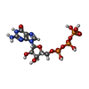
MacromoleculeName: GUANOSINE-5'-TRIPHOSPHATE / type: ligand / ID: 8 / Number of copies: 77 / Formula: GTP
Molecular weightTheoretical: 523.18 Da
Chemical component information

ChemComp-GTP:
GUANOSINE-5'-TRIPHOSPHATE / GTP, energy-carrying molecule*YM

+
Macromolecule #9: GUANOSINE-5'-DIPHOSPHATE
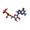
MacromoleculeName: GUANOSINE-5'-DIPHOSPHATE / type: ligand / ID: 9 / Number of copies: 77 / Formula: GDP
Molecular weightTheoretical: 443.201 Da
Chemical component information

ChemComp-GDP:
GUANOSINE-5'-DIPHOSPHATE / GDP, energy-carrying molecule*YM

-
Experimental details

-
Structure determination

Methodcryo EM
Processingsubtomogram averaging
Aggregation statecell

-
Sample preparation

BufferpH: 7.2
VitrificationCryogen name: ETHANE

-
Electron microscopy

MicroscopeTFS KRIOS
Image recordingFilm or detector model: FEI FALCON IV (4k x 4k) / Average electron dose: 4.68 e/Å2
Electron beamAcceleration voltage: 300 kV / Electron source: FIELD EMISSION GUN
Electron opticsIllumination mode: FLOOD BEAM / Imaging mode: BRIGHT FIELD / Nominal defocus max: 4.0 µm / Nominal defocus min: 2.0 µm
Experimental equipment
Model: Titan Krios / Image courtesy: FEI Company

-
Image processing

Final reconstructionResolution.type: BY AUTHOR / Resolution: 7.3 Å / Resolution method: FSC 0.143 CUT-OFF / Software - Name: RELION (ver. 5) / Number subtomograms used: 33196
ExtractionNumber tomograms: 209 / Number images used: 66385 / Software - Name: Warp (ver. 2.0)
CTF correctionType: PHASE FLIPPING AND AMPLITUDE CORRECTION
Final angle assignmentType: MAXIMUM LIKELIHOOD

-
Atomic model buiding 1

DetailsAtomic models were built in Coot. CFAP20 and tubulin models were obtained from PDB: 7UNG and fit as rigid-bodies into the subtomogram average. All other proteins were modeled using AlphaFold3 or Boltz-2 and fit into the density either manually or using DomainSeeker. Initial atomic models were refined into the subtomogram averaging map using rigid-body refinement in phenix.real_space_refine. Geometry outliers were corrected manually in Coot. Final refinement was performed in Phenix using group atomic displacement parameter (ADP) refinement against the subtomogram averages. Side-chain density is not resolved at this resolution, and the model is intended to describe subunit and domain placement rather than atomic-accuracy contacts. The side-chain conformations in the deposited file were inherited from the reference models used to build it rather than determined from our density and should not be interpreted as experimentally determined. The reported contacts trace to a small number of systematic sources rather than many independent modeling errors.
Output model

PDB-11yj:
In situ subtomogram average of the transition zone doublet microtubule in human airway cilia (composite map)

+
About Yorodumi

-
News

-
Feb 9, 2022. New format data for meta-information of EMDB entries

New format data for meta-information of EMDB entries

  • Version 3 of the EMDB header file is now the official format.
  • The previous official version 1.9 will be removed from the archive.

Related info.:EMDB header

External links:wwPDB to switch to version 3 of the EMDB data model

-
Aug 12, 2020. Covid-19 info

Covid-19 info

URL: https://pdbj.org/emnavi/covid19.php

New page: Covid-19 featured information page in EM Navigator.

Related info.:Covid-19 info / Mar 5, 2020. Novel coronavirus structure data

+
Mar 5, 2020. Novel coronavirus structure data

Novel coronavirus structure data

Related info.:Yorodumi Speices / Aug 12, 2020. Covid-19 info

External links:COVID-19 featured content - PDBj / Molecule of the Month (242):Coronavirus Proteases

+
Jan 31, 2019. EMDB accession codes are about to change! (news from PDBe EMDB page)

EMDB accession codes are about to change! (news from PDBe EMDB page)

  • The allocation of 4 digits for EMDB accession codes will soon come to an end. Whilst these codes will remain in use, new EMDB accession codes will include an additional digit and will expand incrementally as the available range of codes is exhausted. The current 4-digit format prefixed with “EMD-” (i.e. EMD-XXXX) will advance to a 5-digit format (i.e. EMD-XXXXX), and so on. It is currently estimated that the 4-digit codes will be depleted around Spring 2019, at which point the 5-digit format will come into force.
  • The EM Navigator/Yorodumi systems omit the EMD- prefix.

Related info.:Q: What is EMD? / ID/Accession-code notation in Yorodumi/EM Navigator

External links:EMDB Accession Codes are Changing Soon! / Contact to PDBj

+
Jul 12, 2017. Major update of PDB

Major update of PDB

  • wwPDB released updated PDB data conforming to the new PDBx/mmCIF dictionary.
  • This is a major update changing the version number from 4 to 5, and with Remediation, in which all the entries are updated.
  • In this update, many items about electron microscopy experimental information are reorganized (e.g. em_software).
  • Now, EM Navigator and Yorodumi are based on the updated data.

External links:wwPDB Remediation / Enriched Model Files Conforming to OneDep Data Standards Now Available in the PDB FTP Archive

-
Yorodumi

Thousand views of thousand structures

  • Yorodumi is a browser for structure data from EMDB, PDB, SASBDB, etc.
  • This page is also the successor to EM Navigator detail page, and also detail information page/front-end page for Omokage search.
  • The word "yorodu" (or yorozu) is an old Japanese word meaning "ten thousand". "mi" (miru) is to see.

Related info.:EMDB / PDB / SASBDB / Comparison of 3 databanks / Yorodumi Search / Aug 31, 2016. New EM Navigator & Yorodumi / Yorodumi Papers / Jmol/JSmol / Function and homology information / Changes in new EM Navigator and Yorodumi

Read more