 Movie
Movie Controller

[English] 日本語
 Yorodumi
- EMDB-76173: In situ subtomogram average of the transition zone linker in huma... -
+ Open data

- Basic information
| Entry |  | ||||||||||||
|---|---|---|---|---|---|---|---|---|---|---|---|---|---|
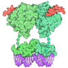
| Title | In situ subtomogram average of the transition zone linker in human airway cilia (composite map) | ||||||||||||
 Map data Map data | A composite map of TZ linker from human airway cilia | ||||||||||||
 Sample Sample |
| ||||||||||||
 Keywords Keywords | Cilia / Transition Zone / TZ linker / Subtomogram Averaging / STRUCTURAL PROTEIN | ||||||||||||
| Function / homology |  Function and homology information Function and homology informationnitric-oxide synthase inhibitor activity / negative regulation of DNA strand resection involved in replication fork processing / eye photoreceptor cell development / negative regulation of phosphorylation / Activation of BIM and translocation to mitochondria / intraciliary retrograde transport / motile cilium assembly / synaptic assembly at neuromuscular junction / F-box domain binding / Intraflagellar transport ...nitric-oxide synthase inhibitor activity / negative regulation of DNA strand resection involved in replication fork processing / eye photoreceptor cell development / negative regulation of phosphorylation / Activation of BIM and translocation to mitochondria / intraciliary retrograde transport / motile cilium assembly / synaptic assembly at neuromuscular junction / F-box domain binding / Intraflagellar transport / positive regulation of intracellular transport / PcG protein complex / regulation of xenophagy / maintenance of protein location in nucleus / Loss of Function of FBXW7 in Cancer and NOTCH1 Signaling / Cul7-RING ubiquitin ligase complex / positive regulation of mitotic cell cycle spindle assembly checkpoint / regulation of cell cycle process / neural crest cell differentiation / COPI-independent Golgi-to-ER retrograde traffic / ubiquitin ligase activator activity / regulation of BMP signaling pathway / regulation of mitophagy / cytoplasmic dynein complex / regulation of centrosome duplication / spermatid development / regulation of TOR signaling / SCF ubiquitin ligase complex / male germ cell nucleus / regulation of DNA damage checkpoint / Macroautophagy / SCF-dependent proteasomal ubiquitin-dependent protein catabolic process / Prolactin receptor signaling / dynein intermediate chain binding / ubiquitin ligase complex scaffold activity / tertiary granule membrane / ciliary tip / limb development / ficolin-1-rich granule membrane / cullin family protein binding / centrosome duplication / cilium assembly / COPI-mediated anterograde transport / ubiquitin-like ligase-substrate adaptor activity / Nuclear events stimulated by ALK signaling in cancer / axon cytoplasm / protein K48-linked ubiquitination / Amplification of signal from unattached kinetochores via a MAD2 inhibitory signal / Loss of Nlp from mitotic centrosomes / Loss of proteins required for interphase microtubule organization from the centrosome / Recruitment of mitotic centrosome proteins and complexes / MHC class II antigen presentation / Recruitment of NuMA to mitotic centrosomes / Anchoring of the basal body to the plasma membrane / substantia nigra development / HSP90 chaperone cycle for steroid hormone receptors (SHR) in the presence of ligand / Mitotic Prometaphase / EML4 and NUDC in mitotic spindle formation / guanyl-nucleotide exchange factor activity / AURKA Activation by TPX2 / Resolution of Sister Chromatid Cohesion / molecular function activator activity / enzyme inhibitor activity / positive regulation of insulin secretion involved in cellular response to glucose stimulus / regulation of mitotic cell cycle / Regulation of BACH1 activity / MAP3K8 (TPL2)-dependent MAPK1/3 activation / G1/S transition of mitotic cell cycle / SCF-beta-TrCP mediated degradation of Emi1 / NIK-->noncanonical NF-kB signaling / Vpu mediated degradation of CD4 / RHO GTPases Activate Formins / Dectin-1 mediated noncanonical NF-kB signaling / Degradation of CRY and PER proteins / Iron uptake and transport / Activation of NF-kappaB in B cells / Degradation of GLI1 by the proteasome / GSK3B and BTRC:CUL1-mediated-degradation of NFE2L2 / Negative regulation of NOTCH4 signaling / mitotic spindle / FBXL7 down-regulates AURKA during mitotic entry and in early mitosis / beta-catenin binding / Degradation of GLI2 by the proteasome / GLI3 is processed to GLI3R by the proteasome / GSK3B-mediated proteasomal degradation of PD-L1(CD274) / NOTCH1 Intracellular Domain Regulates Transcription / regulation of circadian rhythm / Degradation of beta-catenin by the destruction complex / Constitutive Signaling by NOTCH1 PEST Domain Mutants / Constitutive Signaling by NOTCH1 HD+PEST Domain Mutants / CLEC7A (Dectin-1) signaling / SCF(Skp2)-mediated degradation of p27/p21 / FCERI mediated NF-kB activation / kinetochore / protein polyubiquitination / HCMV Early Events / Ubiquitin-Mediated Degradation of Phosphorylated Cdc25A / Interleukin-1 signaling / Orc1 removal from chromatin / Aggrephagy Similarity search - Function | ||||||||||||
| Biological species |  Homo sapiens (human) Homo sapiens (human) | ||||||||||||
| Method | subtomogram averaging / cryo EM / Resolution: 9.6 Å | ||||||||||||
 Authors Authors | Zhou H / Brown A | ||||||||||||
| Funding support |  United States, 3 items United States, 3 items
| ||||||||||||
 Citation Citation | Journal: Acta Crystallogr D Struct Biol / Year: 2019 Title: Macromolecular structure determination using X-rays, neutrons and electrons: recent developments in Phenix. Authors: Dorothee Liebschner / Pavel V Afonine / Matthew L Baker / Gábor Bunkóczi / Vincent B Chen / Tristan I Croll / Bradley Hintze / Li Wei Hung / Swati Jain / Airlie J McCoy / Nigel W Moriarty ...Authors: Dorothee Liebschner / Pavel V Afonine / Matthew L Baker / Gábor Bunkóczi / Vincent B Chen / Tristan I Croll / Bradley Hintze / Li Wei Hung / Swati Jain / Airlie J McCoy / Nigel W Moriarty / Robert D Oeffner / Billy K Poon / Michael G Prisant / Randy J Read / Jane S Richardson / David C Richardson / Massimo D Sammito / Oleg V Sobolev / Duncan H Stockwell / Thomas C Terwilliger / Alexandre G Urzhumtsev / Lizbeth L Videau / Christopher J Williams / Paul D Adams /   Abstract: Diffraction (X-ray, neutron and electron) and electron cryo-microscopy are powerful methods to determine three-dimensional macromolecular structures, which are required to understand biological ...Diffraction (X-ray, neutron and electron) and electron cryo-microscopy are powerful methods to determine three-dimensional macromolecular structures, which are required to understand biological processes and to develop new therapeutics against diseases. The overall structure-solution workflow is similar for these techniques, but nuances exist because the properties of the reduced experimental data are different. Software tools for structure determination should therefore be tailored for each method. Phenix is a comprehensive software package for macromolecular structure determination that handles data from any of these techniques. Tasks performed with Phenix include data-quality assessment, map improvement, model building, the validation/rebuilding/refinement cycle and deposition. Each tool caters to the type of experimental data. The design of Phenix emphasizes the automation of procedures, where possible, to minimize repetitive and time-consuming manual tasks, while default parameters are chosen to encourage best practice. A graphical user interface provides access to many command-line features of Phenix and streamlines the transition between programs, project tracking and re-running of previous tasks. | ||||||||||||
| History |
|
- Structure visualization
| Supplemental images |
|---|

- Downloads & links
-EMDB archive
| Map data | emd_76173.map.gz | 6.1 MB | EMDB map data format | |
|---|---|---|---|---|
| Header (meta data) | emd-76173-v30.xmlemd-76173.xml | 20 KB 20 KB | Display Display | EMDB header |
| Images |  emd_76173.png emd_76173.png | 165.8 KB | ||
| Filedesc metadata | emd-76173.cif.gz | 7.3 KB | ||
| Archive directory |  https://data.pdbj.org/pub/emdb/structures/EMD-76173ftp://data.pdbj.org/pub/emdb/structures/EMD-76173 https://data.pdbj.org/pub/emdb/structures/EMD-76173ftp://data.pdbj.org/pub/emdb/structures/EMD-76173 | HTTPS FTP |
-Related structure data
| Related structure data |  11xuMC  11yjC C: citing same article ( M: atomic model generated by this map |
|---|---|
| Similar structure data |


-Links
| EMDB pages | EMDB (EBI/PDBe) / EMDataResource |
|---|---|
| Related items in Molecule of the Month |













-Map
| File | Download / File: emd_76173.map.gz / Format: CCP4 / Size: 43.9 MB / Type: IMAGE STORED AS FLOATING POINT NUMBER (4 BYTES) | ||||||||||||||||||||||||||||||||||||
|---|---|---|---|---|---|---|---|---|---|---|---|---|---|---|---|---|---|---|---|---|---|---|---|---|---|---|---|---|---|---|---|---|---|---|---|---|---|
| Annotation | A composite map of TZ linker from human airway cilia | ||||||||||||||||||||||||||||||||||||
| Projections & slices | Image control
Images are generated by Spider. generated in cubic-lattice coordinate | ||||||||||||||||||||||||||||||||||||
| Voxel size | X=Y=Z: 1.924 Å | ||||||||||||||||||||||||||||||||||||
| Density |
| ||||||||||||||||||||||||||||||||||||
| Symmetry | Space group: 1 | ||||||||||||||||||||||||||||||||||||
| Details | EMDB XML:
|
 Z (Sec.)
 Y (Row.)
 X (Col.)


-Supplemental data
- Sample components
-Entire : Human airway ciliary transition zone (TZ) linker
| Entire | Name: Human airway ciliary transition zone (TZ) linker |
|---|---|
| Components |
|
-Supramolecule #1: Human airway ciliary transition zone (TZ) linker
| Supramolecule | Name: Human airway ciliary transition zone (TZ) linker / type: organelle_or_cellular_component / ID: 1 / Parent: 0 / Macromolecule list: #2-#4, #1 |
|---|---|
| Source (natural) | Organism: Homo sapiens (human) |
-Macromolecule #1: Epithelial cell-transforming sequence 2 oncogene-like
| Macromolecule | Name: Epithelial cell-transforming sequence 2 oncogene-like / type: protein_or_peptide / ID: 1 / Number of copies: 2 / Enantiomer: LEVO |
|---|---|
| Source (natural) | Organism: Homo sapiens (human) |
| Molecular weight | Theoretical: 105.0175 KDa |
| Sequence | String: MESFHTRFSA WTPFSNKSLN RQLFQERVAL ISHWFDLWTN KQRQEFLFAI FLRCTKSQLR FVQDWFSERM QVAKVDFSTV LPRFISLYI FSFLSPKDLC AAAQVSWPWK FLTEQDCLWM PKCVKFGWFL PYTPTDNEYG AWKRHYIACV SHLDWLTPRE A AATYGTLN ...String: MESFHTRFSA WTPFSNKSLN RQLFQERVAL ISHWFDLWTN KQRQEFLFAI FLRCTKSQLR FVQDWFSERM QVAKVDFSTV LPRFISLYI FSFLSPKDLC AAAQVSWPWK FLTEQDCLWM PKCVKFGWFL PYTPTDNEYG AWKRHYIACV SHLDWLTPRE A AATYGTLN EPKTEDEELL ERQREKCLRK RIWEKIALRK KELFKVRPPW VSGTCCSSVL KPRCQPRLSQ TVRERVGLHE AL EKQLVLT SLETLPKRSN ISGSHSYPLL SKKNWHGVHK NDDRSSYALR PHFMLISSRI PAYEMVMESV KAGVVSVVYE HSV TLESLL YLIEKALDGQ KAQSIGIFSD GDSREINLLQ GYKIGVKNLL RPEVRDFWEK LGSYVATEEE GGHVDFFVPL GASE AGIEV LSQLSQLTGT FFTAPTGIAT GSYQHILSDW LGSQWGKAPS SIYFCESKLQ TWSSFTDFLE ETLKTVRKQL YPFFK ELQK SISGRMIGQF MFDTMGMTNI LNNQDTAQAL ADGLMELSKE DSERNVVEDN SWDTKSRLSK NDLNFEALIN LERILQ KDS AEKRARVVRE LLQSERKYVQ ILEIVRDVYV APLKAALSSN RAILSAANIQ IIFCDILQIL SLNRQFLDNL RDRLQEW GP AHCVGEIVTK FGSQLNTYTN FFNNYPVILK TIEKCREMIP AFRTFLKRHD KTIVTKMLSL PELLLYPSRR FEEYLNLL Y AVRLHTPAEH VDRGDLTTAI DQIKKYKGYI DQMKQNITMK DHLSDIQRII WGCPTLSEVN RYLIRVQDVA QLHCCDEEI SFSLRLYEHI HDLSLFLFND ALLVSSRGTS HTPFERTSKT TYQFIASVAL HRLLIENIPD SKYVKNAFIL QGPKYKWICA TEIEDDKFL WLSVLRNAIK SSMEK UniProtKB: Epithelial cell-transforming sequence 2 oncogene-like |
-Macromolecule #2: S-phase kinase-associated protein 1
| Macromolecule | Name: S-phase kinase-associated protein 1 / type: protein_or_peptide / ID: 2 / Number of copies: 2 / Enantiomer: LEVO |
|---|---|
| Source (natural) | Organism: Homo sapiens (human) |
| Molecular weight | Theoretical: 18.679965 KDa |
| Sequence | String: MPSIKLQSSD GEIFEVDVEI AKQSVTIKTM LEDLGMDDEG DDDPVPLPNV NAAILKKVIQ WCTHHKDDPP PPEDDENKEK RTDDIPVWD QEFLKVDQGT LFELILAANY LDIKGLLDVT CKTVANMIKG KTPEEIRKTF NIKNDFTEEE EAQVRKENQW C EEK UniProtKB: S-phase kinase-associated protein 1 |
-Macromolecule #3: Dynein light chain 1, cytoplasmic
| Macromolecule | Name: Dynein light chain 1, cytoplasmic / type: protein_or_peptide / ID: 3 / Number of copies: 2 / Enantiomer: LEVO |
|---|---|
| Source (natural) | Organism: Homo sapiens (human) |
| Molecular weight | Theoretical: 10.381899 KDa |
| Sequence | String: MCDRKAVIKN ADMSEEMQQD SVECATQALE KYNIEKDIAA HIKKEFDKKY NPTWHCIVGR NFGSYVTHET KHFIYFYLGQ VAILLFKSG UniProtKB: Dynein light chain 1, cytoplasmic |
-Macromolecule #4: Double zinc ribbon and ankyrin repeat-containing protein 1
| Macromolecule | Name: Double zinc ribbon and ankyrin repeat-containing protein 1 type: protein_or_peptide / ID: 4 / Number of copies: 4 / Enantiomer: LEVO |
|---|---|
| Source (natural) | Organism: Homo sapiens (human) |
| Molecular weight | Theoretical: 82.295812 KDa |
| Sequence | String: MTAGSVCVPQ IIPLRVPQPG KANHEIDNNT LLEMKSDTPD VNIYYTLDGS KPEFLKRIGY GENNTFKYIK PITLPDGKIQ VKAIAVSKD CRQSGIVTKV FHVDYEPPNI VSPEDNVENV LKDSSRQEFK NGFVGSKLKK KYKNSENQRS WNVNLRKFPE S PLEIPAYG ...String: MTAGSVCVPQ IIPLRVPQPG KANHEIDNNT LLEMKSDTPD VNIYYTLDGS KPEFLKRIGY GENNTFKYIK PITLPDGKIQ VKAIAVSKD CRQSGIVTKV FHVDYEPPNI VSPEDNVENV LKDSSRQEFK NGFVGSKLKK KYKNSENQRS WNVNLRKFPE S PLEIPAYG GGSGSRPPTR QSQSPGFAHV SGQKCLTSTE IMRIQRETDF LKCAHCLAPR PSDPFARFCQ ECGSPVPPIF GC RLPPPEG AQMGLCAECR SLVPMNTPIC VVCEAPLALQ LQPQASLHLK EKVICRACGT GNPAHLRYCV TCEGALPSSQ ESM CSGDKA PPPPTQKGGT ISCYRCGRWN LWEASFCGWC GAMLGIPAGC SVCPKCGASN HLSARFCGSC GICVKSLVKL SLDR SLALA AEEPRPFSES LNIPLPRSDV GTKRDIGTQT VGLFYPSGKL LAKKEQELAS QKQRQEKMSD HKPLLTAISP GRGYW RRQL DHISAHLRCY AQNNPEFRAL IAEPRMGKLI SATVHEDGCE VSIRLNYSQV SNKNLYLNKA VNFSDHLLSS AAEGDG GLC GSRSSWVSDY SQSTSDTIEK IKRIKNFKTK TFQEKKEQLI PENRLLLKEV GPTGEGRVSV IEQLLDEGAD PNCCDED NR PVITVAVMNK HHEAIPVLVQ RGADIDQQWG PLRNTALHEA TLLGLAGRES TATLLGCNAS IQKKNAGGQT AYDLALNT G DDLVTSLFAA KFGQGLEDQL AQTRSLSLDD C UniProtKB: Double zinc ribbon and ankyrin repeat-containing protein 1 |
-Experimental details
-Structure determination
| Method | cryo EM |
|---|---|
 Processing Processing | subtomogram averaging |
| Aggregation state | cell |
-Sample preparation
| Buffer | pH: 7.2 |
|---|---|
| Vitrification | Cryogen name: ETHANE |
- Electron microscopy
| Microscope | TFS KRIOS |
|---|---|
| Image recording | Film or detector model: FEI FALCON IV (4k x 4k) / Average electron dose: 4.68 e/Å2 |
| Electron beam | Acceleration voltage: 300 kV / Electron source:  FIELD EMISSION GUN FIELD EMISSION GUN |
| Electron optics | Illumination mode: FLOOD BEAM / Imaging mode: BRIGHT FIELD / Nominal defocus max: 4.0 µm / Nominal defocus min: 2.0 µm |
| Experimental equipment |  Model: Titan Krios / Image courtesy: FEI Company |
-Image processing
| Final reconstruction | Resolution.type: BY AUTHOR / Resolution: 9.6 Å / Resolution method: FSC 0.143 CUT-OFF / Software - Name: RELION (ver. 5) Details: The deposited volume is a composite map. The general TZ linker was refined to 9.6 using 12,237 subtomograms. The main part of TZ linker was refined to 8.1 using 6,373 subtomograms. The ...Details: The deposited volume is a composite map. The general TZ linker was refined to 9.6 using 12,237 subtomograms. The main part of TZ linker was refined to 8.1 using 6,373 subtomograms. The region of TZ linker interacting with A tubule was refined to 6.9 using 10,395 subtomograms. The region of TZ linker interacting with B tubule was refined to 7.0 using 12,780 subtomograms. Number subtomograms used: 12237 |
|---|---|
| Extraction | Number tomograms: 209 / Number images used: 66385 / Software - Name: Warp (ver. 2) |
| CTF correction | Type: PHASE FLIPPING AND AMPLITUDE CORRECTION |
| Final angle assignment | Type: MAXIMUM LIKELIHOOD |
