 ムービー
ムービー コントローラー


+ データを開く

- 基本情報
基本情報
| 登録情報 | データベース: EMDB / ID: EMD-7540 | |||||||||
|---|---|---|---|---|---|---|---|---|---|---|
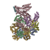
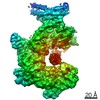
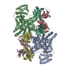
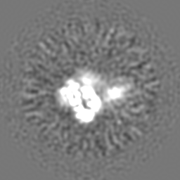
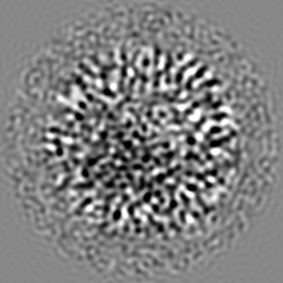
| タイトル | Cryo-EM map of an HIV-1 reverse transcriptase initiation complex in magnesium chloride imaging buffer | |||||||||
 マップデータ マップデータ | HIV-1 RTIC core | |||||||||
 試料 試料 |
| |||||||||
| 生物種 |   Human immunodeficiency virus 1 (ヒト免疫不全ウイルス) / Human immunodeficiency virus 1 (ヒト免疫不全ウイルス) /  Human (ヒト) Human (ヒト) | |||||||||
| 手法 | 単粒子再構成法 / クライオ電子顕微鏡法 / 解像度: 8.2 Å | |||||||||
 データ登録者 データ登録者 | Larsen KP / Chen DH / Puglisi JD / Puglisi EV | |||||||||
 引用 引用 | ジャーナル: Nature / 年: 2018 タイトル: Architecture of an HIV-1 reverse transcriptase initiation complex. 著者: Kevin P Larsen / Yamuna Kalyani Mathiharan / Kalli Kappel / Aaron T Coey / Dong-Hua Chen / Daniel Barrero / Lauren Madigan / Joseph D Puglisi / Georgios Skiniotis / Elisabetta Viani Puglisi /  要旨: Reverse transcription of the HIV-1 RNA genome into double-stranded DNA is a central step in viral infection and a common target of antiretroviral drugs . The reaction is catalysed by viral reverse ...Reverse transcription of the HIV-1 RNA genome into double-stranded DNA is a central step in viral infection and a common target of antiretroviral drugs . The reaction is catalysed by viral reverse transcriptase (RT) that is packaged in an infectious virion with two copies of viral genomic RNA each bound to host lysine 3 transfer RNA (tRNA), which acts as a primer for initiation of reverse transcription. Upon viral entry into cells, initiation is slow and non-processive compared to elongation. Despite extensive efforts, the structural basis of RT function during initiation has remained a mystery. Here we use cryo-electron microscopy to determine a three-dimensional structure of an HIV-1 RT initiation complex. In our structure, RT is in an inactive polymerase conformation with open fingers and thumb and with the nucleic acid primer-template complex shifted away from the active site. The primer binding site (PBS) helix formed between tRNA and HIV-1 RNA lies in the cleft of RT and is extended by additional pairing interactions. The 5' end of the tRNA refolds and stacks on the PBS to create a long helical structure, while the remaining viral RNA forms two helical stems positioned above the RT active site, with a linker that connects these helices to the RNase H region of the PBS. Our results illustrate how RNA structure in the initiation complex alters RT conformation to decrease activity, highlighting a potential target for drug action. | |||||||||
| 履歴 |
|
- 構造の表示
構造の表示
| ムービー |
 ムービービューア ムービービューア |
|---|---|
| 構造ビューア | EMマップ: SurfViewMolmilJmol/JSmol |
| 添付画像 |

- ダウンロードとリンク
-EMDBアーカイブ
| マップデータ | emd_7540.map.gz | 58.7 MB | EMDBマップデータ形式 | |
|---|---|---|---|---|
| ヘッダ (付随情報) | emd-7540-v30.xmlemd-7540.xml | 15.8 KB 15.8 KB | 表示 表示 | EMDBヘッダ |
| 画像 | 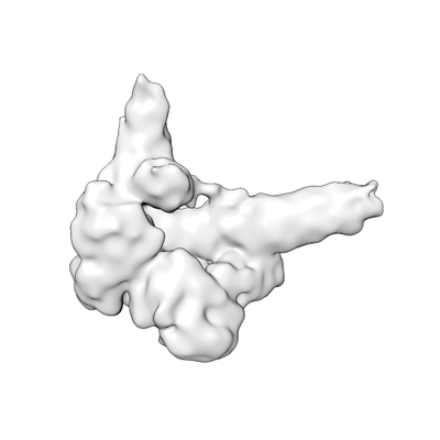 emd_7540.png emd_7540.png | 26.2 KB | ||
| アーカイブディレクトリ |  http://ftp.pdbj.org/pub/emdb/structures/EMD-7540ftp://ftp.pdbj.org/pub/emdb/structures/EMD-7540 http://ftp.pdbj.org/pub/emdb/structures/EMD-7540ftp://ftp.pdbj.org/pub/emdb/structures/EMD-7540 | HTTPS FTP |
-検証レポート
| 文書・要旨 | emd_7540_validation.pdf.gz | 78.6 KB | 表示 | EMDB検証レポート |
|---|---|---|---|---|
| 文書・詳細版 | emd_7540_full_validation.pdf.gz | 77.7 KB | 表示 | |
| XML形式データ | emd_7540_validation.xml.gz | 493 B | 表示 | |
| アーカイブディレクトリ | https://ftp.pdbj.org/pub/emdb/validation_reports/EMD-7540ftp://ftp.pdbj.org/pub/emdb/validation_reports/EMD-7540 | HTTPS FTP |
-関連構造データ


-リンク
| EMDBのページ | EMDB (EBI/PDBe) / EMDataResource |
|---|
-マップ
| ファイル | ダウンロード / ファイル: emd_7540.map.gz / 形式: CCP4 / 大きさ: 64 MB / タイプ: IMAGE STORED AS FLOATING POINT NUMBER (4 BYTES) | ||||||||||||||||||||||||||||||||||||||||||||||||||||||||||||
|---|---|---|---|---|---|---|---|---|---|---|---|---|---|---|---|---|---|---|---|---|---|---|---|---|---|---|---|---|---|---|---|---|---|---|---|---|---|---|---|---|---|---|---|---|---|---|---|---|---|---|---|---|---|---|---|---|---|---|---|---|---|
| 注釈 | HIV-1 RTIC core | ||||||||||||||||||||||||||||||||||||||||||||||||||||||||||||
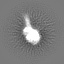
| 投影像・断面図 | 画像のコントロール
画像は Spider により作成 | ||||||||||||||||||||||||||||||||||||||||||||||||||||||||||||
| ボクセルのサイズ | X=Y=Z: 1.286 Å | ||||||||||||||||||||||||||||||||||||||||||||||||||||||||||||
| 密度 |
| ||||||||||||||||||||||||||||||||||||||||||||||||||||||||||||
| 対称性 | 空間群: 1 | ||||||||||||||||||||||||||||||||||||||||||||||||||||||||||||
| 詳細 | EMDB XML:
CCP4マップ ヘッダ情報:
| ||||||||||||||||||||||||||||||||||||||||||||||||||||||||||||
 Z (Sec.)
 Y (Row.)
 X (Col.)


-添付データ
- 試料の構成要素
-全体 : HIV-1 reverse transcriptase initiation complex
| 全体 | 名称: HIV-1 reverse transcriptase initiation complex |
|---|---|
| 要素 |
|
-超分子 #1: HIV-1 reverse transcriptase initiation complex
| 超分子 | 名称: HIV-1 reverse transcriptase initiation complex / タイプ: complex / ID: 1 / 親要素: 0 / 含まれる分子: #1-#4 詳細: Alternate buffer conditions containing magnesium chloride |
|---|---|
| 分子量 | 理論値: 33 KDa |
-超分子 #2: HIV-1 reverse transcriptase
| 超分子 | 名称: HIV-1 reverse transcriptase / タイプ: complex / ID: 2 / 親要素: 1 / 含まれる分子: #1-#2 詳細: A cysteine mutation for crosslinking was introduced into helix H of p66 (Q258C). The protein used in this study also had the C280S mutation, introduced in prior structural work, and the E478Q ...詳細: A cysteine mutation for crosslinking was introduced into helix H of p66 (Q258C). The protein used in this study also had the C280S mutation, introduced in prior structural work, and the E478Q mutation, introduced to eliminate RNase H activity. |
|---|---|
| 由来(天然) | 生物種: Human immunodeficiency virus 1 (ヒト免疫不全ウイルス) |
| 組換発現 | 生物種:  |
-超分子 #3: tRNA lysine3 primer
| 超分子 | 名称: tRNA lysine3 primer / タイプ: complex / ID: 3 / 親要素: 1 / 含まれる分子: #3 詳細: Chemically synthesized and extended tRNA lysine3 primer. Modified nucleotide containing a N2-cystamine was placed at position 71. The tRNA primer has been extended by one ddCTP, bringing its ...詳細: Chemically synthesized and extended tRNA lysine3 primer. Modified nucleotide containing a N2-cystamine was placed at position 71. The tRNA primer has been extended by one ddCTP, bringing its total length in the full complex to 77 nucleotides. |
|---|---|
| 由来(天然) | 生物種: Human (ヒト) |
-超分子 #4: HIV-1 RNA genome fragment
| 超分子 | 名称: HIV-1 RNA genome fragment / タイプ: complex / ID: 4 / 親要素: 1 / 含まれる分子: #4 詳細: HIV-1 RNA genome fragment of 101 nucleotides in length. Contains the primer binding site (PBS), primer activation signal (PAS), A-rich loop, and C-rich region. Generated via T7 transcription and PAGE purified. |
|---|---|
| 由来(天然) | 生物種: Human immunodeficiency virus 1 (ヒト免疫不全ウイルス) |
-実験情報
-構造解析
| 手法 | クライオ電子顕微鏡法 |
|---|---|
 解析 解析 | 単粒子再構成法 |
| 試料の集合状態 | particle |
-試料調製
| 濃度 | 4.5 mg/mL | |||||||||||||||
|---|---|---|---|---|---|---|---|---|---|---|---|---|---|---|---|---|
| 緩衝液 | pH: 8 構成要素:
詳細: Beta-OG was added just prior to freezing. | |||||||||||||||
| グリッド | 材質: COPPER / メッシュ: 200 / 支持フィルム - 材質: CARBON / 支持フィルム - トポロジー: LACEY / 前処理 - タイプ: GLOW DISCHARGE | |||||||||||||||
| 凍結 | 凍結剤: ETHANE / チャンバー内湿度: 85 % / チャンバー内温度: 292 K / 装置: LEICA EM GP 詳細: Blotted for 2.8 sec before plunging into liquid ethane.. | |||||||||||||||
| 詳細 | Sample was monodisperse. |
- 電子顕微鏡法
| 顕微鏡 | FEI TECNAI F20 |
|---|---|
| 撮影 | フィルム・検出器のモデル: GATAN K2 SUMMIT (4k x 4k) 検出モード: COUNTING / デジタル化 - 画像ごとのフレーム数: 2-60 / 実像数: 898 / 平均露光時間: 12.0 sec. / 平均電子線量: 75.0 e/Å2 |
| 電子線 | 加速電圧: 200 kV / 電子線源:  FIELD EMISSION GUN FIELD EMISSION GUN |
| 電子光学系 | 倍率(補正後): 38880 / 照射モード: FLOOD BEAM / 撮影モード: BRIGHT FIELD / Cs: 2.26 mm / 最大 デフォーカス(公称値): 3.0 µm / 最小 デフォーカス(公称値): 2.0 µm / 倍率(公称値): 29000 |
| 試料ステージ | ホルダー冷却材: NITROGEN |
| 実験機器 |  モデル: Tecnai F20 / 画像提供: FEI Company |
