National Institutes of Health/National Institute Of Allergy and Infectious Diseases (NIH/NIAID)
R01 AI127401
United States
Howard Hughes Medical Institute (HHMI)
United States
Citation
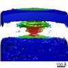
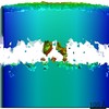
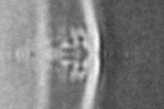
Journal: Cell Rep / Year: 2018 Title: In Vivo Structures of the Helicobacter pylori cag Type IV Secretion System. Authors: Yi-Wei Chang / Carrie L Shaffer / Lee A Rettberg / Debnath Ghosal / Grant J Jensen / Abstract: The type IV secretion system (T4SS) is a versatile nanomachine that translocates diverse effector molecules between microbes and into eukaryotic cells. Here, using electron cryotomography, we reveal ...The type IV secretion system (T4SS) is a versatile nanomachine that translocates diverse effector molecules between microbes and into eukaryotic cells. Here, using electron cryotomography, we reveal the molecular architecture of the Helicobacter pylori cag T4SS. Although most components are unique to H. pylori, the cag T4SS exhibits remarkable architectural similarity to other T4SSs. Our images revealed that, when H. pylori encounters host cells, the bacterium elaborates membranous tubes perforated by lateral ports. Sub-tomogram averaging of the cag T4SS machinery revealed periplasmic densities associated with the outer membrane, a central stalk, and peripheral wing-like densities. Additionally, we resolved pilus-like rod structures extending from the cag T4SS into the inner membrane, as well as densities within the cytoplasmic apparatus corresponding to a short central barrel surrounded by four longer barrels. Collectively, these studies reveal the structure of a dynamic molecular machine that evolved to function in the human gastric niche.
History
Deposition
Feb 21, 2018
-
Header (metadata) release
Apr 18, 2018
-
Map release
Apr 18, 2018
-
Update
Dec 11, 2019
-
Current status
Dec 11, 2019
Processing site: RCSB / Status: Released
-
Structure visualization
Movie
Surface view with section colored by density value
In the structure databanks used in Yorodumi, some data are registered as the other names, "COVID-19 virus" and "2019-nCoV". Here are the details of the virus and the list of structure data.
Jan 31, 2019. EMDB accession codes are about to change! (news from PDBe EMDB page)
EMDB accession codes are about to change! (news from PDBe EMDB page)
The allocation of 4 digits for EMDB accession codes will soon come to an end. Whilst these codes will remain in use, new EMDB accession codes will include an additional digit and will expand incrementally as the available range of codes is exhausted. The current 4-digit format prefixed with “EMD-” (i.e. EMD-XXXX) will advance to a 5-digit format (i.e. EMD-XXXXX), and so on. It is currently estimated that the 4-digit codes will be depleted around Spring 2019, at which point the 5-digit format will come into force.
The EM Navigator/Yorodumi systems omit the EMD- prefix.
Related info.:Q: What is EMD? / ID/Accession-code notation in Yorodumi/EM Navigator
Yorodumi is a browser for structure data from EMDB, PDB, SASBDB, etc.
This page is also the successor to EM Navigator detail page, and also detail information page/front-end page for Omokage search.
The word "yorodu" (or yorozu) is an old Japanese word meaning "ten thousand". "mi" (miru) is to see.
Related info.:EMDB / PDB / SASBDB / Comparison of 3 databanks / Yorodumi Search / Aug 31, 2016. New EM Navigator & Yorodumi / Yorodumi Papers / Jmol/JSmol / Function and homology information / Changes in new EM Navigator and Yorodumi
 Movie
Movie Controller

 Yorodumi
 Open data
 Basic information
 Map data
 Sample
 Helicobacter pylori (bacteria)
 Authors
 United States, 2 items
  Citation
 Structure visualization
 Movie viewer
 Downloads & links
 emd_7474.png
 http://ftp.pdbj.org/pub/emdb/structures/EMD-7474
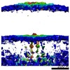
 Z (Sec.)
 Y (Row.)
 X (Col.)


 Sample components
 Processing
 Electron microscopy
 FIELD EMISSION GUN
