[English] 日本語
Yorodumi
- EMDB-7449: Cryo-EM structure of the Gasdermin A3 membrane pore -

+
Open data


ID or keywords:

Loading...

-
Basic information

Entry
Database: EMDB / ID: EMD-7449
TitleCryo-EM structure of the Gasdermin A3 membrane pore
Map dataGasdermin A3 membrane pore
Sample
  • Complex: Gasdermin A3 N domain
    • Protein or peptide: Gasdermin-A3
  • Ligand: CARDIOLIPIN
KeywordsPyroptosis / Pore forming protein / IMMUNE SYSTEM
Function / homology
Function and homology information


sebaceous gland cell differentiation / avascular cornea development in camera-type eye / negative regulation of timing of anagen / hair cycle / wide pore channel activity / mammary gland development / positive regulation of extrinsic apoptotic signaling pathway / phosphatidylinositol-4-phosphate binding / skin development / hair follicle morphogenesis ...sebaceous gland cell differentiation / avascular cornea development in camera-type eye / negative regulation of timing of anagen / hair cycle / wide pore channel activity / mammary gland development / positive regulation of extrinsic apoptotic signaling pathway / phosphatidylinositol-4-phosphate binding / skin development / hair follicle morphogenesis / phosphatidylserine binding / pyroptosis / somatic stem cell population maintenance / hair follicle development / extrinsic apoptotic signaling pathway / phosphatidylinositol-4,5-bisphosphate binding / positive regulation of interleukin-1 beta production / mitochondrial membrane / negative regulation of canonical Wnt signaling pathway / defense response to bacterium / positive regulation of apoptotic process / perinuclear region of cytoplasm / membrane / nucleus / plasma membrane / cytosol
Similarity search - Function
Gasdermin, pore forming domain / Gasdermin pore forming domain / Gasdermin / Gasdermin, PUB domain / Gasdermin PUB domain
Similarity search - Domain/homology
Biological speciesMus musculus (house mouse)
Methodsingle particle reconstruction / cryo EM / Resolution: 3.8 Å
AuthorsRuan J / Wu H
Funding support United States, 3 items
OrganizationGrant numberCountry
National Institutes of Health/National Institute on Minority Health and Health Disparities (NIH/NIMHD)DP1HD087988 United States
National Institutes of Health/National Institute Of Allergy and Infectious Diseases (NIH/NIAID)R01Al124491 United States
National Institutes of Health/National Institute Of Allergy and Infectious Diseases (NIH/NIAID)R01AI123265 United States
Citation
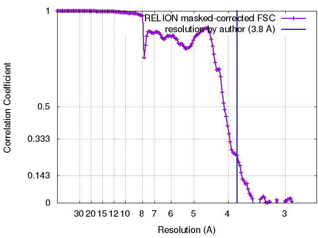
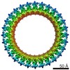
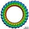
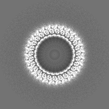
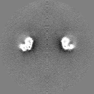
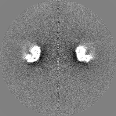
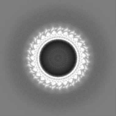
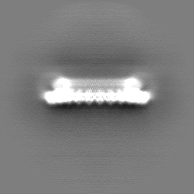
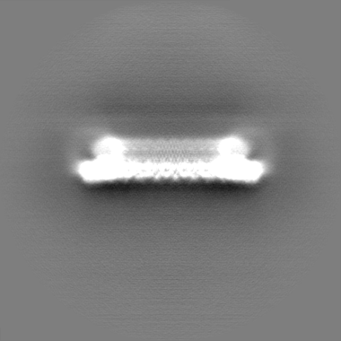
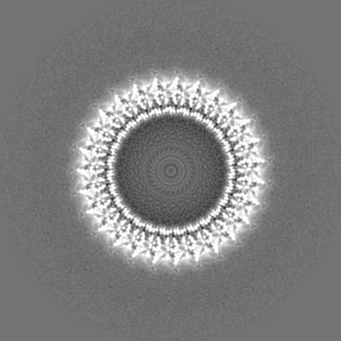
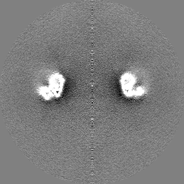
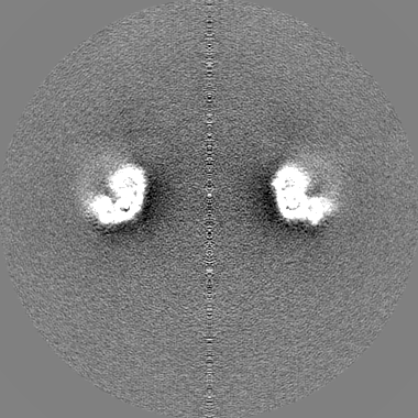
Journal: Nature / Year: 2018
Title: Cryo-EM structure of the gasdermin A3 membrane pore.
Authors: Jianbin Ruan / Shiyu Xia / Xing Liu / Judy Lieberman / Hao Wu /
Abstract: Gasdermins mediate inflammatory cell death after cleavage by caspases or other, unknown enzymes. The cleaved N-terminal fragments bind to acidic membrane lipids to form pores, but the mechanism of ...Gasdermins mediate inflammatory cell death after cleavage by caspases or other, unknown enzymes. The cleaved N-terminal fragments bind to acidic membrane lipids to form pores, but the mechanism of pore formation remains unresolved. Here we present the cryo-electron microscopy structures of the 27-fold and 28-fold single-ring pores formed by the N-terminal fragment of mouse GSDMA3 (GSDMA3-NT) at 3.8 and 4.2 Å resolutions, and of a double-ring pore at 4.6 Å resolution. In the 27-fold pore, a 108-stranded anti-parallel β-barrel is formed by two β-hairpins from each subunit capped by a globular domain. We identify a positively charged helix that interacts with the acidic lipid cardiolipin. GSDMA3-NT undergoes radical conformational changes upon membrane insertion to form long, membrane-spanning β-strands. We also observe an unexpected additional symmetric ring of GSDMA3-NT subunits that does not insert into the membrane in the double-ring pore, which may represent a pre-pore state of GSDMA3-NT. These structures provide a basis that explains the activities of several mutant gasdermins, including defective mutants that are associated with cancer.
History
DepositionFeb 2, 2018-
Header (metadata) releaseMar 28, 2018-
Map releaseApr 25, 2018-
UpdateMar 13, 2024-
Current statusMar 13, 2024Processing site: RCSB / Status: Released

-
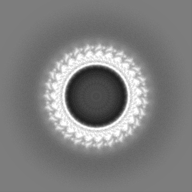
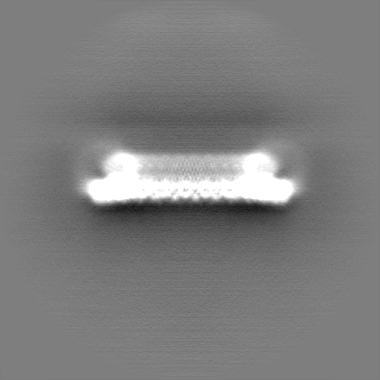
Structure visualization
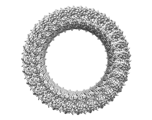
Movie
  • Surface view with section colored by density value
  • Surface level: 0.03
  • Imaged by UCSF Chimera
  • Download
  • Surface view colored by cylindrical radius
  • Surface level: 0.03
  • Imaged by UCSF Chimera
  • Download
  • Surface view with fitted model
  • Atomic models: PDB-6cb8
  • Surface level: 0.03
  • Imaged by UCSF Chimera
  • Download
  • Simplified surface model + fitted atomic model
  • Atomic modelsPDB-6cb8
  • Imaged by Jmol
  • Download
Movie viewer
Structure viewerEM map:
SurfViewMolmilJmol/JSmol
Supplemental images

Downloads & links

-
Map

FileDownload / File: emd_7449.map.gz / Format: CCP4 / Size: 209.3 MB / Type: IMAGE STORED AS FLOATING POINT NUMBER (4 BYTES)
AnnotationGasdermin A3 membrane pore
Voxel sizeX=Y=Z: 1.32 Å
Density
Contour LevelBy EMDB: 0.0119 / Movie #1: 0.03
Minimum - Maximum-0.06966423 - 0.15675774
Average (Standard dev.)0.0002948437 (±0.0036129334)
SymmetrySpace group: 1
Details

EMDB XML:

Map geometry
Axis orderXYZ
Origin-189-189-189
Dimensions380380380
Spacing380380380
CellA=B=C: 501.6 Å
α=β=γ: 90.0 °

CCP4 map header:

modeImage stored as Reals
Å/pix. X/Y/Z1.321.321.32
M x/y/z380380380
origin x/y/z0.0000.0000.000
length x/y/z501.600501.600501.600
α/β/γ90.00090.00090.000
MAP C/R/S123
start NC/NR/NS-189-189-189
NC/NR/NS380380380
D min/max/mean-0.0700.1570.000

-
Supplemental data

-
Additional map: Gasdermin A3 membrane pore

Fileemd_7449_additional_1.map
AnnotationGasdermin A3 membrane pore
Projections & Slices
AxesZYX

Projections

Slices (1/2)
Density Histograms

-
Additional map: Gasdermin A3 membrane pore

Fileemd_7449_additional_2.map
AnnotationGasdermin A3 membrane pore
Projections & Slices
AxesZYX

Projections

Slices (1/2)
Density Histograms

-
Sample components

-
Entire : Gasdermin A3 N domain

EntireName: Gasdermin A3 N domain
Components
  • Complex: Gasdermin A3 N domain
    • Protein or peptide: Gasdermin-A3
  • Ligand: CARDIOLIPIN

-
Supramolecule #1: Gasdermin A3 N domain

SupramoleculeName: Gasdermin A3 N domain / type: complex / ID: 1 / Parent: 0 / Macromolecule list: #1
Source (natural)Organism: Mus musculus (house mouse)
Molecular weightTheoretical: 810 KDa

-
Macromolecule #1: Gasdermin-A3

MacromoleculeName: Gasdermin-A3 / type: protein_or_peptide / ID: 1 / Number of copies: 1 / Enantiomer: LEVO
Source (natural)Organism: Mus musculus (house mouse)
Molecular weightTheoretical: 30.401035 KDa
Recombinant expressionOrganism: Escherichia coli (E. coli)
SequenceString: HMPVFEDVTR ALVRELNPRG DLTPLDSLID FKHFRPFCLV LRKRKSTLFW GARYVRTDYT LLDLLEPGSS PSDLTDSGNF SFKNMLDVQ VQGLVEVPKT VKVKGTAGLS QSSTLEVQTL SVAPSALENL KKERKLSADH SFLNEMRYHE KNLYVVMEAV E AKQEVTVE ...String:
HMPVFEDVTR ALVRELNPRG DLTPLDSLID FKHFRPFCLV LRKRKSTLFW GARYVRTDYT LLDLLEPGSS PSDLTDSGNF SFKNMLDVQ VQGLVEVPKT VKVKGTAGLS QSSTLEVQTL SVAPSALENL KKERKLSADH SFLNEMRYHE KNLYVVMEAV E AKQEVTVE QTGNANAIFS LPSLALLGLQ GSLNNNKAVT IPKGCVLAYR VRLLRVFLFN LWDIPYICND SMQTFPKIRR VP CSAFISP TQMISEEPEE EKLIGELEVL FQ

UniProtKB: Gasdermin-A3

-
Macromolecule #2: CARDIOLIPIN

MacromoleculeName: CARDIOLIPIN / type: ligand / ID: 2 / Number of copies: 1 / Formula: CDL
Molecular weightTheoretical: 1.464043 KDa
Chemical component information

ChemComp-CDL:
CARDIOLIPIN / phospholipid*YM / Cardiolipin

-
Experimental details

-
Structure determination

Methodcryo EM
Processingsingle particle reconstruction
Aggregation stateparticle

-
Sample preparation

Concentration2 mg/mL
BufferpH: 8
VitrificationCryogen name: ETHANE

-
Electron microscopy

MicroscopeFEI TITAN KRIOS
Electron beamAcceleration voltage: 300 kV / Electron source: FIELD EMISSION GUN
Electron opticsIllumination mode: FLOOD BEAM / Imaging mode: BRIGHT FIELDBright-field microscopy
Image recordingFilm or detector model: GATAN K2 SUMMIT (4k x 4k) / Average electron dose: 40.0 e/Å2
Experimental equipment
Model: Titan Krios / Image courtesy: FEI Company

-
Image processing

Startup modelType of model: NONE
Initial angle assignmentType: ANGULAR RECONSTITUTION
Final angle assignmentType: ANGULAR RECONSTITUTION
Final reconstructionApplied symmetry - Point group: C27 (27 fold cyclic) / Resolution.type: BY AUTHOR / Resolution: 3.8 Å / Resolution method: FSC 0.143 CUT-OFF / Software - Name: RELION (ver. 2) / Number images used: 40086
FSC plot (resolution estimation)

+
About Yorodumi

-
News

-
Feb 9, 2022. New format data for meta-information of EMDB entries

New format data for meta-information of EMDB entries

  • Version 3 of the EMDB header file is now the official format.
  • The previous official version 1.9 will be removed from the archive.

Related info.:EMDB header

External links:wwPDB to switch to version 3 of the EMDB data model

-
Aug 12, 2020. Covid-19 info

Covid-19 info

URL: https://pdbj.org/emnavi/covid19.php

New page: Covid-19 featured information page in EM Navigator.

Related info.:Covid-19 info / Mar 5, 2020. Novel coronavirus structure data

+
Mar 5, 2020. Novel coronavirus structure data

Novel coronavirus structure data

Related info.:Yorodumi Speices / Aug 12, 2020. Covid-19 info

External links:COVID-19 featured content - PDBj / Molecule of the Month (242):Coronavirus Proteases

+
Jan 31, 2019. EMDB accession codes are about to change! (news from PDBe EMDB page)

EMDB accession codes are about to change! (news from PDBe EMDB page)

  • The allocation of 4 digits for EMDB accession codes will soon come to an end. Whilst these codes will remain in use, new EMDB accession codes will include an additional digit and will expand incrementally as the available range of codes is exhausted. The current 4-digit format prefixed with “EMD-” (i.e. EMD-XXXX) will advance to a 5-digit format (i.e. EMD-XXXXX), and so on. It is currently estimated that the 4-digit codes will be depleted around Spring 2019, at which point the 5-digit format will come into force.
  • The EM Navigator/Yorodumi systems omit the EMD- prefix.

Related info.:Q: What is EMD? / ID/Accession-code notation in Yorodumi/EM Navigator

External links:EMDB Accession Codes are Changing Soon! / Contact to PDBj

+
Jul 12, 2017. Major update of PDB

Major update of PDB

  • wwPDB released updated PDB data conforming to the new PDBx/mmCIF dictionary.
  • This is a major update changing the version number from 4 to 5, and with Remediation, in which all the entries are updated.
  • In this update, many items about electron microscopy experimental information are reorganized (e.g. em_software).
  • Now, EM Navigator and Yorodumi are based on the updated data.

External links:wwPDB Remediation / Enriched Model Files Conforming to OneDep Data Standards Now Available in the PDB FTP Archive

-
Yorodumi

Thousand views of thousand structures

  • Yorodumi is a browser for structure data from EMDB, PDB, SASBDB, etc.
  • This page is also the successor to EM Navigator detail page, and also detail information page/front-end page for Omokage search.
  • The word "yorodu" (or yorozu) is an old Japanese word meaning "ten thousand". "mi" (miru) is to see.

Related info.:EMDB / PDB / SASBDB / Comparison of 3 databanks / Yorodumi Search / Aug 31, 2016. New EM Navigator & Yorodumi / Yorodumi Papers / Jmol/JSmol / Function and homology information / Changes in new EM Navigator and Yorodumi

Read more