 Movie
Movie Controller


+ Open data

- Basic information
| Entry |  | |||||||||
|---|---|---|---|---|---|---|---|---|---|---|
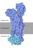
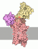
| Title | the structure of ERMA Mg2+ bound form | |||||||||
 Map data Map data | structure of ERMA Mg2+ bound form | |||||||||
 Sample Sample |
| |||||||||
 Keywords Keywords | P-type ATPase like protein / MEMBRANE PROTEIN | |||||||||
| Function / homology | magnesium ion transport from cytosol to endoplasmic reticulum / Transmembrane protein 94 / intracellular magnesium ion homeostasis / P-type magnesium transporter activity / P-type ATPase, transmembrane domain superfamily / endoplasmic reticulum membrane / nucleoplasm / Transmembrane protein 94 Function and homology information Function and homology information | |||||||||
| Biological species |  Homo sapiens (human) Homo sapiens (human) | |||||||||
| Method | single particle reconstruction / cryo EM / Resolution: 2.8 Å | |||||||||
 Authors Authors | Shi N / Jiang Y | |||||||||
| Funding support |  United States, 2 items United States, 2 items
| |||||||||
 Citation Citation | Journal: Sci Adv / Year: 2026 Title: Structural and mutational insights define ERMA as the ER Mg ATPase and reservoir gatekeeper. Authors: Manigandan Venkatesan / Michael L Oldham / Ning Shi / Adhishree Chidambaram / Neelanjan Vishnu / Abitha K Madesh / Kristen Bentz / Peter B Stathopulos / Ravi C Kalathur / Youxing Jiang / Muniswamy Madesh /  Abstract: Magnesium (Mg) is the most abundant divalent cation in cells, yet the mechanisms mediating its organellar transport remain poorly defined. We identify endoplasmic reticulum (ER) Mg adenosine ...Magnesium (Mg) is the most abundant divalent cation in cells, yet the mechanisms mediating its organellar transport remain poorly defined. We identify endoplasmic reticulum (ER) Mg adenosine triphosphatase (ATPase) (ERMA) as the transporter that drives Mg uptake into the ER lumen, establishing the ER as a bi-ionic intracellular reservoir. MagFRET biosensors targeted to the ER demonstrate that ERMA mediates dynamic ER Mg storage and robust adenosine 5'-triphosphate-dependent Mg uptake reaching 15 to 30 millimolar. Cryo-electron microscopy structures of human and mouse ERMA reveal a P-type ATPase fold with an unwound transmembrane 4 (TM4) that coordinates Mg via the unique PILP backbone and the TM5 residue Q1110, whose mutation markedly impairs ERMA-mediated Mg uptake. Functional reconstitution of domain mutants, ERMA-SERCA chimeras, and pathogenic variants confirm ERMA as an ER-resident Mg pump and gatekeeper of ER Mg ionic equilibrium. | |||||||||
| History |
|
- Structure visualization
| Supplemental images |
|---|

- Downloads & links
-EMDB archive
| Map data | emd_73630.map.gz | 82.5 MB | EMDB map data format | |
|---|---|---|---|---|
| Header (meta data) | emd-73630-v30.xmlemd-73630.xml | 16.9 KB 16.9 KB | Display Display | EMDB header |
| FSC (resolution estimation) | emd_73630_fsc.xml | 9.5 KB | Display | FSC data file |
| Images |  emd_73630.png emd_73630.png | 52.3 KB | ||
| Filedesc metadata | emd-73630.cif.gz | 6.7 KB | ||
| Others | emd_73630_half_map_1.map.gzemd_73630_half_map_2.map.gz | 84.7 MB 84.7 MB | ||
| Archive directory |  https://data.pdbj.org/pub/emdb/structures/EMD-73630ftp://data.pdbj.org/pub/emdb/structures/EMD-73630 https://data.pdbj.org/pub/emdb/structures/EMD-73630ftp://data.pdbj.org/pub/emdb/structures/EMD-73630 | HTTPS FTP |
-Related structure data
| Related structure data |  9yydMC  9ywqC M: atomic model generated by this map C: citing same article ( |
|---|---|
| Similar structure data |

-Links
| EMDB pages | EMDB (EBI/PDBe) / EMDataResource |
|---|---|
| Related items in Molecule of the Month |
-Map
| File | Download / File: emd_73630.map.gz / Format: CCP4 / Size: 91.1 MB / Type: IMAGE STORED AS FLOATING POINT NUMBER (4 BYTES) | ||||||||||||||||||||||||||||||||||||
|---|---|---|---|---|---|---|---|---|---|---|---|---|---|---|---|---|---|---|---|---|---|---|---|---|---|---|---|---|---|---|---|---|---|---|---|---|---|
| Annotation | structure of ERMA Mg2+ bound form | ||||||||||||||||||||||||||||||||||||
| Projections & slices | Image control
Images are generated by Spider. | ||||||||||||||||||||||||||||||||||||
| Voxel size | X=Y=Z: 0.827 Å | ||||||||||||||||||||||||||||||||||||
| Density |
| ||||||||||||||||||||||||||||||||||||
| Symmetry | Space group: 1 | ||||||||||||||||||||||||||||||||||||
| Details | EMDB XML:
|
 X (Sec.)
 Y (Row.)
 Z (Col.)


-Supplemental data
-Half map: Half Map B
| File | emd_73630_half_map_1.map | ||||||||||||
|---|---|---|---|---|---|---|---|---|---|---|---|---|---|
| Annotation | Half Map B | ||||||||||||
| Projections & Slices |
| ||||||||||||
| Density Histograms |


-Half map: Half Map A
| File | emd_73630_half_map_2.map | ||||||||||||
|---|---|---|---|---|---|---|---|---|---|---|---|---|---|
| Annotation | Half Map A | ||||||||||||
| Projections & Slices |
| ||||||||||||
| Density Histograms |


- Sample components
-Entire : ERMA monomer Mg++ bound form
| Entire | Name: ERMA monomer Mg++ bound form |
|---|---|
| Components |
|
-Supramolecule #1: ERMA monomer Mg++ bound form
| Supramolecule | Name: ERMA monomer Mg++ bound form / type: complex / ID: 1 / Parent: 0 / Macromolecule list: #1 |
|---|---|
| Source (natural) | Organism: Homo sapiens (human) |
-Macromolecule #1: Isoform 3 of Transmembrane protein 94
| Macromolecule | Name: Isoform 3 of Transmembrane protein 94 / type: protein_or_peptide / ID: 1 / Number of copies: 1 / Enantiomer: LEVO |
|---|---|
| Source (natural) | Organism: Homo sapiens (human) |
| Molecular weight | Theoretical: 153.571797 KDa |
| Recombinant expression | Organism: Homo sapiens (human) |
| Sequence | String: MDYKDDDDKM LFKQAELWMP HQGKGNKGEP PSALGLSTRK ALSVLKEQLE AVLEGHLRER KKCLTWKEVW RSSFLHHSNR CSCFHWPGA SLMLLAVLLL LGCCGGQPAG SRGVGLVNAS ALFLLLLLNL VLIGRQDRLK RREVERRLRG IIDQIQDALR D GREIQWPS ...String: MDYKDDDDKM LFKQAELWMP HQGKGNKGEP PSALGLSTRK ALSVLKEQLE AVLEGHLRER KKCLTWKEVW RSSFLHHSNR CSCFHWPGA SLMLLAVLLL LGCCGGQPAG SRGVGLVNAS ALFLLLLLNL VLIGRQDRLK RREVERRLRG IIDQIQDALR D GREIQWPS AMYPDLHMPF APSWSLHWAY RDGHLVNLPV SLLVEGDIIA LRPGQESFAS LRGIKDDEHI VLEPGDLFPP FS PPPSPRG EVERGPQSPQ QHRLFRVLET PVIDNIRWCL DMALSRPVTA LDNERFTVQS VMLHYAVPVV LAGFLITNAL RFI FSAPGV TSWQYTLLQL QVNGVLPILP LLFPVLWVLA TACGEARVLA QMSKASPSSL LAKFSEDTLS SYTEAVSSQE MLRC IWGHF LRVLGGTSPT LSHSSSLLHS LGSVTVLCCV DKQGILSWPN PSPETVLFFS GKVEPPHSSH EDLTDGLSTR SFCHP EPHE RDALLAGSLN NTLHLSNEQE RGDWPGEAPK PPEPYSHHKA HGRSKHPSGS NVSFSRDTEG GEEEPSKTQP GMESDP YEA EDFVCDYHLE MLSLSQDQQN PSCIQFDDSN WQLHLTSLKP LGLNVLLNLC DASVTERLCR FSDHLCNIAL QESHSAV LP VHVPWGLCEL ARLIGFTPGA KELFKQENHL ALYRLPSAET MKETSLGRLS CVTKRRPPLS HMISLFIKDT TTSTEQML S HGTADVVLEA CTDFWDGADI YPLSGSDRKK VLDFYQRACL SGYCSAFAYK PMNCALSSQL NGKCIELVQV PGQSSIFTM CELPSTIPIK QNARRSSWSS DEGIGEVLEK EDCMQALSGQ IFMGMVSSQY QARLDIVRLI DGLVNACIRF VYFSLEDELK SKVFAEKMG LETGWNCHIS LTPNGDMPGS EIPPSSPSHA GSLHDDLNQV SRDDAEGLLL MEEEGHSDLI SFQPTDSDIP S FLEDSNRA KLPRGIHQVR PHLQNIDNVP LLVPLFTDCT PETMCEMIKI MQEYGEVTCC LGSSANLRNS CLFLQSDISI AL DPLYPSR CSWETFGYAT SISMAQASDG LSPLQLSGQL NSLPCSLTFR QEETISIIRL IEQARHATYG IRKCFLFLLQ CQL TLVVIQ FLSCLVQLPP LLSTTDILWL SCFCYPLLSI SLLGKPPHSS IMSMATGKNL QSIPKKTQHY FLLCFLLKFS LTLS SCLIC FGFTLQSFCD SSRDRNLTNC SSVMLPSNDD RAPAWFEDFA NGLLSAQKLT AALIVLHTVF ISITHVHRTK PLWRK SPLT NLWWAVTVPV VLLGQVVQTA VDLQLWTHRD SHVHFGLEDV PLLTWLLGCL SLVLVVVTNE IVKLHEIRVR VRYQKR QKL QFETKLGMNS PF UniProtKB: Transmembrane protein 94 |
-Macromolecule #2: MAGNESIUM ION
| Macromolecule | Name: MAGNESIUM ION / type: ligand / ID: 2 / Number of copies: 1 / Formula: MG |
|---|---|
| Molecular weight | Theoretical: 24.305 Da |
-Macromolecule #3: (1S)-2-{[(2-AMINOETHOXY)(HYDROXY)PHOSPHORYL]OXY}-1-[(PALMITOYLOXY...
| Macromolecule | Name: (1S)-2-{[(2-AMINOETHOXY)(HYDROXY)PHOSPHORYL]OXY}-1-[(PALMITOYLOXY)METHYL]ETHYL STEARATE type: ligand / ID: 3 / Number of copies: 1 / Formula: PEV |
|---|---|
| Molecular weight | Theoretical: 720.012 Da |
| Chemical component information |  ChemComp-PEV: |
-Experimental details
-Structure determination
| Method | cryo EM |
|---|---|
 Processing Processing | single particle reconstruction |
| Aggregation state | particle |
-Sample preparation
| Buffer | pH: 7.5 |
|---|---|
| Vitrification | Cryogen name: ETHANE |
- Electron microscopy
| Microscope | TFS KRIOS |
|---|---|
| Image recording | Film or detector model: GATAN K3 BIOQUANTUM (6k x 4k) / Average electron dose: 60.0 e/Å2 |
| Electron beam | Acceleration voltage: 300 kV / Electron source:  FIELD EMISSION GUN FIELD EMISSION GUN |
| Electron optics | Illumination mode: FLOOD BEAM / Imaging mode: BRIGHT FIELD / Nominal defocus max: 2.2 µm / Nominal defocus min: 0.9 µm |
| Experimental equipment |  Model: Titan Krios / Image courtesy: FEI Company |

