 Movie
Movie Controller


+ Open data

- Basic information
| Entry |  | |||||||||
|---|---|---|---|---|---|---|---|---|---|---|
| Title | the structure of ERMA complex with ATPrS and Mg++ | |||||||||
 Map data Map data | structure of ERMA complex with ATPrS and Mg | |||||||||
 Sample Sample |
| |||||||||
 Keywords Keywords | P-type ATPase like protein / MEMBRANE PROTEIN | |||||||||
| Function / homology |  Function and homology information Function and homology informationmagnesium ion transport from cytosol to endoplasmic reticulum / intracellular magnesium ion homeostasis / P-type magnesium transporter activity / embryonic heart tube development / embryonic brain development / endoplasmic reticulum membrane / nucleoplasm Similarity search - Function | |||||||||
| Biological species |   Mus musculus (house mouse) Mus musculus (house mouse) | |||||||||
| Method | single particle reconstruction / cryo EM / Resolution: 3.2 Å | |||||||||
 Authors Authors | Shi N / Jiang Y | |||||||||
| Funding support |  United States, 2 items United States, 2 items
| |||||||||
 Citation Citation | Journal: Sci Adv / Year: 2026 Title: Structural and mutational insights define ERMA as the ER Mg ATPase and reservoir gatekeeper. Authors: Manigandan Venkatesan / Michael L Oldham / Ning Shi / Adhishree Chidambaram / Neelanjan Vishnu / Abitha K Madesh / Kristen Bentz / Peter B Stathopulos / Ravi C Kalathur / Youxing Jiang / Muniswamy Madesh /  Abstract: Magnesium (Mg) is the most abundant divalent cation in cells, yet the mechanisms mediating its organellar transport remain poorly defined. We identify endoplasmic reticulum (ER) Mg adenosine ...Magnesium (Mg) is the most abundant divalent cation in cells, yet the mechanisms mediating its organellar transport remain poorly defined. We identify endoplasmic reticulum (ER) Mg adenosine triphosphatase (ATPase) (ERMA) as the transporter that drives Mg uptake into the ER lumen, establishing the ER as a bi-ionic intracellular reservoir. MagFRET biosensors targeted to the ER demonstrate that ERMA mediates dynamic ER Mg storage and robust adenosine 5'-triphosphate-dependent Mg uptake reaching 15 to 30 millimolar. Cryo-electron microscopy structures of human and mouse ERMA reveal a P-type ATPase fold with an unwound transmembrane 4 (TM4) that coordinates Mg via the unique PILP backbone and the TM5 residue Q1110, whose mutation markedly impairs ERMA-mediated Mg uptake. Functional reconstitution of domain mutants, ERMA-SERCA chimeras, and pathogenic variants confirm ERMA as an ER-resident Mg pump and gatekeeper of ER Mg ionic equilibrium. | |||||||||
| History |
|
- Structure visualization
| Supplemental images |
|---|

- Downloads & links
-EMDB archive
| Map data | emd_73556.map.gz | 44.9 MB | EMDB map data format | |
|---|---|---|---|---|
| Header (meta data) | emd-73556-v30.xmlemd-73556.xml | 18 KB 18 KB | Display Display | EMDB header |
| FSC (resolution estimation) | emd_73556_fsc.xml | 11.3 KB | Display | FSC data file |
| Images |  emd_73556.png emd_73556.png | 73.7 KB | ||
| Filedesc metadata | emd-73556.cif.gz | 6.9 KB | ||
| Others | emd_73556_half_map_1.map.gzemd_73556_half_map_2.map.gz | 48.9 MB 48.9 MB | ||
| Archive directory |  https://data.pdbj.org/pub/emdb/structures/EMD-73556ftp://data.pdbj.org/pub/emdb/structures/EMD-73556 https://data.pdbj.org/pub/emdb/structures/EMD-73556ftp://data.pdbj.org/pub/emdb/structures/EMD-73556 | HTTPS FTP |
-Related structure data
| Related structure data |  9ywqMC  9yydC M: atomic model generated by this map C: citing same article ( |
|---|---|
| Similar structure data |

-Links
| EMDB pages | EMDB (EBI/PDBe) / EMDataResource |
|---|---|
| Related items in Molecule of the Month |
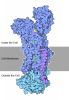
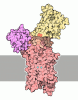
-Map
| File | Download / File: emd_73556.map.gz / Format: CCP4 / Size: 52.7 MB / Type: IMAGE STORED AS FLOATING POINT NUMBER (4 BYTES) | ||||||||||||||||||||||||||||||||||||
|---|---|---|---|---|---|---|---|---|---|---|---|---|---|---|---|---|---|---|---|---|---|---|---|---|---|---|---|---|---|---|---|---|---|---|---|---|---|
| Annotation | structure of ERMA complex with ATPrS and Mg | ||||||||||||||||||||||||||||||||||||
| Projections & slices | Image control
Images are generated by Spider. | ||||||||||||||||||||||||||||||||||||
| Voxel size | X=Y=Z: 1.0392 Å | ||||||||||||||||||||||||||||||||||||
| Density |
| ||||||||||||||||||||||||||||||||||||
| Symmetry | Space group: 1 | ||||||||||||||||||||||||||||||||||||
| Details | EMDB XML:
|
 X (Sec.)
 Y (Row.)
 Z (Col.)


-Supplemental data
-Half map: Half Map B
| File | emd_73556_half_map_1.map | ||||||||||||
|---|---|---|---|---|---|---|---|---|---|---|---|---|---|
| Annotation | Half Map B | ||||||||||||
| Projections & Slices |
| ||||||||||||
| Density Histograms |


-Half map: Half Map A
| File | emd_73556_half_map_2.map | ||||||||||||
|---|---|---|---|---|---|---|---|---|---|---|---|---|---|
| Annotation | Half Map A | ||||||||||||
| Projections & Slices |
| ||||||||||||
| Density Histograms |


- Sample components
-Entire : ERMA
| Entire | Name: ERMA |
|---|---|
| Components |
|
-Supramolecule #1: ERMA
| Supramolecule | Name: ERMA / type: complex / ID: 1 / Parent: 0 / Macromolecule list: #1 |
|---|---|
| Source (natural) | Organism: Mus musculus (house mouse) |
| Molecular weight | Theoretical: 154 kDa/nm |
-Macromolecule #1: Transmembrane protein 94
| Macromolecule | Name: Transmembrane protein 94 / type: protein_or_peptide / ID: 1 / Number of copies: 1 / Enantiomer: LEVO |
|---|---|
| Source (natural) | Organism: Mus musculus (house mouse) |
| Molecular weight | Theoretical: 156.281734 KDa |
| Recombinant expression | Organism:  Homo sapiens (human) Homo sapiens (human) |
| Sequence | String: MDYKDDDDKG GSLETMDLRE KHLGEPPLAL GLSTRKALSV LKEQLEAVLE KHLKERKKSL TWKEAWRSSF LHLSNRCSCF HWPGASLML LAVLLLLCCC GGQPAGSQGV ELVNASALFL LLLLNLVLIG RQDRLKRREV ERRLRGIIDQ IQDALRDGKE I KWPNSMYP ...String: MDYKDDDDKG GSLETMDLRE KHLGEPPLAL GLSTRKALSV LKEQLEAVLE KHLKERKKSL TWKEAWRSSF LHLSNRCSCF HWPGASLML LAVLLLLCCC GGQPAGSQGV ELVNASALFL LLLLNLVLIG RQDRLKRREV ERRLRGIIDQ IQDALRDGKE I KWPNSMYP DLHMPFAPSW SLHWAYRDGH LVNLPVSLLV EGDIIALRPG QESFASLRGI KDDEHIVLEP GDLFPPFSPP PS PRGEVKR GPQNPQQHRL FRVLETPVID NIRWCLDTAL SRPVTALDNE RFTVQSVMLH YAVPVVLAGF LITNALRFMF KAP GVTSWQ YTLLQLQVNG MLPILPLLFP VLWVLATACG EARVLAQMSK ASPSSLLAKF SEDTLSSYTE AVSSQEMLRC IWGH FLRVI QGTSPTLSHS ASLLHSLGSV TVLCCVDKQG ILSWPNPSPE TVLFFSGKVE PPHSSHEDLT DDLSTRSFCH PEVEE EPHE HDALLAGSLN NTLHLSNEQE RSDWLADGPK PSEPYPHHKG HGRSKHPSGS NVSFSRDTEG GEEEPSKAQP GTEGDP YEA EDFVCDYHLE MLSLSQDQQN PSCIQFDDSN WQSHLTSLKP LGLNVLLNLC NASVTERLCR FSDHLCNIAL QESHSAV LP VHVPWGLCEL ARLIGFTPGA KELFKQENHL ALYRLPSAET LKETSLGRPS CVTKRRPPLS HMISLFIKDT ATSTEQML S HGSADVVVEA CTDFWDGADI YPLSGSDRKK VLDFYQRACL SGYCSAFAYK PMNCTLSSQL NGKCIELVQV PGQNSIFTM CELPSTIPIK PNNRRSSWSS DEGIGEVLEK EDCMQALSGQ IFMGMVSSQY QARLDIVRLI DGLVNACIRF VYFSLEDELR SKVFAEKMG LETGWNCHIS LTPNGDMPGS EIPPSSPSHA GSLHDDLNQV SRDDAEGLLL LEEEGHSDLI SFQPTDSDIP S FLEDCNRA KLPRGIHQVR PHLQNIDNVP LLVPLFTDCT PDTMCEMIKI MQEYGEVTCC LGSSANLRNS CLFLQSDVSI AL DPLYPSR CSWETFGYAT STTMAQASDG LSPLQLSGQL NSLPCSLTFR QEESISIIRL IEQARHATYG IRKCFLFLLQ CQL TLVVIQ FLSCLVQLPP LLSTTDILWL SCFCYPLLSI SLLGKPPHSS IMSMATGKNL QSIPKKTQHY FLLCFLLKFS LTIS SCLVC FGFTLQSFCD SARARNLTNC SSVMLCSNDD RAPAWFEDFA NGLLSAQKLT AALIVLHTVF ISITHVHRTK PLWRK SPLT NLWWAVTVPV VLLGQVVQTV VDLQLWTHRD SRVHFGLEDV PLLTWLLGCL SLVLVVVTNE IVKLHEIRVR VRYQKR QKL QFETKLGMNS PFAAATAGIR GTKLNQPLDY KIFSVTE UniProtKB: Transmembrane protein 94 |
-Macromolecule #2: MAGNESIUM ION
| Macromolecule | Name: MAGNESIUM ION / type: ligand / ID: 2 / Number of copies: 1 / Formula: MG |
|---|---|
| Molecular weight | Theoretical: 24.305 Da |
-Macromolecule #3: PHOSPHOTHIOPHOSPHORIC ACID-ADENYLATE ESTER
| Macromolecule | Name: PHOSPHOTHIOPHOSPHORIC ACID-ADENYLATE ESTER / type: ligand / ID: 3 / Number of copies: 1 / Formula: AGS |
|---|---|
| Molecular weight | Theoretical: 523.247 Da |
| Chemical component information | 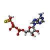 ChemComp-AGS: |
-Macromolecule #4: DECANE
| Macromolecule | Name: DECANE / type: ligand / ID: 4 / Number of copies: 9 / Formula: D10 |
|---|---|
| Molecular weight | Theoretical: 142.282 Da |
| Chemical component information | 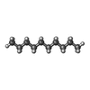 ChemComp-D10: |
-Macromolecule #5: DODECANE
| Macromolecule | Name: DODECANE / type: ligand / ID: 5 / Number of copies: 3 / Formula: D12 |
|---|---|
| Molecular weight | Theoretical: 170.335 Da |
| Chemical component information |  ChemComp-D12: |
-Macromolecule #6: N-OCTANE
| Macromolecule | Name: N-OCTANE / type: ligand / ID: 6 / Number of copies: 1 / Formula: OCT |
|---|---|
| Molecular weight | Theoretical: 114.229 Da |
| Chemical component information |  ChemComp-OCT: |
-Experimental details
-Structure determination
| Method | cryo EM |
|---|---|
 Processing Processing | single particle reconstruction |
| Aggregation state | particle |
-Sample preparation
| Buffer | pH: 7.5 |
|---|---|
| Vitrification | Cryogen name: ETHANE |
- Electron microscopy
| Microscope | TFS KRIOS |
|---|---|
| Image recording | Film or detector model: GATAN K3 BIOQUANTUM (6k x 4k) / Average electron dose: 60.0 e/Å2 |
| Electron beam | Acceleration voltage: 300 kV / Electron source:  FIELD EMISSION GUN FIELD EMISSION GUN |
| Electron optics | Illumination mode: FLOOD BEAM / Imaging mode: BRIGHT FIELD / Nominal defocus max: 2.2 µm / Nominal defocus min: 0.9 µm |
| Sample stage | Cooling holder cryogen: NITROGEN |
| Experimental equipment |  Model: Titan Krios / Image courtesy: FEI Company |

