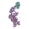
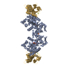
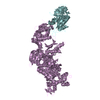
Journal: Science / Year: 2018 Title: Molecular structure of human P-glycoprotein in the ATP-bound, outward-facing conformation. Authors: Youngjin Kim / Jue Chen / Abstract: The multidrug transporter permeability (P)-glycoprotein is an adenosine triphosphate (ATP)-binding cassette exporter responsible for clinical resistance to chemotherapy. P-glycoprotein extrudes toxic ...The multidrug transporter permeability (P)-glycoprotein is an adenosine triphosphate (ATP)-binding cassette exporter responsible for clinical resistance to chemotherapy. P-glycoprotein extrudes toxic molecules and drugs from cells through ATP-powered conformational changes. Despite decades of effort, only the structures of the inward-facing conformation of P-glycoprotein are available. Here we present the structure of human P-glycoprotein in the outward-facing conformation, determined by cryo-electron microscopy at 3.4-angstrom resolution. The two nucleotide-binding domains form a closed dimer occluding two ATP molecules. The drug-binding cavity observed in the inward-facing structures is reorientated toward the extracellular space and compressed to preclude substrate binding. This observation indicates that ATP binding, not hydrolysis, promotes substrate release. The structure evokes a model in which the dynamic nature of P-glycoprotein enables translocation of a large variety of substrates.
History
Deposition
Jan 2, 2018
-
Header (metadata) release
Jan 31, 2018
-
Map release
Jan 31, 2018
-
Update
Mar 13, 2024
-
Current status
Mar 13, 2024
Processing site: RCSB / Status: Released
-
Structure visualization
Movie
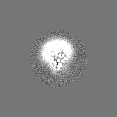
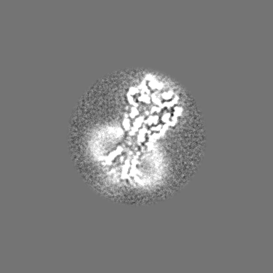
Surface view with section colored by density value
EMPIAR-10803 (Title: Cryo-electron microscopy reconstruction of ATP-bound human P-glycoprotein Data size: 2.0 TB Data #1: Unaligned and uncorrected multiframe movies of human ATP-bound P-glycoprotein [micrographs - multiframe])
In the structure databanks used in Yorodumi, some data are registered as the other names, "COVID-19 virus" and "2019-nCoV". Here are the details of the virus and the list of structure data.
Jan 31, 2019. EMDB accession codes are about to change! (news from PDBe EMDB page)
EMDB accession codes are about to change! (news from PDBe EMDB page)
The allocation of 4 digits for EMDB accession codes will soon come to an end. Whilst these codes will remain in use, new EMDB accession codes will include an additional digit and will expand incrementally as the available range of codes is exhausted. The current 4-digit format prefixed with “EMD-” (i.e. EMD-XXXX) will advance to a 5-digit format (i.e. EMD-XXXXX), and so on. It is currently estimated that the 4-digit codes will be depleted around Spring 2019, at which point the 5-digit format will come into force.
The EM Navigator/Yorodumi systems omit the EMD- prefix.
Related info.:Q: What is EMD? / ID/Accession-code notation in Yorodumi/EM Navigator
Yorodumi is a browser for structure data from EMDB, PDB, SASBDB, etc.
This page is also the successor to EM Navigator detail page, and also detail information page/front-end page for Omokage search.
The word "yorodu" (or yorozu) is an old Japanese word meaning "ten thousand". "mi" (miru) is to see.
Related info.:EMDB / PDB / SASBDB / Comparison of 3 databanks / Yorodumi Search / Aug 31, 2016. New EM Navigator & Yorodumi / Yorodumi Papers / Jmol/JSmol / Function and homology information / Changes in new EM Navigator and Yorodumi
 Movie
Movie Controller

 Yorodumi
 Open data
 Basic information
 Map data
 Sample
 Keywords
 Function and homology information
 Homo sapiens (human)
 Authors
 United States, 1 items
  Citation
 Structure visualization
 Downloads & links
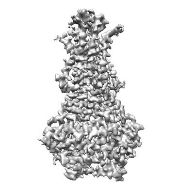 emd_7325.png
 http://ftp.pdbj.org/pub/emdb/structures/EMD-7325


 Z (Sec.)
 Y (Row.)
 X (Col.)


 Sample components
 Processing
 Electron microscopy
 FIELD EMISSION GUN
