Movie
Movie Controller


+ Open data

- Basic information
| Entry | Database: EMDB / ID: EMD-7318 | |||||||||
|---|---|---|---|---|---|---|---|---|---|---|
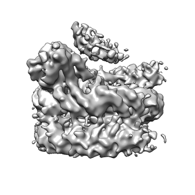
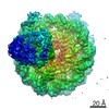
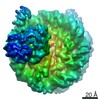
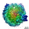
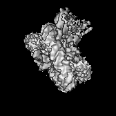
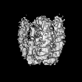
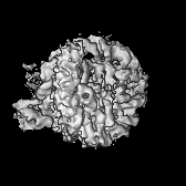
| Title | Cryo-EM structure of CENP-A nucleosome complexed with CENP-LN | |||||||||
 Map data Map data | Cryo-EM structure of CENP-A nucleosome complexed with CENP-LN | |||||||||
 Sample Sample |
| |||||||||
| Biological species |  Homo sapiens (human) Homo sapiens (human) | |||||||||
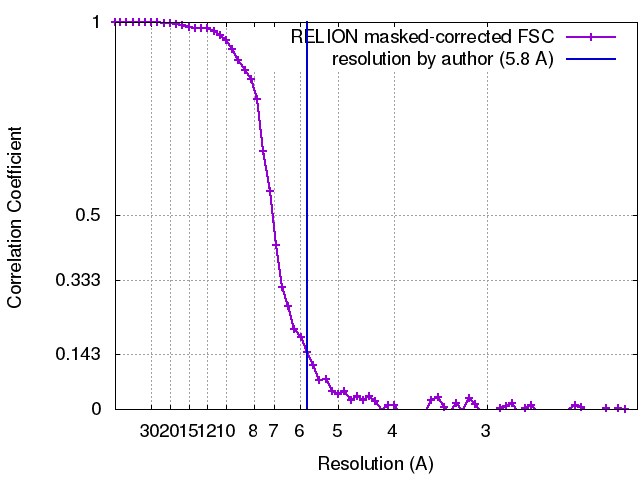
| Method | single particle reconstruction / cryo EM / Resolution: 5.8 Å | |||||||||
 Authors Authors | Li XR / Wang CL / Tian T / Zang JY / Zhou ZH | |||||||||
 Citation Citation | Journal: Cell Res / Year: 2018 Title: Molecular basis for CENP-N recognition of CENP-A nucleosome on the human kinetochore. Authors: Tian Tian / Xiaorun Li / Yingying Liu / Chengliang Wang / Xing Liu / Guoqiang Bi / Xuan Zhang / Xuebiao Yao / Z Hong Zhou / Jianye Zang /   | |||||||||
| History |
|
- Structure visualization
| Movie |
 Movie viewer Movie viewer |
|---|---|
| Structure viewer | EM map: SurfViewMolmilJmol/JSmol |
| Supplemental images |
- Downloads & links
-EMDB archive
| Map data | emd_7318.map.gz | 3 MB | EMDB map data format | |
|---|---|---|---|---|
| Header (meta data) | emd-7318-v30.xmlemd-7318.xml | 19.6 KB 19.6 KB | Display Display | EMDB header |
| FSC (resolution estimation) | emd_7318_fsc.xml | 6.1 KB | Display | FSC data file |
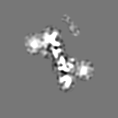
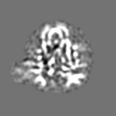
| Images |  emd_7318.png emd_7318.png | 56.7 KB | ||
| Archive directory |  http://ftp.pdbj.org/pub/emdb/structures/EMD-7318ftp://ftp.pdbj.org/pub/emdb/structures/EMD-7318 http://ftp.pdbj.org/pub/emdb/structures/EMD-7318ftp://ftp.pdbj.org/pub/emdb/structures/EMD-7318 | HTTPS FTP |
-Validation report
| Summary document | emd_7318_validation.pdf.gz | 79.2 KB | Display | EMDB validaton report |
|---|---|---|---|---|
| Full document | emd_7318_full_validation.pdf.gz | 78.2 KB | Display | |
| Data in XML | emd_7318_validation.xml.gz | 493 B | Display | |
| Arichive directory | https://ftp.pdbj.org/pub/emdb/validation_reports/EMD-7318ftp://ftp.pdbj.org/pub/emdb/validation_reports/EMD-7318 | HTTPS FTP |
-Related structure data
| Similar structure data |
|---|


-Links
| EMDB pages | EMDB (EBI/PDBe) / EMDataResource |
|---|
-Map
| File | Download / File: emd_7318.map.gz / Format: CCP4 / Size: 18.1 MB / Type: IMAGE STORED AS FLOATING POINT NUMBER (4 BYTES) | ||||||||||||||||||||||||||||||||||||||||||||||||||||||||||||
|---|---|---|---|---|---|---|---|---|---|---|---|---|---|---|---|---|---|---|---|---|---|---|---|---|---|---|---|---|---|---|---|---|---|---|---|---|---|---|---|---|---|---|---|---|---|---|---|---|---|---|---|---|---|---|---|---|---|---|---|---|---|
| Annotation | Cryo-EM structure of CENP-A nucleosome complexed with CENP-LN | ||||||||||||||||||||||||||||||||||||||||||||||||||||||||||||
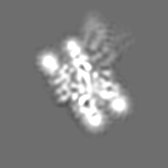
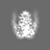
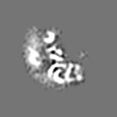
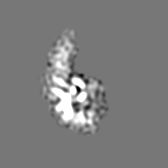
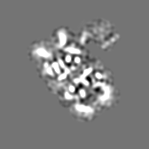
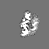
| Projections & slices | Image control
Images are generated by Spider. | ||||||||||||||||||||||||||||||||||||||||||||||||||||||||||||
| Voxel size | X=Y=Z: 1.07 Å | ||||||||||||||||||||||||||||||||||||||||||||||||||||||||||||
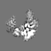
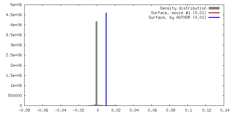
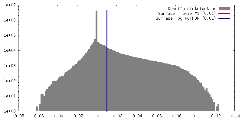
| Density |
| ||||||||||||||||||||||||||||||||||||||||||||||||||||||||||||
| Symmetry | Space group: 1 | ||||||||||||||||||||||||||||||||||||||||||||||||||||||||||||
| Details | EMDB XML:
CCP4 map header:
| ||||||||||||||||||||||||||||||||||||||||||||||||||||||||||||
 Z (Sec.)
 Y (Row.)
 X (Col.)


-Supplemental data
- Sample components
+Entire : CENP-A NCP/CENP-LN complex
+Supramolecule #1: CENP-A NCP/CENP-LN complex
+Supramolecule #2: CENP-A NCP

+Supramolecule #3: CENP-NL complex

 Spodoptera frugiperda (fall armyworm)
+Macromolecule #1: Histone H3-like centromeric protein A (CENP-A)
+Macromolecule #2: Histone H4
+Macromolecule #3: Histone H2A
+Macromolecule #4: Histone H2B
+Macromolecule #11: Centromere protein N (CENP-N)
+Macromolecule #12: Centromere protein L (CENP-L)
+Macromolecule #13: DNA (147-MER)
+Macromolecule #14: DNA (147-MER)
-Experimental details
-Structure determination
| Method | cryo EM |
|---|---|
 Processing Processing | single particle reconstruction |
| Aggregation state | particle |
-Sample preparation
| Concentration | 0.2 mg/mL |
|---|---|
| Buffer | pH: 7.4 |
| Grid | Model: Quantifoil R1.2/1.3 / Material: COPPER / Mesh: 300 / Pretreatment - Type: GLOW DISCHARGE |
| Vitrification | Cryogen name: ETHANE / Chamber humidity: 100 % / Chamber temperature: 293 K / Instrument: FEI VITROBOT MARK II |
- Electron microscopy #1
| Microscopy ID | 1 |
|---|---|
| Microscope | FEI TITAN |
| Image recording | Image recording ID: 1 / Film or detector model: GATAN K2 SUMMIT (4k x 4k) / Detector mode: SUPER-RESOLUTION / Average electron dose: 55.0 e/Å2 |
| Electron beam | Acceleration voltage: 300 kV / Electron source:  FIELD EMISSION GUN FIELD EMISSION GUN |
| Electron optics | Illumination mode: FLOOD BEAM / Imaging mode: BRIGHT FIELD |
-Electron microscopy #1~
| Microscopy ID | 1 |
|---|---|
| Microscope | FEI TECNAI 20 |
| Image recording | Image recording ID: 2 / Film or detector model: GATAN K2 SUMMIT (4k x 4k) / Detector mode: COUNTING / Average electron dose: 46.0 e/Å2 |
| Electron beam | Acceleration voltage: 200 kV / Electron source: FIELD EMISSION GUN |
| Electron optics | Illumination mode: FLOOD BEAM / Imaging mode: BRIGHT FIELD |