+ データを開く
- 基本情報
基本情報
| 登録情報 | データベース: EMDB / ID: EMD-7310 | |||||||||
|---|---|---|---|---|---|---|---|---|---|---|
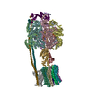
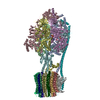
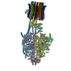
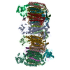
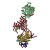
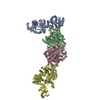
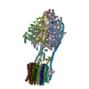
| タイトル | Structure of human PRC2 bound to a hetero-dinucleosome substrate with 35 bp linker DNA. 3D classification of signal subtracted particles lacking the unmodified substrate nucleosome. Example Class3. | |||||||||
 マップデータ マップデータ | Structure of human PRC2 bound to a hetero-dinucleosome substrate with 35 bp linker DNA. 3D classification of signal subtracted particles lacking the unmodified substrate nucleosome. Example Class3. | |||||||||
 試料 試料 |
| |||||||||
| 生物種 |  Homo sapiens (ヒト) Homo sapiens (ヒト) | |||||||||
| 手法 | 単粒子再構成法 / クライオ電子顕微鏡法 / 解像度: 10.1 Å | |||||||||
 データ登録者 データ登録者 | Poepsel S / Kasinath V / Nogales E | |||||||||
 引用 引用 |  ジャーナル: Nat Struct Mol Biol / 年: 2018 ジャーナル: Nat Struct Mol Biol / 年: 2018タイトル: Cryo-EM structures of PRC2 simultaneously engaged with two functionally distinct nucleosomes. 著者: Simon Poepsel / Vignesh Kasinath / Eva Nogales /  要旨: Epigenetic regulation is mediated by protein complexes that couple recognition of chromatin marks to activity or recruitment of chromatin-modifying enzymes. Polycomb repressive complex 2 (PRC2), a ...Epigenetic regulation is mediated by protein complexes that couple recognition of chromatin marks to activity or recruitment of chromatin-modifying enzymes. Polycomb repressive complex 2 (PRC2), a gene silencer that methylates lysine 27 of histone H3, is stimulated upon recognition of its own catalytic product and has been shown to be more active on dinucleosomes than H3 tails or single nucleosomes. These properties probably facilitate local H3K27me2/3 spreading, causing heterochromatin formation and gene repression. Here, cryo-EM reconstructions of human PRC2 bound to bifunctional dinucleosomes show how a single PRC2, via interactions with nucleosomal DNA, positions the H3 tails of the activating and substrate nucleosome to interact with the EED subunit and the SET domain of EZH2, respectively. We show how the geometry of the PRC2-DNA interactions allows PRC2 to accommodate varying lengths of the linker DNA between nucleosomes. Our structures illustrate how an epigenetic regulator engages with a complex chromatin substrate. | |||||||||
| 履歴 |
|
- 構造の表示
構造の表示
| ムービー |
 ムービービューア ムービービューア |
|---|---|
| 構造ビューア | EMマップ:  SurfView SurfView Molmil Molmil Jmol/JSmol Jmol/JSmol |
| 添付画像 |
- ダウンロードとリンク
-EMDBアーカイブ
| マップデータ |  emd_7310.map.gz emd_7310.map.gz | 166.5 MB |  EMDBマップデータ形式 EMDBマップデータ形式 | |
|---|---|---|---|---|
| ヘッダ (付随情報) |  emd-7310-v30.xml emd-7310-v30.xml emd-7310.xml emd-7310.xml | 10.2 KB 10.2 KB | 表示 表示 |  EMDBヘッダ EMDBヘッダ |
| FSC (解像度算出) |  emd_7310_fsc.xml emd_7310_fsc.xml | 12.5 KB | 表示 |  FSCデータファイル FSCデータファイル |
| 画像 |  emd_7310.png emd_7310.png | 42.2 KB | ||
| アーカイブディレクトリ |  http://ftp.pdbj.org/pub/emdb/structures/EMD-7310 http://ftp.pdbj.org/pub/emdb/structures/EMD-7310 ftp://ftp.pdbj.org/pub/emdb/structures/EMD-7310 ftp://ftp.pdbj.org/pub/emdb/structures/EMD-7310 | HTTPS FTP |
-検証レポート
| 文書・要旨 |  emd_7310_validation.pdf.gz emd_7310_validation.pdf.gz | 356.3 KB | 表示 |  EMDB検証レポート EMDB検証レポート |
|---|---|---|---|---|
| 文書・詳細版 |  emd_7310_full_validation.pdf.gz emd_7310_full_validation.pdf.gz | 355.8 KB | 表示 | |
| XML形式データ |  emd_7310_validation.xml.gz emd_7310_validation.xml.gz | 12.7 KB | 表示 | |
| アーカイブディレクトリ |  https://ftp.pdbj.org/pub/emdb/validation_reports/EMD-7310 https://ftp.pdbj.org/pub/emdb/validation_reports/EMD-7310 ftp://ftp.pdbj.org/pub/emdb/validation_reports/EMD-7310 ftp://ftp.pdbj.org/pub/emdb/validation_reports/EMD-7310 | HTTPS FTP |
-関連構造データ
- リンク
リンク
| EMDBのページ |  EMDB (EBI/PDBe) / EMDB (EBI/PDBe) /  EMDataResource EMDataResource |
|---|
- マップ
マップ
| ファイル |  ダウンロード / ファイル: emd_7310.map.gz / 形式: CCP4 / 大きさ: 178 MB / タイプ: IMAGE STORED AS FLOATING POINT NUMBER (4 BYTES) ダウンロード / ファイル: emd_7310.map.gz / 形式: CCP4 / 大きさ: 178 MB / タイプ: IMAGE STORED AS FLOATING POINT NUMBER (4 BYTES) | ||||||||||||||||||||||||||||||||||||||||||||||||||||||||||||||||||||
|---|---|---|---|---|---|---|---|---|---|---|---|---|---|---|---|---|---|---|---|---|---|---|---|---|---|---|---|---|---|---|---|---|---|---|---|---|---|---|---|---|---|---|---|---|---|---|---|---|---|---|---|---|---|---|---|---|---|---|---|---|---|---|---|---|---|---|---|---|---|
| 注釈 | Structure of human PRC2 bound to a hetero-dinucleosome substrate with 35 bp linker DNA. 3D classification of signal subtracted particles lacking the unmodified substrate nucleosome. Example Class3. | ||||||||||||||||||||||||||||||||||||||||||||||||||||||||||||||||||||
| 投影像・断面図 | 画像のコントロール
画像は Spider により作成 | ||||||||||||||||||||||||||||||||||||||||||||||||||||||||||||||||||||
| ボクセルのサイズ | X=Y=Z: 1.32 Å | ||||||||||||||||||||||||||||||||||||||||||||||||||||||||||||||||||||
| 密度 |
| ||||||||||||||||||||||||||||||||||||||||||||||||||||||||||||||||||||
| 対称性 | 空間群: 1 | ||||||||||||||||||||||||||||||||||||||||||||||||||||||||||||||||||||
| 詳細 | EMDB XML:
CCP4マップ ヘッダ情報:
| ||||||||||||||||||||||||||||||||||||||||||||||||||||||||||||||||||||
-添付データ
- 試料の構成要素
-全体 : Human Polycomb Repressive Complex 2 (PRC2) in complex with a hete...
| 全体 | 名称: Human Polycomb Repressive Complex 2 (PRC2) in complex with a heterodinucleosome composed of an unmodified nucleosome and a nucleosome carrying an H3K27 pseudo-trimethyl mark with 35 bp. ...名称: Human Polycomb Repressive Complex 2 (PRC2) in complex with a heterodinucleosome composed of an unmodified nucleosome and a nucleosome carrying an H3K27 pseudo-trimethyl mark with 35 bp. Subclassification of the overall reconstruction after signal subtraction. Example Class3. |
|---|---|
| 要素 |
|
-超分子 #1: Human Polycomb Repressive Complex 2 (PRC2) in complex with a hete...
| 超分子 | 名称: Human Polycomb Repressive Complex 2 (PRC2) in complex with a heterodinucleosome composed of an unmodified nucleosome and a nucleosome carrying an H3K27 pseudo-trimethyl mark with 35 bp. ...名称: Human Polycomb Repressive Complex 2 (PRC2) in complex with a heterodinucleosome composed of an unmodified nucleosome and a nucleosome carrying an H3K27 pseudo-trimethyl mark with 35 bp. Subclassification of the overall reconstruction after signal subtraction. Example Class3. タイプ: complex / ID: 1 / 親要素: 0 / 含まれる分子: #1 |
|---|---|
| 由来(天然) | 生物種:  Homo sapiens (ヒト) Homo sapiens (ヒト) |
| 組換発現 | 生物種:  Trichoplusia ni (イラクサキンウワバ) Trichoplusia ni (イラクサキンウワバ) |
-実験情報
-構造解析
| 手法 | クライオ電子顕微鏡法 |
|---|---|
 解析 解析 | 単粒子再構成法 |
| 試料の集合状態 | particle |
- 試料調製
試料調製
| 濃度 | 1 mg/mL |
|---|---|
| 緩衝液 | pH: 7.9 |
| 凍結 | 凍結剤: ETHANE / チャンバー内湿度: 100 % / チャンバー内温度: 281 K / 装置: FEI VITROBOT MARK IV |
- 電子顕微鏡法
| 顕微鏡 | FEI TITAN |
|---|---|
| 撮影 | フィルム・検出器のモデル: GATAN K2 SUMMIT (4k x 4k) 平均電子線量: 40.0 e/Å2 |
| 電子線 | 加速電圧: 300 kV / 電子線源:  FIELD EMISSION GUN FIELD EMISSION GUN |
| 電子光学系 | 照射モード: FLOOD BEAM / 撮影モード: BRIGHT FIELD |
 ムービー
ムービー コントローラー



 Z (Sec.)
 Y (Row.)
 X (Col.)


