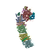
 Movie
Movie Controller


+ Open data

- Basic information
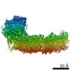
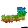
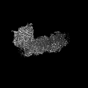
| Entry | Database: EMDB / ID: EMD-0850 | |||||||||
|---|---|---|---|---|---|---|---|---|---|---|
| Title | cryo-EM structure of cyanobacteria NDH-1LdelV complex | |||||||||
 Map data Map data | None | |||||||||
 Sample Sample |
| |||||||||
 Keywords Keywords | photosystem I / cyclic electron transfer / PHOTOSYNTHESIS | |||||||||
| Function / homology |  Function and homology information Function and homology informationTranslocases; Catalysing the translocation of protons; Linked to oxidoreductase reactions / NADH dehydrogenase complex / transmembrane transporter complex / photosynthetic electron transport chain / oxidoreductase activity, acting on NAD(P)H, quinone or similar compound as acceptor / plasma membrane-derived thylakoid membrane / photosynthesis, light reaction / ubiquinone binding / electron transport coupled proton transport / NADH dehydrogenase activity ...Translocases; Catalysing the translocation of protons; Linked to oxidoreductase reactions / NADH dehydrogenase complex / transmembrane transporter complex / photosynthetic electron transport chain / oxidoreductase activity, acting on NAD(P)H, quinone or similar compound as acceptor / plasma membrane-derived thylakoid membrane / photosynthesis, light reaction / ubiquinone binding / electron transport coupled proton transport / NADH dehydrogenase activity / respiratory chain complex I / NADH dehydrogenase (ubiquinone) activity / quinone binding / ATP synthesis coupled electron transport / endomembrane system / aerobic respiration / NAD binding / 4 iron, 4 sulfur cluster binding / iron ion binding / membrane / plasma membrane Similarity search - Function | |||||||||
| Biological species |   Thermosynechococcus elongatus BP-1 (bacteria) Thermosynechococcus elongatus BP-1 (bacteria) | |||||||||
| Method | single particle reconstruction / cryo EM / Resolution: 3.6 Å | |||||||||
 Authors Authors | Zhang C / Shuai J | |||||||||
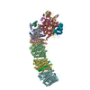
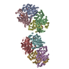
 Citation Citation | Journal: Nat Commun / Year: 2020 Title: Structural insights into NDH-1 mediated cyclic electron transfer. Authors: Chunli Zhang / Jin Shuai / Zhaoxing Ran / Jiaohong Zhao / Zhenfang Wu / Rijing Liao / Jian Wu / Weimin Ma / Ming Lei /  Abstract: NDH-1 is a key component of the cyclic-electron-transfer around photosystem I (PSI CET) pathway, an important antioxidant mechanism for efficient photosynthesis. Here, we report a 3.2-Å-resolution ...NDH-1 is a key component of the cyclic-electron-transfer around photosystem I (PSI CET) pathway, an important antioxidant mechanism for efficient photosynthesis. Here, we report a 3.2-Å-resolution cryo-EM structure of the ferredoxin (Fd)-NDH-1L complex from the cyanobacterium Thermosynechococcus elongatus. The structure reveals three β-carotene and fifteen lipid molecules in the membrane arm of NDH-1L. Regulatory oxygenic photosynthesis-specific (OPS) subunits NdhV, NdhS and NdhO are close to the Fd-binding site whilst NdhL is adjacent to the plastoquinone (PQ) cavity, and they play different roles in PSI CET under high-light stress. NdhV assists in the binding of Fd to NDH-1L and accelerates PSI CET in response to short-term high-light exposure. In contrast, prolonged high-light irradiation switches on the expression and assembly of the NDH-1MS complex, which likely contains no NdhO to further accelerate PSI CET and reduce ROS production. We propose that this hierarchical mechanism is necessary for the survival of cyanobacteria in an aerobic environment. | |||||||||
| History |
|
- Structure visualization
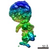
| Movie |
Movie viewer |
|---|---|
| Structure viewer | EM map: SurfViewMolmilJmol/JSmol |
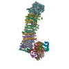
| Supplemental images |

- Downloads & links
-EMDB archive
| Map data | emd_0850.map.gz | 96.4 MB | EMDB map data format | |
|---|---|---|---|---|
| Header (meta data) | emd-0850-v30.xmlemd-0850.xml | 31.1 KB 31.1 KB | Display Display | EMDB header |
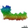
| Images | 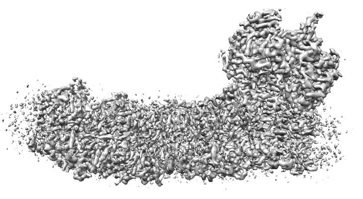 emd_0850.png emd_0850.png | 131.8 KB | ||
| Filedesc metadata | emd-0850.cif.gz | 8.6 KB | ||
| Archive directory |  http://ftp.pdbj.org/pub/emdb/structures/EMD-0850ftp://ftp.pdbj.org/pub/emdb/structures/EMD-0850 http://ftp.pdbj.org/pub/emdb/structures/EMD-0850ftp://ftp.pdbj.org/pub/emdb/structures/EMD-0850 | HTTPS FTP |
-Related structure data
| Related structure data |  6l7pMC  0849C 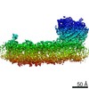 0851C  6l7oC C: citing same article ( M: atomic model generated by this map |
|---|---|
| Similar structure data |

-Links
| EMDB pages | EMDB (EBI/PDBe) / EMDataResource |
|---|---|
| Related items in Molecule of the Month |


-Map
| File | Download / File: emd_0850.map.gz / Format: CCP4 / Size: 103 MB / Type: IMAGE STORED AS FLOATING POINT NUMBER (4 BYTES) | ||||||||||||||||||||||||||||||||||||||||||||||||||||||||||||
|---|---|---|---|---|---|---|---|---|---|---|---|---|---|---|---|---|---|---|---|---|---|---|---|---|---|---|---|---|---|---|---|---|---|---|---|---|---|---|---|---|---|---|---|---|---|---|---|---|---|---|---|---|---|---|---|---|---|---|---|---|---|
| Annotation | None | ||||||||||||||||||||||||||||||||||||||||||||||||||||||||||||
| Projections & slices | Image control
Images are generated by Spider. | ||||||||||||||||||||||||||||||||||||||||||||||||||||||||||||
| Voxel size | X=Y=Z: 1.09 Å | ||||||||||||||||||||||||||||||||||||||||||||||||||||||||||||
| Density |
| ||||||||||||||||||||||||||||||||||||||||||||||||||||||||||||
| Symmetry | Space group: 1 | ||||||||||||||||||||||||||||||||||||||||||||||||||||||||||||
| Details | EMDB XML:
CCP4 map header:
| ||||||||||||||||||||||||||||||||||||||||||||||||||||||||||||
 Z (Sec.)
 Y (Row.)
 X (Col.)


-Supplemental data
- Sample components
+Entire : NDH-1LDelV
+Supramolecule #1: NDH-1LDelV
+Macromolecule #1: NAD(P)H-quinone oxidoreductase subunit 1
+Macromolecule #2: NAD(P)H-quinone oxidoreductase subunit 2
+Macromolecule #3: NAD(P)H-quinone oxidoreductase subunit 3
+Macromolecule #4: NAD(P)H-quinone oxidoreductase chain 4 1
+Macromolecule #5: NAD(P)H-quinone oxidoreductase subunit 4L
+Macromolecule #6: NADH dehydrogenase subunit 5
+Macromolecule #7: NADH-quinone oxidoreductase subunit J
+Macromolecule #8: NAD(P)H-quinone oxidoreductase subunit H
+Macromolecule #9: NAD(P)H-quinone oxidoreductase subunit I
+Macromolecule #10: NAD(P)H-quinone oxidoreductase subunit J
+Macromolecule #11: NAD(P)H-quinone oxidoreductase subunit K
+Macromolecule #12: NAD(P)H-quinone oxidoreductase subunit L
+Macromolecule #13: NAD(P)H-quinone oxidoreductase subunit M
+Macromolecule #14: NAD(P)H-quinone oxidoreductase subunit N
+Macromolecule #15: NAD(P)H-quinone oxidoreductase subunit O
+Macromolecule #16: NAD(P)H-quinone oxidoreductase subunit P
+Macromolecule #17: NAD(P)H-quinone oxidoreductase subunit Q
+Macromolecule #18: Tlr0636 protein
+Macromolecule #19: BETA-CAROTENE

+Macromolecule #20: 1,2-DIPALMITOYL-PHOSPHATIDYL-GLYCEROLE

+Macromolecule #21: DIGALACTOSYL DIACYL GLYCEROL (DGDG)

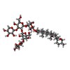
+Macromolecule #22: Digitonin
+Macromolecule #23: PHYLLOQUINONE

+Macromolecule #24: 1,2-DI-O-ACYL-3-O-[6-DEOXY-6-SULFO-ALPHA-D-GLUCOPYRANOSYL]-SN-GLYCEROL

+Macromolecule #25: IRON/SULFUR CLUSTER

-Experimental details
-Structure determination
| Method | cryo EM |
|---|---|
 Processing Processing | single particle reconstruction |
| Aggregation state | particle |
-Sample preparation
| Buffer | pH: 6 |
|---|---|
| Vitrification | Cryogen name: ETHANE |
- Electron microscopy
| Microscope | FEI TITAN KRIOS |
|---|---|
| Image recording | Film or detector model: FEI FALCON III (4k x 4k) / Average electron dose: 40.0 e/Å2 |
| Electron beam | Acceleration voltage: 300 kV / Electron source:  FIELD EMISSION GUN FIELD EMISSION GUN |
| Electron optics | Illumination mode: FLOOD BEAM / Imaging mode: BRIGHT FIELD |
| Experimental equipment |  Model: Titan Krios / Image courtesy: FEI Company |
-Image processing
| Startup model | Type of model: OTHER |
|---|---|
| Final reconstruction | Resolution.type: BY AUTHOR / Resolution: 3.6 Å / Resolution method: FSC 0.143 CUT-OFF / Number images used: 439946 |
| Initial angle assignment | Type: COMMON LINE |
| Final angle assignment | Type: MAXIMUM LIKELIHOOD |
