 Movie
Movie Controller


+ Open data

- Basic information
| Entry | Database: EMDB / ID: EMD-7298 | |||||||||
|---|---|---|---|---|---|---|---|---|---|---|
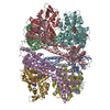
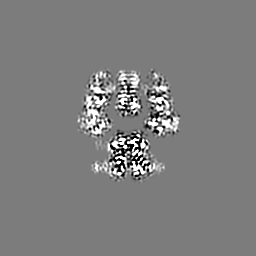
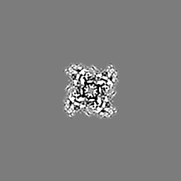
| Title | 4.1 angstrom Mg2+-unbound structure of mouse TRPM7 | |||||||||
 Map data Map data | ||||||||||
 Sample Sample |
| |||||||||
 Keywords Keywords | CryoEM / Truncated mouse TRPM7 / MEMBRANE PROTEIN | |||||||||
| Function / homology |  Function and homology information Function and homology informationcalcium-dependent cell-matrix adhesion / intracellular magnesium ion homeostasis / varicosity / magnesium ion transport / magnesium ion transmembrane transport / magnesium ion homeostasis / zinc ion transport / zinc ion transmembrane transporter activity / magnesium ion transmembrane transporter activity / TRP channels ...calcium-dependent cell-matrix adhesion / intracellular magnesium ion homeostasis / varicosity / magnesium ion transport / magnesium ion transmembrane transport / magnesium ion homeostasis / zinc ion transport / zinc ion transmembrane transporter activity / magnesium ion transmembrane transporter activity / TRP channels / actomyosin structure organization / necroptotic process / myosin binding / monoatomic cation transmembrane transport / monoatomic cation channel activity / ruffle / memory / calcium channel activity / calcium ion transmembrane transport / kinase activity / calcium ion transport / synaptic vesicle membrane / actin binding / cytoplasmic vesicle / protein homotetramerization / protein kinase activity / non-specific serine/threonine protein kinase / positive regulation of apoptotic process / protein serine kinase activity / protein serine/threonine kinase activity / neuronal cell body / ATP binding / metal ion binding / nucleus / plasma membrane Similarity search - Function | |||||||||
| Biological species |  | |||||||||
| Method | single particle reconstruction / cryo EM / Resolution: 4.1 Å | |||||||||
 Authors Authors | Zhang J / Li Z | |||||||||
 Citation Citation | Journal: Proc Natl Acad Sci U S A / Year: 2018 Title: Structure of the mammalian TRPM7, a magnesium channel required during embryonic development. Authors: Jingjing Duan / Zongli Li / Jian Li / Raymond E Hulse / Ana Santa-Cruz / William C Valinsky / Sunday A Abiria / Grigory Krapivinsky / Jin Zhang / David E Clapham /   Abstract: The transient receptor potential ion channel subfamily M, member 7 (TRPM7), is a ubiquitously expressed protein that is required for mouse embryonic development. TRPM7 contains both an ion channel ...The transient receptor potential ion channel subfamily M, member 7 (TRPM7), is a ubiquitously expressed protein that is required for mouse embryonic development. TRPM7 contains both an ion channel and an α-kinase. The channel domain comprises a nonselective cation channel with notable permeability to Mg and Zn Here, we report the closed state structures of the mouse TRPM7 channel domain in three different ionic conditions to overall resolutions of 3.3, 3.7, and 4.1 Å. The structures reveal key residues for an ion binding site in the selectivity filter, with proposed partially hydrated Mg ions occupying the center of the conduction pore. In high [Mg], a prominent external disulfide bond is found in the pore helix, which is essential for ion channel function. Our results provide a structural framework for understanding the TRPM1/3/6/7 subfamily and extend the knowledge base upon which to study the diversity and evolution of TRP channels. | |||||||||
| History |
|
- Structure visualization
| Movie |
Movie viewer |
|---|---|
| Structure viewer | EM map: SurfViewMolmilJmol/JSmol |
| Supplemental images |

- Downloads & links
-EMDB archive
| Map data | emd_7298.map.gz | 5.7 MB | EMDB map data format | |
|---|---|---|---|---|
| Header (meta data) | emd-7298-v30.xmlemd-7298.xml | 14.6 KB 14.6 KB | Display Display | EMDB header |
| Images | 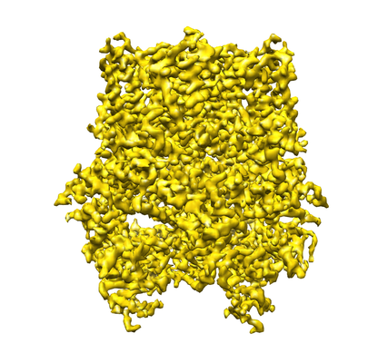 emd_7298.png emd_7298.png | 169.8 KB | ||
| Filedesc metadata | emd-7298.cif.gz | 6.1 KB | ||
| Archive directory |  http://ftp.pdbj.org/pub/emdb/structures/EMD-7298ftp://ftp.pdbj.org/pub/emdb/structures/EMD-7298 http://ftp.pdbj.org/pub/emdb/structures/EMD-7298ftp://ftp.pdbj.org/pub/emdb/structures/EMD-7298 | HTTPS FTP |
-Related structure data
| Related structure data |  6bwfMC  6975C  7297C  5zx5C  6bwdC M: atomic model generated by this map C: citing same article ( |
|---|---|
| Similar structure data |
-Links
| EMDB pages | EMDB (EBI/PDBe) / EMDataResource |
|---|
-Map
| File | Download / File: emd_7298.map.gz / Format: CCP4 / Size: 64 MB / Type: IMAGE STORED AS FLOATING POINT NUMBER (4 BYTES) | ||||||||||||||||||||||||||||||||||||||||||||||||||||||||||||
|---|---|---|---|---|---|---|---|---|---|---|---|---|---|---|---|---|---|---|---|---|---|---|---|---|---|---|---|---|---|---|---|---|---|---|---|---|---|---|---|---|---|---|---|---|---|---|---|---|---|---|---|---|---|---|---|---|---|---|---|---|---|
| Projections & slices | Image control
Images are generated by Spider. | ||||||||||||||||||||||||||||||||||||||||||||||||||||||||||||
| Voxel size | X=Y=Z: 1.23 Å | ||||||||||||||||||||||||||||||||||||||||||||||||||||||||||||
| Density |
| ||||||||||||||||||||||||||||||||||||||||||||||||||||||||||||
| Symmetry | Space group: 1 | ||||||||||||||||||||||||||||||||||||||||||||||||||||||||||||
| Details | EMDB XML:
CCP4 map header:
| ||||||||||||||||||||||||||||||||||||||||||||||||||||||||||||
 Z (Sec.)
 Y (Row.)
 X (Col.)


-Supplemental data
- Sample components
-Entire : Membrane Protein, Transient Receptor Potential ion channel
| Entire | Name: Membrane Protein, Transient Receptor Potential ion channel |
|---|---|
| Components |
|
-Supramolecule #1: Membrane Protein, Transient Receptor Potential ion channel
| Supramolecule | Name: Membrane Protein, Transient Receptor Potential ion channel type: cell / ID: 1 / Parent: 0 / Macromolecule list: all |
|---|---|
| Source (natural) | Organism: |
-Macromolecule #1: TRPM7
| Macromolecule | Name: TRPM7 / type: protein_or_peptide / ID: 1 / Number of copies: 4 / Enantiomer: LEVO |
|---|---|
| Source (natural) | Organism: |
| Molecular weight | Theoretical: 107.475609 KDa |
| Recombinant expression | Organism:   Spodoptera frugiperda (fall armyworm) Spodoptera frugiperda (fall armyworm) |
| Sequence | String: (UNK)(UNK)(UNK)(UNK)(UNK)(UNK)(UNK)(UNK)(UNK)(UNK) (UNK)(UNK)(UNK)(UNK)(UNK)(UNK) (UNK)(UNK)(UNK) (UNK)(UNK)(UNK)(UNK)(UNK)(UNK)(UNK)(UNK)(UNK)(UNK) (UNK)(UNK)(UNK) (UNK)(UNK)(UNK)(UNK)(UNK) ...String: (UNK)(UNK)(UNK)(UNK)(UNK)(UNK)(UNK)(UNK)(UNK)(UNK) (UNK)(UNK)(UNK)(UNK)(UNK)(UNK) (UNK)(UNK)(UNK) (UNK)(UNK)(UNK)(UNK)(UNK)(UNK)(UNK)(UNK)(UNK)(UNK) (UNK)(UNK)(UNK) (UNK)(UNK)(UNK)(UNK)(UNK)(UNK) (UNK)(UNK)(UNK)(UNK)(UNK)(UNK)(UNK)(UNK)(UNK)(UNK) (UNK)(UNK)(UNK)(UNK)(UNK)(UNK)(UNK)(UNK)(UNK) (UNK)(UNK)(UNK)(UNK)(UNK)(UNK)(UNK) (UNK)(UNK) (UNK)(UNK)(UNK)(UNK)(UNK)(UNK)(UNK)(UNK)(UNK)(UNK) (UNK)(UNK)(UNK)(UNK) (UNK)(UNK)(UNK)(UNK)(UNK) (UNK)(UNK)(UNK)(UNK)(UNK)(UNK)(UNK)(UNK)(UNK)(UNK) (UNK) (UNK)(UNK)(UNK)(UNK)(UNK)(UNK)(UNK)(UNK) (UNK)(UNK)(UNK)(UNK)(UNK)(UNK)(UNK)(UNK) (UNK) (UNK)(UNK)(UNK)(UNK)(UNK)(UNK)(UNK)KFL TIPRLEELYN TKQGPTNPML FHLIRDVKQG NLPPGYK IT LIDIGLVIEY LMGGTYRCTY TRKRFRLIYN SLGGNNRRSG RNTSSSTPQL RKSHETFGNR ADKKEKMRHN HFIKTAQP Y RPKMDASMEE GKKKRTKDEI VDIDDPETKR FPYPLNELLI WACLMKRQVM ARFLWQHGEE SMAKALVACK IYRSMAYEA KQSDLVDDTS EELKQYSNDF GQLAVELLEQ SFRQDETMAM KLLTYELKNW SNSTCLKLAV SSRLRPFVAH TCTQMLLSDM WMGRLNMRK NSWYKVILSI LVPPAILMLE YKTKAEMSHI PQSQDAHQMT MEDSENNFHN ITEEIPMEVF KEVKILDSSD G KNEMEIHI KSKKLPITRK FYAFYHAPIV KFWFNTLAYL GFLMLYTFVV LVKMEQLPSV QEWIVIAYIF TYAIEKVREV FM SEAGKIS QKIKVWFSDY FNVSDTIAII SFFVGFGLRF GAKWNYINAY DNHVFVAGRL IYCLNIIFWY VRLLDFLAVN QQA GPYVMM IGKMVANMFY IVVIMALVLL SFGVPRKAIL YPHEEPSWSL AKDIVFHPYW MIFGEVYAYE IDVCANDSTL PTIC GPGTW LTPFLQAVYL FVQYIIMVNL LIAFFNNVYL QVKAISNIVW KYQRYHFIMA YHEKPVLPPP LIILSHIVSL FCCVC KRRK KDKTSDGPKL FLTEEDQKKL HDFEEQCVEM YFDEKDDKFN SGSEERIRVT FERVEQMSIQ IKEVGDRVNY IKRSLQ SLD SQIGHLQDLS ALTVDTLKTL TAQKASEASK VHNEITRELS ISKHLAQNLI DDVPVRPLWK KPSAVNTLSS S |
-Experimental details
-Structure determination
| Method | cryo EM |
|---|---|
 Processing Processing | single particle reconstruction |
| Aggregation state | particle |
-Sample preparation
| Buffer | pH: 7.5 |
|---|---|
| Vitrification | Cryogen name: ETHANE / Instrument: FEI VITROBOT MARK I |
- Electron microscopy
| Microscope | FEI POLARA 300 |
|---|---|
| Image recording | Film or detector model: GATAN K2 BASE (4k x 4k) / Average electron dose: 56.0 e/Å2 |
| Electron beam | Acceleration voltage: 300 kV / Electron source:  FIELD EMISSION GUN FIELD EMISSION GUN |
| Electron optics | Illumination mode: FLOOD BEAM / Imaging mode: BRIGHT FIELD |
| Experimental equipment |  Model: Tecnai Polara / Image courtesy: FEI Company |
