 Movie
Movie Controller


+ Open data

- Basic information
| Entry | Database: PDB / ID: 6bwf | |||||||||||||||||||||||||||||||||||||||||||||||||||
|---|---|---|---|---|---|---|---|---|---|---|---|---|---|---|---|---|---|---|---|---|---|---|---|---|---|---|---|---|---|---|---|---|---|---|---|---|---|---|---|---|---|---|---|---|---|---|---|---|---|---|---|---|
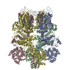
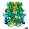
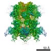
| Title | 4.1 angstrom Mg2+-unbound structure of mouse TRPM7 | |||||||||||||||||||||||||||||||||||||||||||||||||||
 Components Components | TRPM7 | |||||||||||||||||||||||||||||||||||||||||||||||||||
 Keywords Keywords | MEMBRANE PROTEIN / CryoEM / Truncated mouse TRPM7 | |||||||||||||||||||||||||||||||||||||||||||||||||||
| Function / homology |  Function and homology information Function and homology informationcalcium-dependent cell-matrix adhesion / intracellular magnesium ion homeostasis / varicosity / magnesium ion transport / magnesium ion transmembrane transport / magnesium ion homeostasis / zinc ion transport / zinc ion transmembrane transporter activity / magnesium ion transmembrane transporter activity / TRP channels ...calcium-dependent cell-matrix adhesion / intracellular magnesium ion homeostasis / varicosity / magnesium ion transport / magnesium ion transmembrane transport / magnesium ion homeostasis / zinc ion transport / zinc ion transmembrane transporter activity / magnesium ion transmembrane transporter activity / TRP channels / actomyosin structure organization / myosin binding / necroptotic process / monoatomic cation transmembrane transport / monoatomic cation channel activity / ruffle / calcium ion transmembrane transport / calcium channel activity / memory / kinase activity / calcium ion transport / synaptic vesicle membrane / actin binding / cytoplasmic vesicle / protein homotetramerization / protein kinase activity / non-specific serine/threonine protein kinase / positive regulation of apoptotic process / protein serine kinase activity / protein serine/threonine kinase activity / neuronal cell body / ATP binding / metal ion binding / nucleus / plasma membrane Similarity search - Function | |||||||||||||||||||||||||||||||||||||||||||||||||||
| Biological species |  | |||||||||||||||||||||||||||||||||||||||||||||||||||
| Method | ELECTRON MICROSCOPY / single particle reconstruction / cryo EM / Resolution: 4.1 Å | |||||||||||||||||||||||||||||||||||||||||||||||||||
 Authors Authors | Zhang, J. / Li, Z. / Duan, J. / Abiria, S.A. / Clapham, D.E. | |||||||||||||||||||||||||||||||||||||||||||||||||||
 Citation Citation | Journal: Proc Natl Acad Sci U S A / Year: 2018 Title: Structure of the mammalian TRPM7, a magnesium channel required during embryonic development. Authors: Jingjing Duan / Zongli Li / Jian Li / Raymond E Hulse / Ana Santa-Cruz / William C Valinsky / Sunday A Abiria / Grigory Krapivinsky / Jin Zhang / David E Clapham /   Abstract: The transient receptor potential ion channel subfamily M, member 7 (TRPM7), is a ubiquitously expressed protein that is required for mouse embryonic development. TRPM7 contains both an ion channel ...The transient receptor potential ion channel subfamily M, member 7 (TRPM7), is a ubiquitously expressed protein that is required for mouse embryonic development. TRPM7 contains both an ion channel and an α-kinase. The channel domain comprises a nonselective cation channel with notable permeability to Mg and Zn Here, we report the closed state structures of the mouse TRPM7 channel domain in three different ionic conditions to overall resolutions of 3.3, 3.7, and 4.1 Å. The structures reveal key residues for an ion binding site in the selectivity filter, with proposed partially hydrated Mg ions occupying the center of the conduction pore. In high [Mg], a prominent external disulfide bond is found in the pore helix, which is essential for ion channel function. Our results provide a structural framework for understanding the TRPM1/3/6/7 subfamily and extend the knowledge base upon which to study the diversity and evolution of TRP channels. | |||||||||||||||||||||||||||||||||||||||||||||||||||
| History |
|
- Structure visualization
| Movie |
Movie viewer |
|---|---|
| Structure viewer | Molecule: MolmilJmol/JSmol |
- Downloads & links
-Download
| PDBx/mmCIF format | 6bwf.cif.gz | 516.6 KB | Display | PDBx/mmCIF format |
|---|---|---|---|---|
| PDB format | pdb6bwf.ent.gz | 405.4 KB | Display | PDB format |
| PDBx/mmJSON format | 6bwf.json.gz | Tree view | PDBx/mmJSON format | |
| Others |  Other downloads Other downloads |
-Validation report
| Arichive directory | https://data.pdbj.org/pub/pdb/validation_reports/bw/6bwfftp://data.pdbj.org/pub/pdb/validation_reports/bw/6bwf | HTTPS FTP |
|---|
-Related structure data
| Related structure data |  7298MC  6975C  7297C  5zx5C  6bwdC M: map data used to model this data C: citing same article ( |
|---|---|
| Similar structure data |

-Links
 PDBj
PDBj- Assembly
| Deposited unit | 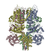
|
|---|---|
| 1 |
|
-Components
| #1: Protein | Mass: 107475.609 Da / Num. of mol.: 4 Source method: isolated from a genetically manipulated source Source: (gene. exp.)   Spodoptera frugiperda (fall armyworm) / References: UniProt: Q923J1*PLUS Spodoptera frugiperda (fall armyworm) / References: UniProt: Q923J1*PLUSHas protein modification | N | Sequence details | The full sample sequence is MSQKSWIESTLTKRECVYIIPSSKDPHRCLPGCQICQQLVRCFCGRLVKQHACFTASLAMKYSDV ...The full sample sequence is MSQKSWIEST | |
|---|
-Experimental details
-Experiment
| Experiment | Method: ELECTRON MICROSCOPY |
|---|---|
| EM experiment | Aggregation state: PARTICLE / 3D reconstruction method: single particle reconstruction |
- Sample preparation
| Component | Name: Membrane Protein, Transient Receptor Potential ion channel Type: CELL / Entity ID: all / Source: RECOMBINANT |
|---|---|
| Source (natural) | Organism: |
| Source (recombinant) | Organism: Spodoptera frugiperda (fall armyworm) |
| Buffer solution | pH: 7.5 |
| Specimen | Embedding applied: NO / Shadowing applied: NO / Staining applied: NO / Vitrification applied: YES |
| Vitrification | Instrument: FEI VITROBOT MARK I / Cryogen name: ETHANE |
- Electron microscopy imaging
| Experimental equipment |  Model: Tecnai Polara / Image courtesy: FEI Company |
|---|---|
| Microscopy | Model: FEI POLARA 300 |
| Electron gun | Electron source:  FIELD EMISSION GUN / Accelerating voltage: 300 kV / Illumination mode: FLOOD BEAM FIELD EMISSION GUN / Accelerating voltage: 300 kV / Illumination mode: FLOOD BEAM |
| Electron lens | Mode: BRIGHT FIELD |
| Image recording | Electron dose: 56 e/Å2 / Film or detector model: GATAN K2 BASE (4k x 4k) |
- Processing
| Software | Name: PHENIX / Version: 1.11.1_2575: / Classification: refinement | ||||||||||||||||||||||||
|---|---|---|---|---|---|---|---|---|---|---|---|---|---|---|---|---|---|---|---|---|---|---|---|---|---|
| EM software | Name: PHENIX / Category: model refinement | ||||||||||||||||||||||||
| CTF correction | Type: PHASE FLIPPING AND AMPLITUDE CORRECTION | ||||||||||||||||||||||||
| 3D reconstruction | Resolution: 4.1 Å / Resolution method: FSC 0.143 CUT-OFF / Num. of particles: 206032 / Symmetry type: POINT | ||||||||||||||||||||||||
| Refine LS restraints |
|
