[English] 日本語
Yorodumi
- EMDB-72497: V-shaped (channel-formed), ATP-bound, VX809-bound conformation of... -

+
Open data


ID or keywords:

Loading...

-
Basic information

Entry
Database: EMDB / ID: EMD-72497
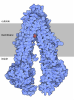
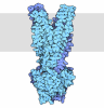
TitleV-shaped (channel-formed), ATP-bound, VX809-bound conformation of wild-type human CFTR (composite map from PHENIX based on consensus and local refinement maps from cryoSPARC)
Map data
Sample
  • Complex: Wild-type human CFTR solubilized in digitonin and cholesterol-hemisuccinate in the presence of 2 mM MgATP
    • Organelle or cellular component: Wild type human Cystic Fibrosis Transmembrane Conductance Regulator (hCFTR)
      • Protein or peptide: x 1 types
  • Ligand: x 14 types
Keywordscystic fibrosis / CFTR / nanobody / protein folding / MEMBRANE PROTEIN
Function / homology
Function and homology information


Sec61 translocon complex binding / channel-conductance-controlling ATPase / intracellularly ATP-gated chloride channel activity / positive regulation of enamel mineralization / RHO GTPases regulate CFTR trafficking / transepithelial water transport / intracellular pH elevation / amelogenesis / chloride channel inhibitor activity / multicellular organismal-level water homeostasis ...Sec61 translocon complex binding / channel-conductance-controlling ATPase / intracellularly ATP-gated chloride channel activity / positive regulation of enamel mineralization / RHO GTPases regulate CFTR trafficking / transepithelial water transport / intracellular pH elevation / amelogenesis / chloride channel inhibitor activity / multicellular organismal-level water homeostasis / water transport / chloride channel regulator activity / Golgi-associated vesicle membrane / bicarbonate transmembrane transporter activity / membrane hyperpolarization / bicarbonate transport / chloride transmembrane transporter activity / sperm capacitation / RHOQ GTPase cycle / chloride channel activity / ATPase-coupled transmembrane transporter activity / ABC-type transporter activity / 14-3-3 protein binding / cellular response to cAMP / response to endoplasmic reticulum stress / cellular response to forskolin / chloride transmembrane transport / Developmental Lineage of Pancreatic Ductal Cells / PDZ domain binding / clathrin-coated endocytic vesicle membrane / Late endosomal microautophagy / Defective CFTR causes cystic fibrosis / recycling endosome / transmembrane transport / ABC-family protein mediated transport / recycling endosome membrane / Chaperone Mediated Autophagy / Aggrephagy / Cargo recognition for clathrin-mediated endocytosis / Clathrin-mediated endocytosis / protein-folding chaperone binding / early endosome membrane / basolateral plasma membrane / early endosome / apical plasma membrane / endosome membrane / Ub-specific processing proteases / lysosomal membrane / endoplasmic reticulum membrane / enzyme binding / cell surface / ATP hydrolysis activity / protein-containing complex / ATP binding / membrane / nucleus / plasma membrane / cytosol / cytoplasm
Similarity search - Function
: / CFTR regulator domain / Cystic fibrosis TM conductance regulator (CFTR), regulator domain / Cystic fibrosis transmembrane conductance regulator / : / ABC transporter transmembrane region / ABC transporter type 1, transmembrane domain / ABC transporter integral membrane type-1 fused domain profile. / ABC transporter type 1, transmembrane domain superfamily / ABC transporter-like, conserved site ...: / CFTR regulator domain / Cystic fibrosis TM conductance regulator (CFTR), regulator domain / Cystic fibrosis transmembrane conductance regulator / : / ABC transporter transmembrane region / ABC transporter type 1, transmembrane domain / ABC transporter integral membrane type-1 fused domain profile. / ABC transporter type 1, transmembrane domain superfamily / ABC transporter-like, conserved site / ABC transporters family signature. / ABC transporter / ABC transporter-like, ATP-binding domain / ATP-binding cassette, ABC transporter-type domain profile. / ATPases associated with a variety of cellular activities / AAA+ ATPase domain / P-loop containing nucleoside triphosphate hydrolase
Similarity search - Domain/homology
Cystic fibrosis transmembrane conductance regulator
Similarity search - Component
Biological speciesHomo sapiens (human)
Methodsingle particle reconstruction / cryo EM / Resolution: 3.14 Å
AuthorsHunt JF / Paige AS / Baranwal J / Cohen BM / Goldberg PM / Wang C / Loughlin BJ / Kappes JC / Yang Z / Jiang F ...Hunt JF / Paige AS / Baranwal J / Cohen BM / Goldberg PM / Wang C / Loughlin BJ / Kappes JC / Yang Z / Jiang F / Govaerts C / Overtus M / Rich Z
Funding support United States, 1 items
OrganizationGrant numberCountry
Cystic Fibrosis FoundationHUNT13XX0, HUNT18G0, HUNT20G0, 004400G222-Hunt, 007997G224-Hunt, FRANK16XX0, FRANK18G0 United States
CitationJournal: To Be Published
Title: Nanobody-Driven Stabilization Synergistically Rescues F508del-CFTR and Reveals an Alternative Active State of the Channel
Authors: Hunt JF / Paige AS / Baranwal J / Cohen BM / Goldberg PM / Wang C / Loughlin BJ / Kappes JC / Yang Z / Jiang F / Govaerts C / Overtus M / Rich Z
History
DepositionSep 3, 2025-
Header (metadata) releaseSep 16, 2026-
Map releaseSep 16, 2026-
UpdateSep 16, 2026-
Current statusSep 16, 2026Processing site: RCSB / Status: Released

-
Structure visualization

Supplemental images

Downloads & links

-
Map

FileDownload / File: emd_72497.map.gz / Format: CCP4 / Size: 125 MB / Type: IMAGE STORED AS FLOATING POINT NUMBER (4 BYTES)
Projections & slices

Image control

Size
Brightness
Contrast
Others
AxesX (Sec.)Y (Row.)Z (Col.)
0.83 Å/pix.
x 320 pix.
= 265.6 Å
0.83 Å/pix.
x 320 pix.
= 265.6 Å
0.83 Å/pix.
x 320 pix.
= 265.6 Å

Surface

Projections

Slices (1/3)

Slices (1/2)

Slices (2/3)

Images are generated by Spider.

Voxel sizeX=Y=Z: 0.83 Å
Density
Contour LevelBy AUTHOR: 7.567
Minimum - Maximum-46.664290000000001 - 60.676093999999999
Average (Standard dev.)0.0006388041 (±1.2159761)
SymmetrySpace group: 1
Details

EMDB XML:

Map geometry
Axis orderZYX
Origin000
Dimensions320320320
Spacing320320320
CellA=B=C: 265.6 Å
α=β=γ: 90.0 °

-
Supplemental data

-
Half map: #2

Fileemd_72497_half_map_1.map
Projections & Slices
AxesZYX

Projections

Slices (1/2)
Density Histograms

-
Half map: #1

Fileemd_72497_half_map_2.map
Projections & Slices
AxesZYX

Projections

Slices (1/2)
Density Histograms

-
Sample components

+
Entire : Wild-type human CFTR solubilized in digitonin and cholesterol-hem...

EntireName: Wild-type human CFTR solubilized in digitonin and cholesterol-hemisuccinate in the presence of 2 mM MgATP
Components
  • Complex: Wild-type human CFTR solubilized in digitonin and cholesterol-hemisuccinate in the presence of 2 mM MgATP
    • Organelle or cellular component: Wild type human Cystic Fibrosis Transmembrane Conductance Regulator (hCFTR)
      • Protein or peptide: Cystic fibrosis transmembrane conductance regulator
  • Ligand: Digitonin
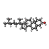
  • Ligand: CHOLESTEROL
  • Ligand: O-[(R)-{[(2R)-2,3-bis(octadecanoyloxy)propyl]oxy}(hydroxy)phosphoryl]-L-serine
  • Ligand: PHOSPHATIDYLETHANOLAMINE
  • Ligand: OLEIC ACID
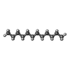
  • Ligand: PENTADECANE
  • Ligand: PALMITIC ACID
  • Ligand: DODECANE
  • Ligand: 1-PALMITOYL-2-LINOLEOYL-SN-GLYCERO-3-PHOSPHOCHOLINE
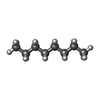
  • Ligand: N-OCTANE
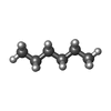
  • Ligand: HEXANE
  • Ligand: ADENOSINE-5'-TRIPHOSPHATE
  • Ligand: MAGNESIUM ION
  • Ligand: Lumacaftor

+
Supramolecule #1: Wild-type human CFTR solubilized in digitonin and cholesterol-hem...

SupramoleculeName: Wild-type human CFTR solubilized in digitonin and cholesterol-hemisuccinate in the presence of 2 mM MgATP
type: complex / ID: 1 / Parent: 0 / Macromolecule list: #1
Molecular weightTheoretical: 172.5 KDa

+
Supramolecule #2: Wild type human Cystic Fibrosis Transmembrane Conductance Regulat...

SupramoleculeName: Wild type human Cystic Fibrosis Transmembrane Conductance Regulator (hCFTR)
type: organelle_or_cellular_component / ID: 2 / Parent: 1 / Macromolecule list: #1
Source (natural)Organism: Homo sapiens (human)

+
Macromolecule #1: Cystic fibrosis transmembrane conductance regulator

MacromoleculeName: Cystic fibrosis transmembrane conductance regulator / type: protein_or_peptide / ID: 1
Details: The sequence assignment for residues 637-640, which was guided by an AlphaFold3 model, is tentative. The lipid model is based on inspection of density features exceeding the size of a water ...Details: The sequence assignment for residues 637-640, which was guided by an AlphaFold3 model, is tentative. The lipid model is based on inspection of density features exceeding the size of a water molecule at a 6.0 sigma contour level, which were modeled using the map at approximately a 4.5 sigma contour level. All those chemical and chemical fragment assignments are hypotheses guided by inspection of the density and the chemical environment, and none of them are supported by definitive evidence. The residue numbers of the modeled lipids are coded to indicate the confidence level of the chemical assignment and the extent of conservation between models for different hCFTR conformations. Residue numbers in the 4000, 5000, and 6000 series correspond to higher, lower, and lowest levels of confidence in the chemical assignments. Residue numbers in each of those series ending 1-299 designate sites at which binding of an equivalent lipid species is conserved in the model for at least one additional hCFTR conformation, while those ending 301-599 designate sites at which a different lipid species is bound in at least one additional hCFTR conformation. Residue numbers ending 601-899 designate sites not occupied by a lipid species in any other hCFTR conformations structurally characterized to date. Digitonin molecule L5001 is bound in a site at the C-terminus of NBD1 previously demonstrated to interact with hydrophobic drugs in a solution NMR study of the isolated domain (Hudson et al., 2017, Mol. Pharmacol. 92:124). This tentative assignment is supported by the observation of convincing density for digitonin at the same site in NBD-associated hCFTR structures, although the density in this structure, which exceeds the size of a water molecule at 6.5 sigma contour level, only approximately matches digitonin. All density features exceeding the size of a water molecule at a 6.0 sigma contour level have been modeled except for thirteen features proximal to the Alpha Helical Subdomains in NBD1 and NBD2 and the C-terminus of NBD1; these features exceed the size of a water molecule at contour levels from 6.0-8.5 sigma. The features proximal to the C-terminus of NBD1 likely represents residual density from partially ordered conformations of residues 641-668 at the N-terminus of the R Region. This inference is based on the observation that filtering the map to low resolution shows continuous density at this site that matches the shape of an alpha-helical hairpin formed by this protein segment in AlphaFold3 models. The features proximal to the Alpha Helical Subdomains in NBD1 and NBD2 seem likely to represent a combination of residual density from other disordered segments of hCFTR and noise from the local refinement algorithm used to generate the final composite map used for modeling.
Number of copies: 1 / Enantiomer: LEVO / EC number: channel-conductance-controlling ATPase
Source (natural)Organism: Homo sapiens (human)
Molecular weightTheoretical: 172.734984 KDa
Recombinant expressionOrganism: Cricetulus griseus (Chinese hamster)
SequenceString: MQRSPLEKAS VVSKLFFSWT RPILRKGYRQ RLELSDIYQI PSVDSADNLS EKLEREWDRE LASKKNPKLI NALRRCFFWR FMFYGIFLY LGEVTKAVQP LLLGRIIASY DPDNKEERSI AIYLGIGLCL LFIVRTLLLH PAIFGLHHIG MQMRIAMFSL I YKKTLKLS ...String:
MQRSPLEKAS VVSKLFFSWT RPILRKGYRQ RLELSDIYQI PSVDSADNLS EKLEREWDRE LASKKNPKLI NALRRCFFWR FMFYGIFLY LGEVTKAVQP LLLGRIIASY DPDNKEERSI AIYLGIGLCL LFIVRTLLLH PAIFGLHHIG MQMRIAMFSL I YKKTLKLS SRVLDKISIG QLVSLLSNNL NKFDEGLALA HFVWIAPLQV ALLMGLIWEL LQASAFCGLG FLIVLALFQA GL GRMMMKY RDQRAGKISE RLVITSEMIE NIQSVKAYCW EEAMEKMIEN LRQTELKLTR KAAYVRYFNS SAFFFSGFFV VFL SVLPYA LIKGIILRKI FTTISFCIVL RMAVTRQFPW AVQTWYDSLG AINKIQDFLQ KQEYKTLEYN LTTTEVVMEN VTAF WEEGF GELFEKAKQN NNNRKTSNGD DSLFFSNFSL LGTPVLKDIN FKIERGQLLA VAGSTGAGKT SLLMMIMGEL EPSEG KIKH SGRISFCSQF SWIMPGTIKE NIIFGVSYDE YRYRSVIKAC QLEEDISKFA EKDNIVLGEG GITLSGGQRA RISLAR AVY KDADLYLLDS PFGYLDVLTE KEIFESCVCK LMANKTRILV TSKMEHLKKA DKILILHEGS SYFYGTFSEL QNLQPDF SS KLMGCDSFDQ FSAERRNSIL TETLHRFSLE GDAPVSWTET KKQSFKQTGE FGEKRKNSIL NPINSIRKFS IVQKTPLQ M NGIEEDSDEP LERRLSLVPD SEQGEAILPR ISVISTGPTL QARRRQSVLN LMTHSVNQGQ NIHRKTTAST RKVSLAPQA NLTELDIYSR RLSQETGLEI SEEINEEDLK ECFFDDMESI PAVTTWNTYL RYITVHKSLI FVLIWCLVIF LAEVAASLVV LWLLGNTPL QDKGNSTHSR NNSYAVIITS TSSYYVFYIY VGVADTLLAM GFFRGLPLVH TLITVSKILH HKMLHSVLQA P MSTLNTLK AGGILNRFSK DIAILDDLLP LTIFDFIQLL LIVIGAIAVV AVLQPYIFVA TVPVIVAFIM LRAYFLQTSQ QL KQLESEG RSPIFTHLVT SLKGLWTLRA FGRQPYFETL FHKALNLHTA NWFLYLSTLR WFQMRIEMIF VIFFIAVTFI SIL TTGEGE GRVGIILTLA MNIMSTLQWA VNSSIDVDSL MRSVSRVFKF IDMPTEGKPT KSTKPYKNGQ LSKVMIIENS HVKK DDIWP SGGQMTVKDL TAKYTEGGNA ILENISFSIS PGQRVGLLGR TGSGKSTLLS AFLRLLNTEG EIQIDGVSWD SITLQ QWRK AFGVIPQKVF IFSGTFRKNL DPYEQWSDQE IWKVADEVGL RSVIEQFPGK LDFVLVDGGC VLSHGHKQLM CLARSV LSK AKILLLDEPS AHLDPVTYQI IRRTLKQAFA DCTVILCEHR IEAMLECQQF LVIEENKVRQ YDSIQKLLNE RSLFRQA IS PSDRVKLFPH RNSSKCKSKP QIAALKEETE EEVQDTRLLE ENLYFQGGGG SGGSWSHPQF EKAAAGGGSG GGSWSHPQ F EK

UniProtKB: Cystic fibrosis transmembrane conductance regulator

+
Macromolecule #2: Digitonin

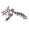
MacromoleculeName: Digitonin / type: ligand / ID: 2
Details: The lipid model is based on inspection of density features in the transmembrane region exceeding the size of a water molecule at a 5.5 sigma contour level, which were modeled using the map ...Details: The lipid model is based on inspection of density features in the transmembrane region exceeding the size of a water molecule at a 5.5 sigma contour level, which were modeled using the map at a 4.0 sigma contour level. The chemical and chemical fragment assignments at these sites are hypotheses guided by inspection of the density and the chemical environment. These assignments are not supported by any other experimental evidence, and they should all be considered placeholders for significant density features. Modeled lipids and lipid fragments were given residue numbers encoding to the confidence level of the chemical assignment (5000 series for high, 6000 series for medium, and 7000 series for low). A putative digitonin molecule modeled as residue 8001 is located in an extramembranous cavity on the surface of NBD1 that was previously shown by NMR to interact with hydrophobic drugs (https://pubmed.ncbi.nlm.nih.gov/28546419/). This prominent density feature was modeled because a similar feature is generally observed at the same site in maps for other conformations of human CFTR that have well-defined density for NBD1. Five smaller density features proximal to the surfaces of the NBDs that exceeded the 5.5 sigma threshold were not modeled because similar features are not observed in other conformations, and they likely represent noise in the local refinements of the subdomains of the NBDs that were used to generate the composite map used for modeling. A density feature proximal to the protein surface modeled as peptide chain P seems likely to represent a segment of the disordered glycosylated extracellular surface loop spanning residues 887-910.
Number of copies: 2 / Formula: AJP
Source (natural)Organism: Homo sapiens (human)
Molecular weightTheoretical: 1.229312 KDa
Chemical component information

ChemComp-AJP:
Digitonin / detergent*YM

+
Macromolecule #3: CHOLESTEROL

MacromoleculeName: CHOLESTEROL / type: ligand / ID: 3 / Number of copies: 10 / Formula: CLR
Molecular weightTheoretical: 386.654 Da
Chemical component information

ChemComp-CLR:
CHOLESTEROL

+
Macromolecule #4: O-[(R)-{[(2R)-2,3-bis(octadecanoyloxy)propyl]oxy}(hydroxy)phospho...
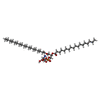
MacromoleculeName: O-[(R)-{[(2R)-2,3-bis(octadecanoyloxy)propyl]oxy}(hydroxy)phosphoryl]-L-serine
type: ligand / ID: 4 / Number of copies: 2 / Formula: P5S
Molecular weightTheoretical: 792.075 Da
Chemical component information

ChemComp-P5S:
O-[(R)-{[(2R)-2,3-bis(octadecanoyloxy)propyl]oxy}(hydroxy)phosphoryl]-L-serine

+
Macromolecule #5: PHOSPHATIDYLETHANOLAMINE
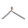
MacromoleculeName: PHOSPHATIDYLETHANOLAMINE / type: ligand / ID: 5 / Number of copies: 2 / Formula: PTY
Molecular weightTheoretical: 734.039 Da
Chemical component information

ChemComp-PTY:
PHOSPHATIDYLETHANOLAMINE / phospholipid*YM

+
Macromolecule #6: OLEIC ACID

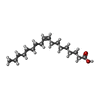
MacromoleculeName: OLEIC ACID / type: ligand / ID: 6 / Number of copies: 1 / Formula: OLA
Molecular weightTheoretical: 282.461 Da
Chemical component information

ChemComp-OLA:
OLEIC ACID

+
Macromolecule #7: PENTADECANE

MacromoleculeName: PENTADECANE / type: ligand / ID: 7 / Number of copies: 1 / Formula: MYS
Molecular weightTheoretical: 212.415 Da
Chemical component information

ChemComp-MYS:
PENTADECANE

+
Macromolecule #8: PALMITIC ACID

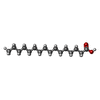
MacromoleculeName: PALMITIC ACID / type: ligand / ID: 8 / Number of copies: 1 / Formula: PLM
Molecular weightTheoretical: 256.424 Da
Chemical component information

ChemComp-PLM:
PALMITIC ACID

+
Macromolecule #9: DODECANE

MacromoleculeName: DODECANE / type: ligand / ID: 9 / Number of copies: 2 / Formula: D12
Molecular weightTheoretical: 170.335 Da
Chemical component information

ChemComp-D12:
DODECANE

+
Macromolecule #10: 1-PALMITOYL-2-LINOLEOYL-SN-GLYCERO-3-PHOSPHOCHOLINE
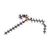
MacromoleculeName: 1-PALMITOYL-2-LINOLEOYL-SN-GLYCERO-3-PHOSPHOCHOLINE / type: ligand / ID: 10 / Number of copies: 1 / Formula: CPL
Molecular weightTheoretical: 758.06 Da
Chemical component information

ChemComp-CPL:
1-PALMITOYL-2-LINOLEOYL-SN-GLYCERO-3-PHOSPHOCHOLINE / phospholipid*YM

+
Macromolecule #11: N-OCTANE

MacromoleculeName: N-OCTANE / type: ligand / ID: 11 / Number of copies: 1 / Formula: OCT
Molecular weightTheoretical: 114.229 Da
Chemical component information

ChemComp-OCT:
N-OCTANE

+
Macromolecule #12: HEXANE

MacromoleculeName: HEXANE / type: ligand / ID: 12 / Number of copies: 1 / Formula: HEX
Molecular weightTheoretical: 86.175 Da
Chemical component information

ChemComp-HEX:
HEXANE

+
Macromolecule #13: ADENOSINE-5'-TRIPHOSPHATE
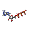
MacromoleculeName: ADENOSINE-5'-TRIPHOSPHATE / type: ligand / ID: 13 / Number of copies: 2 / Formula: ATP
Molecular weightTheoretical: 507.181 Da
Chemical component information

ChemComp-ATP:
ADENOSINE-5'-TRIPHOSPHATE / ATP, energy-carrying molecule*YM

+
Macromolecule #14: MAGNESIUM ION

MacromoleculeName: MAGNESIUM ION / type: ligand / ID: 14 / Number of copies: 2 / Formula: MG
Molecular weightTheoretical: 24.305 Da

+
Macromolecule #15: Lumacaftor

MacromoleculeName: Lumacaftor / type: ligand / ID: 15 / Number of copies: 1 / Formula: VX8
Molecular weightTheoretical: 452.407 Da
Chemical component information

ChemComp-VX8:
Lumacaftor

-
Experimental details

-
Structure determination

Methodcryo EM
Processingsingle particle reconstruction
Aggregation stateparticle

-
Sample preparation

Concentration1.5 mg/mL
BufferpH: 7.5
Component:
ConcentrationFormulaName
200.0 mMNaClSodium Chloride
3.0 mMMgCl2Magnesium Chloride
50.0 mMC4H11NO3Tris buffer
0.06 % (w/v)C56H92O29Digitonin
2.0 mMC10H16N5O13P3ATP
GridModel: Quantifoil R0.6/1 / Material: GOLD / Mesh: 300 / Support film - Material: GOLD / Support film - topology: HOLEY / Support film - Film thickness: 50 / Pretreatment - Type: PLASMA CLEANING / Pretreatment - Time: 25 sec. / Pretreatment - Atmosphere: OTHER
Details: The grid was treated in a Solarus Plasma Cleaner 950 (Gatan Inc., USA) for 25 sec with O2/H2 flow-rates of 27.5/6.4 sccm and 15 W cleaning power.
VitrificationCryogen name: ETHANE / Chamber humidity: 100 % / Chamber temperature: 277 K / Instrument: FEI VITROBOT MARK IV

-
Electron microscopy

MicroscopeTFS KRIOS
TemperatureMin: 85.0 K / Max: 90.0 K
Specialist opticsEnergy filter - Name: GIF Quantum ER / Energy filter - Slit width: 20 eV
Image recordingFilm or detector model: GATAN K3 BIOQUANTUM (6k x 4k) / Digitization - Dimensions - Width: 5760 pixel / Digitization - Dimensions - Height: 4092 pixel / Number grids imaged: 1 / Number real images: 11110 / Average exposure time: 2.5 sec. / Average electron dose: 58.0 e/Å2
Details: Movies comprised 40 frames collected in 2.5 seconds.
Electron beamAcceleration voltage: 300 kV / Electron source: FIELD EMISSION GUN
Electron opticsC2 aperture diameter: 100.0 µm / Illumination mode: FLOOD BEAM / Imaging mode: BRIGHT FIELD / Cs: 2.7 mm / Nominal defocus max: 2.9 µm / Nominal defocus min: 0.2 µm / Nominal magnification: 105000
Sample stageSpecimen holder model: FEI TITAN KRIOS AUTOGRID HOLDER / Cooling holder cryogen: NITROGEN
Experimental equipment
Model: Titan Krios / Image courtesy: FEI Company

+
Image processing

Particle selectionNumber selected: 2493670
Details: Particle count after low-resolution template picking, which was followed by light 2D cleaning. That process, which included all 2D classes roughly resembling views of CFTR, yielded 702,919 ...Details: Particle count after low-resolution template picking, which was followed by light 2D cleaning. That process, which included all 2D classes roughly resembling views of CFTR, yielded 702,919 particles that were input into iterative heterorefinement against a set of protein classes and 6 decoy volumes, resulting in 252,534 particles (42%) being assigned to the final set of protein classes.
CTF correctionSoftware - Name: cryoSPARC (ver. 4.3.1) / Software - details: Patch CTF correction / Type: PHASE FLIPPING AND AMPLITUDE CORRECTION
Startup modelType of model: NONE
Final reconstructionNumber classes used: 1 / Algorithm: BACK PROJECTION / Resolution.type: BY AUTHOR / Resolution: 3.14 Å / Resolution method: FSC 0.143 CUT-OFF / Software - Name: cryoSPARC (ver. 4.7.0) / Number images used: 81674
Initial angle assignmentType: MAXIMUM LIKELIHOOD / Software - Name: cryoSPARC (ver. 4.3.1)
Final angle assignmentType: MAXIMUM LIKELIHOOD / Software - Name: cryoSPARC (ver. 4.3.1)
Final 3D classificationNumber classes: 4 / Avg.num./class: 63346 / Software - Name: cryoSPARC (ver. 4.7.0)
FSC plot (resolution estimation)

-
Atomic model buiding 1

DetailsReal space refinement in PHENIX v 2.1_6048
Output model

PDB-9y4t:
V-shaped (channel-formed), ATP-bound, VX809-bound conformation of wild-type human CFTR (composite map from PHENIX based on consensus and local refinement maps from cryoSPARC)

+
About Yorodumi

-
News

-
Feb 9, 2022. New format data for meta-information of EMDB entries

New format data for meta-information of EMDB entries

  • Version 3 of the EMDB header file is now the official format.
  • The previous official version 1.9 will be removed from the archive.

Related info.:EMDB header

External links:wwPDB to switch to version 3 of the EMDB data model

-
Aug 12, 2020. Covid-19 info

Covid-19 info

URL: https://pdbj.org/emnavi/covid19.php

New page: Covid-19 featured information page in EM Navigator.

Related info.:Covid-19 info / Mar 5, 2020. Novel coronavirus structure data

+
Mar 5, 2020. Novel coronavirus structure data

Novel coronavirus structure data

Related info.:Yorodumi Speices / Aug 12, 2020. Covid-19 info

External links:COVID-19 featured content - PDBj / Molecule of the Month (242):Coronavirus Proteases

+
Jan 31, 2019. EMDB accession codes are about to change! (news from PDBe EMDB page)

EMDB accession codes are about to change! (news from PDBe EMDB page)

  • The allocation of 4 digits for EMDB accession codes will soon come to an end. Whilst these codes will remain in use, new EMDB accession codes will include an additional digit and will expand incrementally as the available range of codes is exhausted. The current 4-digit format prefixed with “EMD-” (i.e. EMD-XXXX) will advance to a 5-digit format (i.e. EMD-XXXXX), and so on. It is currently estimated that the 4-digit codes will be depleted around Spring 2019, at which point the 5-digit format will come into force.
  • The EM Navigator/Yorodumi systems omit the EMD- prefix.

Related info.:Q: What is EMD? / ID/Accession-code notation in Yorodumi/EM Navigator

External links:EMDB Accession Codes are Changing Soon! / Contact to PDBj

+
Jul 12, 2017. Major update of PDB

Major update of PDB

  • wwPDB released updated PDB data conforming to the new PDBx/mmCIF dictionary.
  • This is a major update changing the version number from 4 to 5, and with Remediation, in which all the entries are updated.
  • In this update, many items about electron microscopy experimental information are reorganized (e.g. em_software).
  • Now, EM Navigator and Yorodumi are based on the updated data.

External links:wwPDB Remediation / Enriched Model Files Conforming to OneDep Data Standards Now Available in the PDB FTP Archive

-
Yorodumi

Thousand views of thousand structures

  • Yorodumi is a browser for structure data from EMDB, PDB, SASBDB, etc.
  • This page is also the successor to EM Navigator detail page, and also detail information page/front-end page for Omokage search.
  • The word "yorodu" (or yorozu) is an old Japanese word meaning "ten thousand". "mi" (miru) is to see.

Related info.:EMDB / PDB / SASBDB / Comparison of 3 databanks / Yorodumi Search / Aug 31, 2016. New EM Navigator & Yorodumi / Yorodumi Papers / Jmol/JSmol / Function and homology information / Changes in new EM Navigator and Yorodumi

Read more