Parkin-FBXW7-Cul1 ubiquitin ligase complex / negative regulation of beige fat cell differentiation / cullin-RING-type E3 NEDD8 transferase / NEDD8 transferase activity / cullin-RING ubiquitin ligase complex / Cul7-RING ubiquitin ligase complex / cellular response to chemical stress / Loss of Function of FBXW7 in Cancer and NOTCH1 Signaling / positive regulation of protein autoubiquitination / RNA polymerase II transcription initiation surveillance ...Parkin-FBXW7-Cul1 ubiquitin ligase complex / negative regulation of beige fat cell differentiation / cullin-RING-type E3 NEDD8 transferase / NEDD8 transferase activity / cullin-RING ubiquitin ligase complex / Cul7-RING ubiquitin ligase complex / cellular response to chemical stress / Loss of Function of FBXW7 in Cancer and NOTCH1 Signaling / positive regulation of protein autoubiquitination / RNA polymerase II transcription initiation surveillance / protein neddylation / NEDD8 ligase activity / protein K27-linked ubiquitination / negative regulation of response to oxidative stress / VCB complex / Cul5-RING ubiquitin ligase complex / ubiquitin-ubiquitin ligase activity / ubiquitin-dependent protein catabolic process via the C-end degron rule pathway / SCF ubiquitin ligase complex / Cul2-RING ubiquitin ligase complex / Cul3-RING ubiquitin ligase complex / negative regulation of type I interferon production / SCF-dependent proteasomal ubiquitin-dependent protein catabolic process / Prolactin receptor signaling / negative regulation of mitophagy / Cul4A-RING E3 ubiquitin ligase complex / Cul4-RING E3 ubiquitin ligase complex / Cul4B-RING E3 ubiquitin ligase complex / ubiquitin ligase complex scaffold activity / cullin family protein binding / protein monoubiquitination / site of DNA damage / signal transduction in response to DNA damage / Nuclear events stimulated by ALK signaling in cancer / protein K48-linked ubiquitination / negative regulation of insulin receptor signaling pathway / regulation of cellular response to insulin stimulus / positive regulation of TORC1 signaling / transcription-coupled nucleotide-excision repair / post-translational protein modification / intrinsic apoptotic signaling pathway / animal organ morphogenesis / T cell activation / Regulation of BACH1 activity / MAP3K8 (TPL2)-dependent MAPK1/3 activation / negative regulation of canonical NF-kappaB signal transduction / cellular response to amino acid stimulus / SCF-beta-TrCP mediated degradation of Emi1 / NIK-->noncanonical NF-kB signaling / negative regulation of canonical Wnt signaling pathway / G1/S transition of mitotic cell cycle / Dectin-1 mediated noncanonical NF-kB signaling / Degradation of DVL / Degradation of CRY and PER proteins / Iron uptake and transport / Activation of NF-kappaB in B cells / Degradation of GLI1 by the proteasome / Recognition of DNA damage by PCNA-containing replication complex / RING-type E3 ubiquitin transferase / GSK3B and BTRC:CUL1-mediated-degradation of NFE2L2 / Negative regulation of NOTCH4 signaling / Hedgehog 'on' state / FBXL7 down-regulates AURKA during mitotic entry and in early mitosis / Vif-mediated degradation of APOBEC3G / Degradation of GLI2 by the proteasome / GLI3 is processed to GLI3R by the proteasome / Ubiquitin-Mediated Degradation of Phosphorylated Cdc25A / NOTCH1 Intracellular Domain Regulates Transcription / Evasion by RSV of host interferon responses / Degradation of beta-catenin by the destruction complex / DNA Damage Recognition in GG-NER / Oxygen-dependent proline hydroxylation of Hypoxia-inducible Factor Alpha / Constitutive Signaling by NOTCH1 PEST Domain Mutants / Constitutive Signaling by NOTCH1 HD+PEST Domain Mutants / CLEC7A (Dectin-1) signaling / Dual Incision in GG-NER / Transcription-Coupled Nucleotide Excision Repair (TC-NER) / SCF(Skp2)-mediated degradation of p27/p21 / Formation of TC-NER Pre-Incision Complex / FCERI mediated NF-kB activation / Regulation of expression of SLITs and ROBOs / Formation of Incision Complex in GG-NER / Interleukin-1 signaling / Orc1 removal from chromatin / protein polyubiquitination / ubiquitin-protein transferase activity / Cyclin D associated events in G1 / Dual incision in TC-NER / positive regulation of protein catabolic process / Gap-filling DNA repair synthesis and ligation in TC-NER / Regulation of RUNX2 expression and activity / Regulation of RAS by GAPs / cellular response to UV / ubiquitin protein ligase activity / Regulation of PLK1 Activity at G2/M Transition / KEAP1-NFE2L2 pathway / MAPK cascade / Downstream TCR signaling / positive regulation of proteasomal ubiquitin-dependent protein catabolic process / Antigen processing: Ubiquitination & Proteasome degradation Similarity search - Function
National Institutes of Health/National Institute of General Medical Sciences (NIH/NIGMS)
R35 GM136401
United States
National Institutes of Health/National Cancer Institute (NIH/NCI)
P01 CA092584
United States
Citation
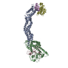
Journal: Nat Commun / Year: 2026 Title: Structural basis for fork reversal and RAD51 regulation by the SCF ubiquitin ligase complex of F-box helicase 1. Authors: Briana H Greer / Javier Mendia-Garcia / Elwood A Mullins / Emma M Peacock / Sander K Haigh / Carl J Schiltz / Clara Aicart-Ramos / Miaw-Sheue Tsai / David Cortez / Fernando Moreno-Herrero / Brandt F Eichman / Abstract: Replication fork reversal helps maintain genomic stability during replication stress. F-box helicase 1 (FBH1) catalyzes fork reversal and is an SCF (SKP-CUL1-F-box) E3 ubiquitin ligase that limits ...Replication fork reversal helps maintain genomic stability during replication stress. F-box helicase 1 (FBH1) catalyzes fork reversal and is an SCF (SKP-CUL1-F-box) E3 ubiquitin ligase that limits RAD51 association with chromatin. Here, we show that preferential binding of SCF to the lagging strand template at DNA fork structures stimulates helicase activity and is required for fork reversal. A cryo-EM structure of SCF bound to DNA representing a stalled fork reveals an intimate interaction between FBH1 and the fork junction. Disruption of this interface severely curtails fork reversal in vitro and replication progression in cells, providing a model for how ssDNA translocation by FBH1 facilitates annealing of parental DNA by a fundamentally different mechanism than the fork remodelers SMARCAL, HLTF, and ZRANB3. The structure provides a model for SCF disassembly of RAD51 filaments through translocation and ubiquitination, and implies that RAD51 is associated with the lagging strand at stalled forks.
In the structure databanks used in Yorodumi, some data are registered as the other names, "COVID-19 virus" and "2019-nCoV". Here are the details of the virus and the list of structure data.
Jan 31, 2019. EMDB accession codes are about to change! (news from PDBe EMDB page)
EMDB accession codes are about to change! (news from PDBe EMDB page)
The allocation of 4 digits for EMDB accession codes will soon come to an end. Whilst these codes will remain in use, new EMDB accession codes will include an additional digit and will expand incrementally as the available range of codes is exhausted. The current 4-digit format prefixed with “EMD-” (i.e. EMD-XXXX) will advance to a 5-digit format (i.e. EMD-XXXXX), and so on. It is currently estimated that the 4-digit codes will be depleted around Spring 2019, at which point the 5-digit format will come into force.
The EM Navigator/Yorodumi systems omit the EMD- prefix.
Related info.:Q: What is EMD? / ID/Accession-code notation in Yorodumi/EM Navigator
Yorodumi is a browser for structure data from EMDB, PDB, SASBDB, etc.
This page is also the successor to EM Navigator detail page, and also detail information page/front-end page for Omokage search.
The word "yorodu" (or yorozu) is an old Japanese word meaning "ten thousand". "mi" (miru) is to see.
Related info.:EMDB / PDB / SASBDB / Comparison of 3 databanks / Yorodumi Search / Aug 31, 2016. New EM Navigator & Yorodumi / Yorodumi Papers / Jmol/JSmol / Function and homology information / Changes in new EM Navigator and Yorodumi
 Movie
Movie Controller

 Yorodumi
 Open data
 Basic information
 Map data
 Sample
 Keywords
 Function and homology information
 Homo sapiens (human) / synthetic construct (others)
 Authors
 United States, 2 items
  Citation
 Structure visualization
 Downloads & links
 emd_72359.png
 http://ftp.pdbj.org/pub/emdb/structures/EMD-72359


 Z (Sec.)
 Y (Row.)
 X (Col.)


 Sample components
 Spodoptera frugiperda (fall armyworm)
 Processing
 Electron microscopy
 FIELD EMISSION GUN