 Movie
Movie Controller

[English] 日本語
 Yorodumi
- EMDB-72130: Negative Stain EM map of KSHV glycoprotein gHgL in complex with M... -
+ Open data

- Basic information
| Entry |  | |||||||||
|---|---|---|---|---|---|---|---|---|---|---|
| Title | Negative Stain EM map of KSHV glycoprotein gHgL in complex with MLKH1 FAB | |||||||||
 Map data Map data | ||||||||||
 Sample Sample |
| |||||||||
 Keywords Keywords | Viral protein / Complex / Antibody / KSHV / IMMUNE SYSTEM | |||||||||
| Function / homology |  Function and homology information Function and homology informationhost cell endosome membrane / host cell Golgi apparatus / fusion of virus membrane with host plasma membrane / viral envelope / symbiont entry into host cell / host cell plasma membrane / virion membrane Similarity search - Function | |||||||||
| Biological species |   Human gammaherpesvirus 8 / Human gammaherpesvirus 8 /  | |||||||||
| Method | single particle reconstruction / negative staining / Resolution: 15.6 Å | |||||||||
 Authors Authors | Kher G / Aldridge NT / Lang K / Pancera M | |||||||||
| Funding support |  United States, 1 items United States, 1 items
| |||||||||
 Citation Citation | Journal: PLoS Pathog / Year: 2026 Title: Monoclonal neutralizing antibodies elicited by infection with Kaposi sarcoma-associated herpesvirus reveal critical sites of vulnerability on gH/gL. Authors: Yu-Hsin Wan / Sara Pernikoff / Nicholas T Aldridge / Kevin Lang / Holly M Dudley / Samuel C Scharffenberger / Gargi Kher / Warren Phipps / Marie Pancera / Jim Boonyaratanakornkit / Andrew T McGuire / Abstract: Kaposi sarcoma-associated herpesvirus (KSHV) is an oncogenic virus that causes Kaposi sarcoma, primary effusion lymphoma and multicentric Castleman disease. A vaccine that prevents KSHV infection or ...Kaposi sarcoma-associated herpesvirus (KSHV) is an oncogenic virus that causes Kaposi sarcoma, primary effusion lymphoma and multicentric Castleman disease. A vaccine that prevents KSHV infection or serves in the treatment of KSHV-related diseases represents a critical unmet need, however, the types of immune responses a vaccine should elicit have not been well defined. The gH/gL glycoprotein complex is an important target of KSHV-neutralizing antibodies, but the epitope specificities targeted by these antibodies remain unknown. Here, we isolated 12 gH/gL-specific monoclonal antibodies (mAbs) from KSHV-infected donors and performed structure/function analyses. These mAbs bind recombinant gH/gL with nanomolar affinities and epitope binning analyses revealed that the mAbs bind to 5 epitope clusters on gH/gL. Seven mAbs were able to neutralize KSHV infection of epithelial cell lines. Two potent neutralizing mAbs mapped to the EphA2 binding site as determined by inhibition of the receptor-ligand interaction and negative stain electron microscopy (nsEM) of the mAb/gH/gL complex. The epitopes of other neutralizing mAbs targeting novel sites of vulnerability were determined by a combination of cryogenic electron microscopy and nsEM. Together, these mAbs help to define the relevant epitope targets for KSHV vaccine design, have utility in understanding the role of antibodies in preventing KSHV infection, enable the development of immunotherapy approaches, and provide valuable tools to understand the molecular details of the KSHV entry process. | |||||||||
| History |
|
- Structure visualization
| Supplemental images |
|---|

- Downloads & links
-EMDB archive
| Map data | emd_72130.map.gz | 403.9 KB | EMDB map data format | |
|---|---|---|---|---|
| Header (meta data) | emd-72130-v30.xmlemd-72130.xml | 18 KB 18 KB | Display Display | EMDB header |
| FSC (resolution estimation) | emd_72130_fsc.xml | 8.6 KB | Display | FSC data file |
| Images |  emd_72130.png emd_72130.png | 20.2 KB | ||
| Filedesc metadata | emd-72130.cif.gz | 5.6 KB | ||
| Others | emd_72130_half_map_1.map.gzemd_72130_half_map_2.map.gz | 59.3 MB 59.3 MB | ||
| Archive directory |  http://ftp.pdbj.org/pub/emdb/structures/EMD-72130ftp://ftp.pdbj.org/pub/emdb/structures/EMD-72130 http://ftp.pdbj.org/pub/emdb/structures/EMD-72130ftp://ftp.pdbj.org/pub/emdb/structures/EMD-72130 | HTTPS FTP |
-Related structure data


-Links
| EMDB pages | EMDB (EBI/PDBe) / EMDataResource |
|---|
-Map
| File | Download / File: emd_72130.map.gz / Format: CCP4 / Size: 432.6 KB / Type: IMAGE STORED AS FLOATING POINT NUMBER (4 BYTES) | ||||||||||||||||||||||||||||||||||||
|---|---|---|---|---|---|---|---|---|---|---|---|---|---|---|---|---|---|---|---|---|---|---|---|---|---|---|---|---|---|---|---|---|---|---|---|---|---|
| Projections & slices | Image control
Images are generated by Spider. | ||||||||||||||||||||||||||||||||||||
| Voxel size | X=Y=Z: 8.42667 Å | ||||||||||||||||||||||||||||||||||||
| Density |
| ||||||||||||||||||||||||||||||||||||
| Symmetry | Space group: 1 | ||||||||||||||||||||||||||||||||||||
| Details | EMDB XML:
|
 Z (Sec.)
 Y (Row.)
 X (Col.)


-Supplemental data
-Half map: #1
| File | emd_72130_half_map_1.map | ||||||||||||
|---|---|---|---|---|---|---|---|---|---|---|---|---|---|
| Projections & Slices |
| ||||||||||||
| Density Histograms |


-Half map: #2
| File | emd_72130_half_map_2.map | ||||||||||||
|---|---|---|---|---|---|---|---|---|---|---|---|---|---|
| Projections & Slices |
| ||||||||||||
| Density Histograms |


- Sample components
-Entire : KSHV glycoprotein Heterodimer gHgl 1:1 MLKH1 FAB
| Entire | Name: KSHV glycoprotein Heterodimer gHgl 1:1 MLKH1 FAB |
|---|---|
| Components |
|
-Supramolecule #1: KSHV glycoprotein Heterodimer gHgl 1:1 MLKH1 FAB
| Supramolecule | Name: KSHV glycoprotein Heterodimer gHgl 1:1 MLKH1 FAB / type: complex / ID: 1 / Parent: 0 / Macromolecule list: all |
|---|---|
| Molecular weight | Theoretical: 145 KDa |
-Macromolecule #1: Glycoprotein gH
| Macromolecule | Name: Glycoprotein gH / type: protein_or_peptide / ID: 1 / Enantiomer: LEVO |
|---|---|
| Source (natural) | Organism: Human gammaherpesvirus 8 / Strain: GK18 |
| Recombinant expression | Organism:  Homo sapiens (human) Homo sapiens (human) |
| Sequence | String: MDAMKRGLCC VLLLCGAVFV SPSASATGAL PTTATTITRS ATQLINGRTN LSIELEFNGT SFFLNWQNLL NVITEPALTE LWTSAEVAED LRVTLKKRQS LFFPNKTVVI SGDGHRYTCE VPTSSQTYNI TKGFNYSALP GHLGGFGINA RLVLGDIFAS KWSLFARDTP ...String: MDAMKRGLCC VLLLCGAVFV SPSASATGAL PTTATTITRS ATQLINGRTN LSIELEFNGT SFFLNWQNLL NVITEPALTE LWTSAEVAED LRVTLKKRQS LFFPNKTVVI SGDGHRYTCE VPTSSQTYNI TKGFNYSALP GHLGGFGINA RLVLGDIFAS KWSLFARDTP EYRVFYPMNV MAVKFSISIG NNESGVALYG VVSEDFVVVT LHNRSKEANE TASHLLFGLP DSLPSLKGHA TYDELTFARN AKYALVAILP KDSYQTLLTE NYTRIFLNMT ESTPLEFTRT IQTRIVSIEA RRACAAQEAA PDIFLVLFQM LVAHFLVARG IAEHRFVEVD CVCRQYAELY FLRRISRLCM PTFTTVGYNH TTLGAVAATQ IARVSATKLA SLPRSSQETV LAMVQLGARD GAVPSSILEG IAMVVEHMYT AYTYVYTLGD TERKLMLDIH TVLTDSCPPK DSGVSEKLLR TYLMFTSMCT NIELGEMIAR FSKPDSLNIY RAFSPCFLGL RYDLHPAKLR AEAPQSSALT RTAVARGTSG FAELLHALHL DSLNLIPAIN CSKITADKII ATVPLPHVTY IISSEALSNA VVYEVSEIFL KSAMFISAIK PDCSGFNFSQ IDRHIPIVYN ISTPRRGCPL CDSVIMSYDE SDGLQSLMYV TNERVQTNLF LDKSPFFDNN NLHIHYLWLR DNGTVVEIRG MYGSGSGHHH HHH UniProtKB: Envelope glycoprotein H |
-Macromolecule #2: Glycoprotein gL
| Macromolecule | Name: Glycoprotein gL / type: protein_or_peptide / ID: 2 / Enantiomer: LEVO |
|---|---|
| Source (natural) | Organism: Human gammaherpesvirus 8 / Strain: GK18 |
| Recombinant expression | Organism: Homo sapiens (human) |
| Sequence | String: MDAMKRGLCC VLLLCGAVFV SPSASDGIQY VALPCCAIQA SAASTLPLFF AVHSIHFADP NHCNGVCIAK LRSKTGDITV ETCVNGFNLR SFLVAVVRRL GSWASQENLR LLWYLQRSLT AYTVGFNATT ADSSIHNVNI IIISVGKAMN RTGSVSGSQT RAKSSSRRAH AGQKGK UniProtKB: Envelope glycoprotein L |
-Macromolecule #3: MLKH1 FAB Ligth Chain
| Macromolecule | Name: MLKH1 FAB Ligth Chain / type: protein_or_peptide / ID: 3 / Enantiomer: LEVO |
|---|---|
| Source (natural) | Organism: |
| Recombinant expression | Organism: Homo sapiens (human) |
| Sequence | String: MGWSCIILFL VATATGVHSE IVLTQSPATL SVSPGERATL SCRASQSVSG YLAWYQQKPG QAPRLLIYAA STRATGVPG RFSGSGSGTE FTLTISSLQS EDFAVYYCQQ YTDWPPRLTF GGGTKVEIKR TVAAPSVFIF P PSDEQLKS GTASVVCLLN NFYPREAKVQ ...String: MGWSCIILFL VATATGVHSE IVLTQSPATL SVSPGERATL SCRASQSVSG YLAWYQQKPG QAPRLLIYAA STRATGVPG RFSGSGSGTE FTLTISSLQS EDFAVYYCQQ YTDWPPRLTF GGGTKVEIKR TVAAPSVFIF P PSDEQLKS GTASVVCLLN NFYPREAKVQ WKVDNALQSG NSQESVTEQD SKDSTYSLSS TLTLSKADYE KH KVYACEV THQGLSSPVT KSFNRGEC |
-Macromolecule #4: MLKH1 FAB Heavy Chain
| Macromolecule | Name: MLKH1 FAB Heavy Chain / type: protein_or_peptide / ID: 4 / Enantiomer: LEVO |
|---|---|
| Source (natural) | Organism: |
| Recombinant expression | Organism: Homo sapiens (human) |
| Sequence | String: MGWSCIILFL VATATGVHSQ VQLQESGPGL VKPSQTLSLT CTVSGGSISS GDYYWNWIRQ HPVKGLEWIG YIYSSGDTH YNPSLKSRVT ISQDTSTNQF SLSLTSVTAA DTAVYYCARV NPTAAPRWYF FDSWGQGTLV A VSSASTKG PSVFPLAPSS KSTSGGTAAL ...String: MGWSCIILFL VATATGVHSQ VQLQESGPGL VKPSQTLSLT CTVSGGSISS GDYYWNWIRQ HPVKGLEWIG YIYSSGDTH YNPSLKSRVT ISQDTSTNQF SLSLTSVTAA DTAVYYCARV NPTAAPRWYF FDSWGQGTLV A VSSASTKG PSVFPLAPSS KSTSGGTAAL GCLVKDYFPE PVTVSWNSGA LTSGVHTFPA VLQSSGLYSL SS VVTVPSS SLGTQTYICN VNHKPSNTKV DKRVEPKSCD KTHHHHHH |
-Experimental details
-Structure determination
| Method | negative staining |
|---|---|
 Processing Processing | single particle reconstruction |
| Aggregation state | particle |
-Sample preparation
| Buffer | pH: 7.5 / Details: 1x TBS |
|---|---|
| Staining | Type: NEGATIVE / Material: Uranyl Formate / Details: 2% Uranyl Formate |
- Electron microscopy
| Microscope | TFS TALOS L120C |
|---|---|
| Image recording | Film or detector model: FEI CETA (4k x 4k) / Average electron dose: 40.0 e/Å2 |
| Electron beam | Acceleration voltage: 120 kV / Electron source: LAB6 |
| Electron optics | C2 aperture diameter: 70.0 µm / Illumination mode: FLOOD BEAM / Imaging mode: BRIGHT FIELD / Cs: 2.0 mm / Nominal defocus max: 2.0 µm / Nominal defocus min: 0.3 µm / Nominal magnification: 96000 |
| Sample stage | Cooling holder cryogen: NITROGEN |
| Experimental equipment | 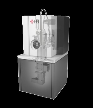 Model: Talos L120C / Image courtesy: FEI Company |

