 Movie
Movie Controller

[English] 日本語
 Yorodumi
- EMDB-71445: Antibody (1B2) Bound Rifamycin Synthetase Module 1 in the Transac... -
+ Open data

- Basic information
| Entry |  | |||||||||
|---|---|---|---|---|---|---|---|---|---|---|
| Title | Antibody (1B2) Bound Rifamycin Synthetase Module 1 in the Transacylation Mode | |||||||||
 Map data Map data | Antibody (1B2) Bound Rifamycin Synthetase Module 1 in the Transacylation Mode | |||||||||
 Sample Sample |
| |||||||||
 Keywords Keywords | Polyketide Synthase Module / Antibody (Fab) / Transferase-Immune System complex | |||||||||
| Function / homology |  Function and homology information Function and homology information6-deoxyerythronolide-B synthase / erythronolide synthase activity / fatty acid synthase activity / phosphopantetheine binding / 3-oxoacyl-[acyl-carrier-protein] synthase activity / fatty acid biosynthetic process Similarity search - Function | |||||||||
| Biological species |  Amycolatopsis mediterranei (bacteria) / Amycolatopsis mediterranei (bacteria) /  Homo sapiens (human) Homo sapiens (human) | |||||||||
| Method | single particle reconstruction / cryo EM / Resolution: 3.96 Å | |||||||||
 Authors Authors | Cogan DP / Liu C / West RC / Chen M | |||||||||
| Funding support |  United States, 1 items United States, 1 items
| |||||||||
 Citation Citation | Journal: bioRxiv / Year: 2025 Title: Molecular Basis for Asynchronous Chain Elongation During Rifamycin Antibiotic Biosynthesis. Authors: Chengli Liu / Ryan C West / Muyuan Chen / Whitaker Cohn / George Wang / Aryan M Mandot / Selena Kim / Dillon P Cogan / Abstract: The rifamycin synthetase (RIFS) from the bacterium is a large (3.5 MDa) multienzyme system that catalyzes over 40 chemical reactions to generate a complex precursor to the antibiotic rifamycin B. It ...The rifamycin synthetase (RIFS) from the bacterium is a large (3.5 MDa) multienzyme system that catalyzes over 40 chemical reactions to generate a complex precursor to the antibiotic rifamycin B. It is considered a hybrid enzymatic assembly line and consists of an N-terminal nonribosomal peptide synthetase loading module followed by a decamodular polyketide synthase (PKS). While the biosynthetic functions are known for each enzymatic domain of RIFS, structural and biochemical analyses of this system from purified components are relatively scarce. Here, we examine the biosynthetic mechanism of RIFS through complementary crosslinking, kinetic, and structural analyses of its first PKS module (M1). Thiol-selective crosslinking of M1 provided a plausible molecular basis for previously observed conformational asymmetry with respect to ketosynthase (KS)-substrate carrier protein (CP) interactions during polyketide chain elongation. Our data suggest that C-terminal dimeric interfaces-which are ubiquitous in bacterial PKSs-force their adjacent CP domains to co-migrate between two equivalent KS active site chambers. Cryogenic electron microscopy analysis of M1 further supported this observation while uncovering its unique architecture. Single-turnover kinetic analysis of M1 indicated that although removal of C-terminal dimeric interfaces supported 2-fold greater KS-CP interactions, it did not increase the partial product occupancy of the homodimeric protein. Our findings cast light on molecular details of natural antibiotic biosynthesis that will aid in the design of artificial megasynth(et)ases with untold product structures and bioactivities. | |||||||||
| History |
|
- Structure visualization
| Supplemental images |
|---|

- Downloads & links
-EMDB archive
| Map data | emd_71445.map.gz | 171.6 MB | EMDB map data format | |
|---|---|---|---|---|
| Header (meta data) | emd-71445-v30.xmlemd-71445.xml | 31.1 KB 31.1 KB | Display Display | EMDB header |
| Images |  emd_71445.png emd_71445.png | 24.5 KB | ||
| Filedesc metadata | emd-71445.cif.gz | 8.6 KB | ||
| Others | emd_71445_additional_1.map.gzemd_71445_half_map_1.map.gzemd_71445_half_map_2.map.gz | 10.7 MB 317.8 MB 317.8 MB | ||
| Archive directory |  http://ftp.pdbj.org/pub/emdb/structures/EMD-71445ftp://ftp.pdbj.org/pub/emdb/structures/EMD-71445 http://ftp.pdbj.org/pub/emdb/structures/EMD-71445ftp://ftp.pdbj.org/pub/emdb/structures/EMD-71445 | HTTPS FTP |
-Related structure data
| Related structure data |  9patMC  9pavC 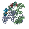 9pc6C M: atomic model generated by this map C: citing same article ( |
|---|---|
| Similar structure data |


-Links
| EMDB pages | EMDB (EBI/PDBe) / EMDataResource |
|---|---|
| Related items in Molecule of the Month |


-Map
| File | Download / File: emd_71445.map.gz / Format: CCP4 / Size: 343 MB / Type: IMAGE STORED AS FLOATING POINT NUMBER (4 BYTES) | ||||||||||||||||||||||||||||||||||||
|---|---|---|---|---|---|---|---|---|---|---|---|---|---|---|---|---|---|---|---|---|---|---|---|---|---|---|---|---|---|---|---|---|---|---|---|---|---|
| Annotation | Antibody (1B2) Bound Rifamycin Synthetase Module 1 in the Transacylation Mode | ||||||||||||||||||||||||||||||||||||
| Projections & slices | Image control
Images are generated by Spider. | ||||||||||||||||||||||||||||||||||||
| Voxel size | X=Y=Z: 1.1 Å | ||||||||||||||||||||||||||||||||||||
| Density |
| ||||||||||||||||||||||||||||||||||||
| Symmetry | Space group: 1 | ||||||||||||||||||||||||||||||||||||
| Details | EMDB XML:
|
 Z (Sec.)
 Y (Row.)
 X (Col.)


-Supplemental data
-Additional map: Additional Map
| File | emd_71445_additional_1.map | ||||||||||||
|---|---|---|---|---|---|---|---|---|---|---|---|---|---|
| Annotation | Additional Map | ||||||||||||
| Projections & Slices |
| ||||||||||||
| Density Histograms |


-Half map: Half Map B
| File | emd_71445_half_map_1.map | ||||||||||||
|---|---|---|---|---|---|---|---|---|---|---|---|---|---|
| Annotation | Half Map B | ||||||||||||
| Projections & Slices |
| ||||||||||||
| Density Histograms |


-Half map: Half Map A
| File | emd_71445_half_map_2.map | ||||||||||||
|---|---|---|---|---|---|---|---|---|---|---|---|---|---|
| Annotation | Half Map A | ||||||||||||
| Projections & Slices |
| ||||||||||||
| Density Histograms |


- Sample components
-Entire : Two antibody fragments (Fabs) in complex with homodimeric polyket...
| Entire | Name: Two antibody fragments (Fabs) in complex with homodimeric polyketide synthase module 1 of the rifamycin synthetase |
|---|---|
| Components |
|
-Supramolecule #1: Two antibody fragments (Fabs) in complex with homodimeric polyket...
| Supramolecule | Name: Two antibody fragments (Fabs) in complex with homodimeric polyketide synthase module 1 of the rifamycin synthetase type: complex / ID: 1 / Parent: 0 / Macromolecule list: all |
|---|---|
| Source (natural) | Organism: Amycolatopsis mediterranei (bacteria) / Strain: NRRL B-3240 |
| Molecular weight | Theoretical: 454.49469 KDa |
-Macromolecule #1: 6-deoxyerythronolide-B synthase
| Macromolecule | Name: 6-deoxyerythronolide-B synthase / type: protein_or_peptide / ID: 1 / Number of copies: 2 / Enantiomer: LEVO / EC number: 6-deoxyerythronolide-B synthase |
|---|---|
| Source (natural) | Organism: Amycolatopsis mediterranei (bacteria) / Strain: NRRL B-3240 |
| Molecular weight | Theoretical: 175.359844 KDa |
| Recombinant expression | Organism: |
| Sequence | String: MASTDSEKVA EYLRRATLDL RAARQRIREL EGEPIAIVGM ACRLPGGVAS PEDLWRLVAE RVDAVSEFPG DRGWDLDSLI DPDRERAGT SYVGQGGFLH DAGEFDAGFF GISPREAVAM DPQQRLLLET SWEALENAGV DPIALKGTDT GVFSGLMGQG Y GSGAVAPE ...String: MASTDSEKVA EYLRRATLDL RAARQRIREL EGEPIAIVGM ACRLPGGVAS PEDLWRLVAE RVDAVSEFPG DRGWDLDSLI DPDRERAGT SYVGQGGFLH DAGEFDAGFF GISPREAVAM DPQQRLLLET SWEALENAGV DPIALKGTDT GVFSGLMGQG Y GSGAVAPE LEGFVTTGVA SSVASGRVSY VLGLEGPAVT VDTACSSSLV AMHLAAQALR QGECSMALAG GVTVMATPGS FV EFSRQRA LAPDGRCKAF AAAADGTGWS EGVGVVVLER LSVARERGHR ILAVLRGSAV NQDGASNGLT APNGLSQQRV IRR ALAAAG LAPSDVDVVE AHGTGTTLGD PIEAQALLAT YGQERKQPLW LGSLKSNIGH AQAAAGVAGV IKMVQALRHE TLPP TLHVD KPTLEVDWSA GAIELLTEAR AWPRNGRPRR AGVSSFGVSG TNAHLILEEA PAEEPVAAPE LPVVPLVVSA RSTES LSGQ AERLASLLEG DVSLTEVAGA LVSRRAVLDE RAVVVAGSRE EAVTGLRALN TAGSGTPGKV VWVFPGQGTQ WAGMGR ELL AESPVFAERI AECAAALAPW IDWSLVDVLR GEGDLGRVDV LQPACFAVMV GLAAVWESVG VRPDAVVGHS QGEIAAA CV SGALSLEDAA KVVALRSQAI AAELSGRGGM ASVALGEDDV VSRLVDGVEV AAVNGPSSVV IAGDAHALDA TLEILSGE G IRVRRVAVDY ASHTRHVEDI RDTLAETLAG ISAQAPAVPF YSTVTSEWVR DAGVLDGGYW YRNLRNQVRF GAAATALLE QGHTVFVEVS AHPVTVQPLS ELTGDAIGTL RREDGGLRRL LASMGELFVR GIDVDWTAMV PAAGWVDLPT YAFEHRHYWL EPAEPASAG DPLLGTVVST PGSDRLTAVA QWSRRAQPWA VDGLVPNAAL VEAAIRLGDL AGTPVVGELV VDAPVVLPRR G SREVQLIV GEPGEQRRRP IEVFSREADE PWTRHAHGTL APAAAAVPEP AAAGDATDVT VAGLRDADRY GIHPALLDAA VR TVVGDDL LPSVWTGVSL LASGATAVTV TPTATGLRLT DPAGQPVLTV ESVRGTPFVA EQGTTDALFR VDWPEIPLPT AET ADFLPY EATSAEATLS ALQAWLADPA ETRLAVVTGD CTEPGAAAIW GLVRSAQSEH PGRIVLADLD DPAVLPAVVA SGEP QVRVR NGVASVPRLT RVTPRQDARP LDPEGTVLIT GGTGTLGALT ARHLVTAHGV RHLVLVSRRG EAPELQEELT ALGAS VAIA ACDVADRAQL EAVLRAIPAE HPLTAVIHTA GVLDDGVVTE LTPDRLATVR RPKVDAARLL DELTREADLA AFVLFS SAA GVLGNPGQAG YAAANAELDA LARQRNSLDL PAVSIAWGYW ATVSGMTEHL GDADLRRNQR IGMSGLPADE GMALLDA AI ATGGTLVAAK FDVAALRATA KAGGPVPPLL RGLAPLPRRA AAKTASLTER LAGLAETEQA AALLDLVRRH AAEVLGHS G AESVHSGRTF KDAGFDSLTA VELRNRLAAA TGLTLSPAMI FDYPKPPALA DHLRAKLFGT EVRGEAPSAL AGLDALEAA LPEVPATERE ELVQRLERML AALRPVAQAA DASGTGANPS GDDLGEAGVD ELLEALGREL DGDGNSSSVD KLAAALEHHH HHH UniProtKB: 6-deoxyerythronolide-B synthase |
-Macromolecule #2: 6-deoxyerythronolide-B synthase
| Macromolecule | Name: 6-deoxyerythronolide-B synthase / type: protein_or_peptide / ID: 2 / Number of copies: 1 / Enantiomer: LEVO / EC number: 6-deoxyerythronolide-B synthase |
|---|---|
| Source (natural) | Organism: Amycolatopsis mediterranei (bacteria) / Strain: NRRL B-3240 |
| Molecular weight | Theoretical: 175.700172 KDa |
| Recombinant expression | Organism: |
| Sequence | String: MASTDSEKVA EYLRRATLDL RAARQRIREL EGEPIAIVGM ACRLPGGVAS PEDLWRLVAE RVDAVSEFPG DRGWDLDSLI DPDRERAGT SYVGQGGFLH DAGEFDAGFF GISPREAVAM DPQQRLLLET SWEALENAGV DPIALKGTDT GVFSGLMGQG Y GSGAVAPE ...String: MASTDSEKVA EYLRRATLDL RAARQRIREL EGEPIAIVGM ACRLPGGVAS PEDLWRLVAE RVDAVSEFPG DRGWDLDSLI DPDRERAGT SYVGQGGFLH DAGEFDAGFF GISPREAVAM DPQQRLLLET SWEALENAGV DPIALKGTDT GVFSGLMGQG Y GSGAVAPE LEGFVTTGVA SSVASGRVSY VLGLEGPAVT VDTACSSSLV AMHLAAQALR QGECSMALAG GVTVMATPGS FV EFSRQRA LAPDGRCKAF AAAADGTGWS EGVGVVVLER LSVARERGHR ILAVLRGSAV NQDGASNGLT APNGLSQQRV IRR ALAAAG LAPSDVDVVE AHGTGTTLGD PIEAQALLAT YGQERKQPLW LGSLKSNIGH AQAAAGVAGV IKMVQALRHE TLPP TLHVD KPTLEVDWSA GAIELLTEAR AWPRNGRPRR AGVSSFGVSG TNAHLILEEA PAEEPVAAPE LPVVPLVVSA RSTES LSGQ AERLASLLEG DVSLTEVAGA LVSRRAVLDE RAVVVAGSRE EAVTGLRALN TAGSGTPGKV VWVFPGQGTQ WAGMGR ELL AESPVFAERI AECAAALAPW IDWSLVDVLR GEGDLGRVDV LQPACFAVMV GLAAVWESVG VRPDAVVGHS QGEIAAA CV SGALSLEDAA KVVALRSQAI AAELSGRGGM ASVALGEDDV VSRLVDGVEV AAVNGPSSVV IAGDAHALDA TLEILSGE G IRVRRVAVDY ASHTRHVEDI RDTLAETLAG ISAQAPAVPF YSTVTSEWVR DAGVLDGGYW YRNLRNQVRF GAAATALLE QGHTVFVEVS AHPVTVQPLS ELTGDAIGTL RREDGGLRRL LASMGELFVR GIDVDWTAMV PAAGWVDLPT YAFEHRHYWL EPAEPASAG DPLLGTVVST PGSDRLTAVA QWSRRAQPWA VDGLVPNAAL VEAAIRLGDL AGTPVVGELV VDAPVVLPRR G SREVQLIV GEPGEQRRRP IEVFSREADE PWTRHAHGTL APAAAAVPEP AAAGDATDVT VAGLRDADRY GIHPALLDAA VR TVVGDDL LPSVWTGVSL LASGATAVTV TPTATGLRLT DPAGQPVLTV ESVRGTPFVA EQGTTDALFR VDWPEIPLPT AET ADFLPY EATSAEATLS ALQAWLADPA ETRLAVVTGD CTEPGAAAIW GLVRSAQSEH PGRIVLADLD DPAVLPAVVA SGEP QVRVR NGVASVPRLT RVTPRQDARP LDPEGTVLIT GGTGTLGALT ARHLVTAHGV RHLVLVSRRG EAPELQEELT ALGAS VAIA ACDVADRAQL EAVLRAIPAE HPLTAVIHTA GVLDDGVVTE LTPDRLATVR RPKVDAARLL DELTREADLA AFVLFS SAA GVLGNPGQAG YAAANAELDA LARQRNSLDL PAVSIAWGYW ATVSGMTEHL GDADLRRNQR IGMSGLPADE GMALLDA AI ATGGTLVAAK FDVAALRATA KAGGPVPPLL RGLAPLPRRA AAKTASLTER LAGLAETEQA AALLDLVRRH AAEVLGHS G AESVHSGRTF KDAGFD(4HH)LTA VELRNRLAAA TGLTLSPAMI FDYPKPPALA DHLRAKLFGT EVRGEAPSAL AGLDA LEAA LPEVPATERE ELVQRLERML AALRPVAQAA DASGTGANPS GDDLGEAGVD ELLEALGREL DGDGNSSSVD KLAAAL EHH HHHH UniProtKB: 6-deoxyerythronolide-B synthase |
-Macromolecule #3: Antibody Fragment 1B2 Heavy Chain
| Macromolecule | Name: Antibody Fragment 1B2 Heavy Chain / type: protein_or_peptide / ID: 3 / Number of copies: 2 / Enantiomer: LEVO |
|---|---|
| Source (natural) | Organism: Homo sapiens (human) |
| Molecular weight | Theoretical: 26.447611 KDa |
| Recombinant expression | Organism: |
| Sequence | String: MAEVQLVQSG GGLVQPGRSL RLSCTASGFT FGDYAMSWVR QAPGKGLEWV GFIRSKAYGG TTEYAASVKG RFTISRDDSK SIAYLQMNS LKTEDTAVYY CTRGGTLFDY WGQGTLVTVS SASTKGPSVF PLAPSSKSTS GGTAALGCLV KDYFPEPVTV S WNSGALTS ...String: MAEVQLVQSG GGLVQPGRSL RLSCTASGFT FGDYAMSWVR QAPGKGLEWV GFIRSKAYGG TTEYAASVKG RFTISRDDSK SIAYLQMNS LKTEDTAVYY CTRGGTLFDY WGQGTLVTVS SASTKGPSVF PLAPSSKSTS GGTAALGCLV KDYFPEPVTV S WNSGALTS GVHTFPAVLQ SSGLYSLSSV VTVPSSSLGT QTYICNVNHK PSNTKVDKKV EPKSCAALVP RGSAHHHHHH AA DYKDDDD KA |
-Macromolecule #4: Antibody Fragment 1B2 Light Chain
| Macromolecule | Name: Antibody Fragment 1B2 Light Chain / type: protein_or_peptide / ID: 4 / Number of copies: 2 / Enantiomer: LEVO |
|---|---|
| Source (natural) | Organism: Homo sapiens (human) |
| Molecular weight | Theoretical: 25.715832 KDa |
| Recombinant expression | Organism: |
| Sequence | String: LFAIPLVVPF YSHSALDVVM TQSPLSLPVT PGEPASISCR SSQSLLHSNG YNYLDWYLQK PGQSPQLLIY LGSNRASGVP DRFSGSGSG TDFTLKISRV EAEDVGVYYC MQSLQTPRLT FGPGTKVDIK RTVAAPSVFI FPPSDEQLKS GTASVVCLLN N FYPRGAKV ...String: LFAIPLVVPF YSHSALDVVM TQSPLSLPVT PGEPASISCR SSQSLLHSNG YNYLDWYLQK PGQSPQLLIY LGSNRASGVP DRFSGSGSG TDFTLKISRV EAEDVGVYYC MQSLQTPRLT FGPGTKVDIK RTVAAPSVFI FPPSDEQLKS GTASVVCLLN N FYPRGAKV QWKVDNALQS GNSQESVTEQ DSKDSTYSLS STLTLSKADY EKHKVYACEV THQGLSSPVT KSFNRGEC |
-Experimental details
-Structure determination
| Method | cryo EM |
|---|---|
 Processing Processing | single particle reconstruction |
| Aggregation state | particle |
-Sample preparation
| Concentration | 10 mg/mL | |||||||||
|---|---|---|---|---|---|---|---|---|---|---|
| Buffer | pH: 7.2 Component:
Details: 100 mM citric acid, 10 mM HEPES, pH 7.2 (NaOH) | |||||||||
| Grid | Model: Quantifoil R2/1 / Material: COPPER / Mesh: 300 / Support film - Material: CARBON / Support film - topology: HOLEY / Pretreatment - Type: GLOW DISCHARGE / Pretreatment - Time: 30 sec. / Pretreatment - Atmosphere: AIR / Pretreatment - Pressure: 0.036000000000000004 kPa | |||||||||
| Vitrification | Cryogen name: ETHANE / Chamber humidity: 100 % / Chamber temperature: 277.15 K / Instrument: FEI VITROBOT MARK IV |
- Electron microscopy
| Microscope | TFS KRIOS |
|---|---|
| Image recording | Film or detector model: GATAN K3 BIOQUANTUM (6k x 4k) / Number grids imaged: 1 / Number real images: 10005 / Average exposure time: 5.72 sec. / Average electron dose: 50.0 e/Å2 |
| Electron beam | Acceleration voltage: 300 kV / Electron source:  FIELD EMISSION GUN FIELD EMISSION GUN |
| Electron optics | Illumination mode: FLOOD BEAM / Imaging mode: BRIGHT FIELD / Cs: 2.7 mm / Nominal defocus max: 3.988 µm / Nominal defocus min: 1.188 µm |
| Sample stage | Specimen holder model: FEI TITAN KRIOS AUTOGRID HOLDER / Cooling holder cryogen: NITROGEN |
| Experimental equipment |  Model: Titan Krios / Image courtesy: FEI Company |
+Image processing
-Atomic model buiding 1
| Initial model | Chain - Source name: AlphaFold / Chain - Initial model type: in silico model |
|---|---|
| Refinement | Space: REAL / Protocol: RIGID BODY FIT |
| Output model | PDB-9pat: |
