 Movie
Movie Controller


+ Open data

- Basic information
| Entry |  | |||||||||
|---|---|---|---|---|---|---|---|---|---|---|
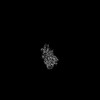
| Title | Structure of Mycobacterium smegmatis EtfD | |||||||||
 Map data Map data | CryoEM map of Mycobacterium smegmatis EtfD (sharpened) | |||||||||
 Sample Sample |
| |||||||||
 Keywords Keywords | oxidoreductase / membrane protein / electron transport chain / beta oxidation | |||||||||
| Function / homology |  Function and homology information Function and homology information4 iron, 4 sulfur cluster binding / oxidoreductase activity / metal ion binding / plasma membrane Similarity search - Function | |||||||||
| Biological species |  Mycolicibacterium smegmatis MC2 155 (bacteria) Mycolicibacterium smegmatis MC2 155 (bacteria) | |||||||||
| Method | single particle reconstruction / cryo EM / Resolution: 3.2 Å | |||||||||
 Authors Authors | Courbon GM / Rubinstein JL | |||||||||
| Funding support |  Canada, 1 items Canada, 1 items
| |||||||||
 Citation Citation | Journal: EMBO J / Year: 2026 Title: Structural basis for EtfD-mediated coupling of β-oxidation and the respiratory chain in mycobacteria. Authors: Gautier M Courbon / Vadim Makarov / Stewart T Cole / Dirk Schnapinger / Sabine Ehrt / John L Rubinstein /    Abstract: Targeting β-oxidation has been proposed as a strategy for shortening tuberculosis (TB) treatment by killing non-replicating Mycobacterium tuberculosis within granulomas where the pathogen relies on ...Targeting β-oxidation has been proposed as a strategy for shortening tuberculosis (TB) treatment by killing non-replicating Mycobacterium tuberculosis within granulomas where the pathogen relies on host-derived lipids. The protein EtfD is thought to couple β-oxidation of fatty acids with the respiratory chain in mycobacteria. However, the structure of EtfD is not known and, as the presumed link between two complex processes, its activity has been difficult to measure, impeding its exploitation as a drug target. Here we show that Mycobacterium smegmatis, a fast growing and nonpathogenic model for M. tuberculosis, relies on EtfD for extracting energy from β-oxidation. The electron cryomicroscopy structure of M. smegmatis EtfD reveals an unusual linear [3Fe-4S] cluster that has not been seen in other protein structures, and suggests how EtfD transfers electrons from β-oxidation to the respiratory chain. We devised an assay that couples EtfD activity to a fluorescent readout of proton pumping by the respiratory chain, which can be used to identify compounds that block mycobacteria from using β-oxidation to power oxidative phosphorylation. | |||||||||
| History |
|
- Structure visualization
| Supplemental images |
|---|

- Downloads & links
-EMDB archive
| Map data | emd_70545.map.gz | 118.1 MB | EMDB map data format | |
|---|---|---|---|---|
| Header (meta data) | emd-70545-v30.xmlemd-70545.xml | 24.5 KB 24.5 KB | Display Display | EMDB header |
| FSC (resolution estimation) | emd_70545_fsc.xml | 10.5 KB | Display | FSC data file |
| Images |  emd_70545.png emd_70545.png | 68.2 KB | ||
| Filedesc metadata | emd-70545.cif.gz | 6.8 KB | ||
| Others | emd_70545_additional_1.map.gzemd_70545_half_map_1.map.gzemd_70545_half_map_2.map.gz | 63 MB 116.2 MB 116.2 MB | ||
| Archive directory |  http://ftp.pdbj.org/pub/emdb/structures/EMD-70545ftp://ftp.pdbj.org/pub/emdb/structures/EMD-70545 http://ftp.pdbj.org/pub/emdb/structures/EMD-70545ftp://ftp.pdbj.org/pub/emdb/structures/EMD-70545 | HTTPS FTP |
-Related structure data
| Related structure data |  9ojnMC M: atomic model generated by this map C: citing same article ( |
|---|---|
| Similar structure data |
-Links
| EMDB pages | EMDB (EBI/PDBe) / EMDataResource |
|---|---|
| Related items in Molecule of the Month |


-Map
| File | Download / File: emd_70545.map.gz / Format: CCP4 / Size: 125 MB / Type: IMAGE STORED AS FLOATING POINT NUMBER (4 BYTES) | ||||||||||||||||||||||||||||||||||||
|---|---|---|---|---|---|---|---|---|---|---|---|---|---|---|---|---|---|---|---|---|---|---|---|---|---|---|---|---|---|---|---|---|---|---|---|---|---|
| Annotation | CryoEM map of Mycobacterium smegmatis EtfD (sharpened) | ||||||||||||||||||||||||||||||||||||
| Projections & slices | Image control
Images are generated by Spider. | ||||||||||||||||||||||||||||||||||||
| Voxel size | X=Y=Z: 1.024 Å | ||||||||||||||||||||||||||||||||||||
| Density |
| ||||||||||||||||||||||||||||||||||||
| Symmetry | Space group: 1 | ||||||||||||||||||||||||||||||||||||
| Details | EMDB XML:
|
 Z (Sec.)
 Y (Row.)
 X (Col.)


-Supplemental data
-Additional map: CryoEM map of Mycobacterium smegmatis EtfD (unsharpened)
| File | emd_70545_additional_1.map | ||||||||||||
|---|---|---|---|---|---|---|---|---|---|---|---|---|---|
| Annotation | CryoEM map of Mycobacterium smegmatis EtfD (unsharpened) | ||||||||||||
| Projections & Slices |
| ||||||||||||
| Density Histograms |


-Half map: CryoEM map of Mycobacterium smegmatis EtfD (Half B)
| File | emd_70545_half_map_1.map | ||||||||||||
|---|---|---|---|---|---|---|---|---|---|---|---|---|---|
| Annotation | CryoEM map of Mycobacterium smegmatis EtfD (Half B) | ||||||||||||
| Projections & Slices |
| ||||||||||||
| Density Histograms |


-Half map: CryoEM map of Mycobacterium smegmatis EtfD (Half A)
| File | emd_70545_half_map_2.map | ||||||||||||
|---|---|---|---|---|---|---|---|---|---|---|---|---|---|
| Annotation | CryoEM map of Mycobacterium smegmatis EtfD (Half A) | ||||||||||||
| Projections & Slices |
| ||||||||||||
| Density Histograms |


- Sample components
-Entire : Structure of Mycobacterium smegmatis EtfD
| Entire | Name: Structure of Mycobacterium smegmatis EtfD |
|---|---|
| Components |
|
-Supramolecule #1: Structure of Mycobacterium smegmatis EtfD
| Supramolecule | Name: Structure of Mycobacterium smegmatis EtfD / type: complex / ID: 1 / Parent: 0 / Macromolecule list: #1 |
|---|---|
| Source (natural) | Organism: Mycolicibacterium smegmatis MC2 155 (bacteria) |
-Macromolecule #1: Iron-sulfur-binding reductase
| Macromolecule | Name: Iron-sulfur-binding reductase / type: protein_or_peptide / ID: 1 / Number of copies: 1 / Enantiomer: LEVO |
|---|---|
| Source (natural) | Organism: Mycolicibacterium smegmatis MC2 155 (bacteria) |
| Molecular weight | Theoretical: 111.045625 KDa |
| Sequence | String: MAHTLEVSRL IIGLLMTAIV LVFAAKRVLW LTKLIRSGQK TLDENGRKND LQKRITTQIT EVFGQTRLLR WSVPGIAHFF TMWGFFVLA SVYLEAYGVL FDPEFHIPFI GRWPVLGFLQ DFFAVAVLLG IIVFAIIRVV REPKKIGRDS RFYGSHTGGA W EILFMIFL ...String: MAHTLEVSRL IIGLLMTAIV LVFAAKRVLW LTKLIRSGQK TLDENGRKND LQKRITTQIT EVFGQTRLLR WSVPGIAHFF TMWGFFVLA SVYLEAYGVL FDPEFHIPFI GRWPVLGFLQ DFFAVAVLLG IIVFAIIRVV REPKKIGRDS RFYGSHTGGA W EILFMIFL VIATYALFRG AAVNTLGERF PYQSGAFFSD FMAWILRPLG ATANMWIETV ALMGHIGVML VFLLIVLHSK HL HIGLAPI NVTFKRLPNG LGPLLPMESN GEYIDFEDPA EDAVFGKGKI EDFTWKGYLD FTTCTECGRC QSQCPAWNTG KPL SPKLVI MNLRDHMFAK APYILGEKPS PLESTPEGGL GEKARGEKHE QKHAHDHVPE SGFERILGSG PEQALRPLVG TEEQ GGVID PDVLWSCTNC GACVEQCPVD IEHIDHIVDM RRYQVMVESE FPGELGVLFK NLETKGNPWG QNAKDRTNWI DEVDF DVPV YGEDVDSFDG FEYLFWVGCA GAYEDRAKKT TKAVAELLAT AGVKFLVLGT GETCTGDSAR RSGNEFLFQQ LAAQNV ETI NELFEGVETV DRKIVVTCPH CFNTIGREYP QLGANYSVVH HTQLLNRLVR DKKLVPVKSV SEQNGQPVTY HDPCFLG RH NKVYEAPREL VEASGVTLKE MPRHADRGLC CGAGGARMWM EEHIGKRVNV ERTEEAMDTA STIATGCPFC RVMITDGV D DVAASRNVEK AEVLDVAQLL LNSLDTSKVT LPEKGTAAKE SEKRAAARAE AEAKAEAAAP PVEEAAPEAE APAAPAAGG AEAKPVTGLG MAGAAKRPGA KKAAPAAEAS AAPAAAPAPA KGLGLAGGAK RPGAKKAAAP AAEAPAAPAS DAPPVKGLGL AGGAKRPGA KKTAAAAPAE KPAATEAPEA SATPAAPAAP VKGLGLAAGA KRPGAKKTAA APAEKPAAAE TEAPAPAETA A PAEPAKPE PPVVGLGIAA GARRPGAKKA AAKPAAAPAP AAEKPAEQAA EPEKPAEKPA EPEKPEPPVV GLGIKPGAKR PG KR UniProtKB: Iron-sulfur-binding reductase |
-Macromolecule #2: DODECYL-BETA-D-MALTOSIDE
| Macromolecule | Name: DODECYL-BETA-D-MALTOSIDE / type: ligand / ID: 2 / Number of copies: 1 / Formula: LMT |
|---|---|
| Molecular weight | Theoretical: 510.615 Da |
| Chemical component information |  ChemComp-LMT: |
-Macromolecule #3: MENAQUINONE-9
| Macromolecule | Name: MENAQUINONE-9 / type: ligand / ID: 3 / Number of copies: 1 / Formula: MQ9 |
|---|---|
| Molecular weight | Theoretical: 785.233 Da |
| Chemical component information | 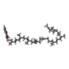 ChemComp-MQ9: |
-Macromolecule #4: PROTOPORPHYRIN IX CONTAINING FE
| Macromolecule | Name: PROTOPORPHYRIN IX CONTAINING FE / type: ligand / ID: 4 / Number of copies: 1 / Formula: HEM |
|---|---|
| Molecular weight | Theoretical: 616.487 Da |
| Chemical component information |  ChemComp-HEM: |
-Macromolecule #5: IRON/SULFUR CLUSTER
| Macromolecule | Name: IRON/SULFUR CLUSTER / type: ligand / ID: 5 / Number of copies: 2 / Formula: SF4 |
|---|---|
| Molecular weight | Theoretical: 351.64 Da |
| Chemical component information |  ChemComp-FS1: |
-Macromolecule #6: Non-cubane [4Fe-4S]-cluster
| Macromolecule | Name: Non-cubane [4Fe-4S]-cluster / type: ligand / ID: 6 / Number of copies: 1 / Formula: 9S8 |
|---|---|
| Molecular weight | Theoretical: 351.64 Da |
| Chemical component information | 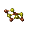 ChemComp-9S8: |
-Macromolecule #7: 1,3,5,7-tetrathia-2$l^{2},4$l^{4},6$l^{2}-triferraspiro[3.3]heptane
| Macromolecule | Name: 1,3,5,7-tetrathia-2$l^{2},4$l^{4},6$l^{2}-triferraspiro[3.3]heptane type: ligand / ID: 7 / Number of copies: 1 / Formula: A1CBX |
|---|---|
| Molecular weight | Theoretical: 295.795 Da |
-Experimental details
-Structure determination
| Method | cryo EM |
|---|---|
 Processing Processing | single particle reconstruction |
| Aggregation state | particle |
-Sample preparation
| Buffer | pH: 7.4 |
|---|---|
| Vitrification | Cryogen name: ETHANE |
- Electron microscopy
| Microscope | TFS KRIOS |
|---|---|
| Image recording | Film or detector model: TFS FALCON 4i (4k x 4k) / Average electron dose: 70.0 e/Å2 |
| Electron beam | Acceleration voltage: 300 kV / Electron source:  FIELD EMISSION GUN FIELD EMISSION GUN |
| Electron optics | Illumination mode: FLOOD BEAM / Imaging mode: BRIGHT FIELD / Nominal defocus max: 2.8000000000000003 µm / Nominal defocus min: 0.4 µm |
| Experimental equipment |  Model: Titan Krios / Image courtesy: FEI Company |

