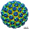
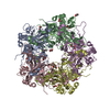
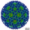
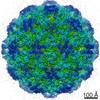
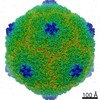
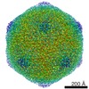
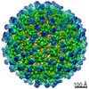
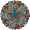
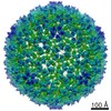
Journal: mBio / Year: 2017 Title: Crystal Structures of Two Immune Complexes Identify Determinants for Viral Infectivity and Type-Specific Neutralization of Human Papillomavirus. Authors: Zhihai Li / Daning Wang / Ying Gu / Shuo Song / Maozhou He / Jingjie Shi / Xinlin Liu / Shuangping Wei / Jinjin Li / Hai Yu / Qingbing Zheng / Xiaodong Yan / Timothy S Baker / Jun Zhang / ...Authors: Zhihai Li / Daning Wang / Ying Gu / Shuo Song / Maozhou He / Jingjie Shi / Xinlin Liu / Shuangping Wei / Jinjin Li / Hai Yu / Qingbing Zheng / Xiaodong Yan / Timothy S Baker / Jun Zhang / Jason S McLellan / Shaowei Li / Ningshao Xia / Abstract: Persistent, high-risk human papillomavirus (HPV) infection is the primary cause of cervical cancer. Neutralizing antibodies elicited by L1-only virus-like particles (VLPs) can block HPV infection; ...Persistent, high-risk human papillomavirus (HPV) infection is the primary cause of cervical cancer. Neutralizing antibodies elicited by L1-only virus-like particles (VLPs) can block HPV infection; however, the lack of high-resolution structures has limited our understanding of the mode of virus infection and the requirement for type specificity at the molecular level. Here, we describe two antibodies, A12A3 and 28F10, that specifically bind to and neutralize HPV58 and HPV59, respectively, through two distinct binding stoichiometries. We show that the epitopes of A12A3 are clustered in the DE loops of two adjacent HPV58 L1 monomers, whereas 28F10 recognizes the HPV59 FG loop of a single monomer. Via structure-based mutagenesis and analysis of antibody binding, we further identified the residues HPV58 D154, S168, and N170 and HPV59 M267, Q270, E273, Y276, K278, and R283, which play critical roles in virus infection. By substituting these strategic epitope residues into other HPV genotypes, we could then redirect the type-specific binding of the antibodies to these genotypes, thus highlighting the importance of these specific residues, HPV58 R161, S168, and N308 and HPV59 Q270, E273, and D281. Overall, our findings provide molecular insights into potential structural determinants of HPV required for infectivity and type specificity. High-risk human papillomaviruses (HPVs) are considered the major causative pathogens of cancers that affect epithelial mucosa, such as cervical cancer. However, because of the lack of high-resolution structural information on the sites of neutralization, we have yet to determine the precise mode of HPV infection and how different types of HPV cause infection. Our crystal structures in this study have uncovered discrete binding stoichiometries for two different antibodies. We show that one A12A3 Fab binds to the center of one HPV58 pentamer, whereas five 28F10 Fabs bind along the top fringe of one HPV59 pentamer. Furthermore, through targeted epitope analysis, we show that 6 to 7 discontinuous residues of the L1 major capsid protein of HPV are determinants, at least in part, for virus infection and type specificity. This knowledge will help us to unravel the process of HPV infection and can potentially be used to drive the development of therapeutics that target neutralization-sensitive sites.
History
Deposition
Aug 10, 2017
-
Header (metadata) release
Oct 25, 2017
-
Map release
Oct 25, 2017
-
Update
Oct 25, 2017
-
Current status
Oct 25, 2017
Processing site: PDBj / Status: Released
-
Structure visualization
Movie
Surface view with section colored by density value
In the structure databanks used in Yorodumi, some data are registered as the other names, "COVID-19 virus" and "2019-nCoV". Here are the details of the virus and the list of structure data.
Jan 31, 2019. EMDB accession codes are about to change! (news from PDBe EMDB page)
EMDB accession codes are about to change! (news from PDBe EMDB page)
The allocation of 4 digits for EMDB accession codes will soon come to an end. Whilst these codes will remain in use, new EMDB accession codes will include an additional digit and will expand incrementally as the available range of codes is exhausted. The current 4-digit format prefixed with “EMD-” (i.e. EMD-XXXX) will advance to a 5-digit format (i.e. EMD-XXXXX), and so on. It is currently estimated that the 4-digit codes will be depleted around Spring 2019, at which point the 5-digit format will come into force.
The EM Navigator/Yorodumi systems omit the EMD- prefix.
Related info.:Q: What is EMD? / ID/Accession-code notation in Yorodumi/EM Navigator
Yorodumi is a browser for structure data from EMDB, PDB, SASBDB, etc.
This page is also the successor to EM Navigator detail page, and also detail information page/front-end page for Omokage search.
The word "yorodu" (or yorozu) is an old Japanese word meaning "ten thousand". "mi" (miru) is to see.
Related info.:EMDB / PDB / SASBDB / Comparison of 3 databanks / Yorodumi Search / Aug 31, 2016. New EM Navigator & Yorodumi / Yorodumi Papers / Jmol/JSmol / Function and homology information / Changes in new EM Navigator and Yorodumi
 Movie
Movie Controller

 Yorodumi
 Open data
 Basic information
 Map data
 Sample
 Human papillomavirus
 Authors
 Citation

 Structure visualization
 Movie viewer
 Downloads & links
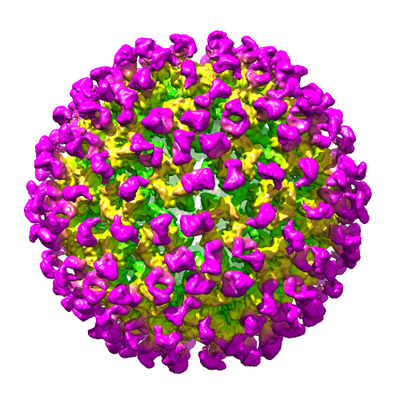 emd_6814.png
 http://ftp.pdbj.org/pub/emdb/structures/EMD-6814

 Z (Sec.)
 Y (Row.)
 X (Col.)


 Sample components
 Processing
 Electron microscopy
 FIELD EMISSION GUN
