 Movie
Movie Controller

[English] 日本語
 Yorodumi
- EMDB-6745: Cryo-EM structure of complex of HBsAg and Fab fragment of therape... -
+ Open data

- Basic information
| Entry | Database: EMDB / ID: EMD-6745 | |||||||||
|---|---|---|---|---|---|---|---|---|---|---|
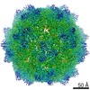
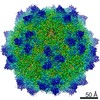
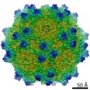
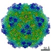
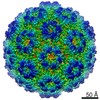
| Title | Cryo-EM structure of complex of HBsAg and Fab fragment of therapeutic mAb-E6F6. | |||||||||
 Map data Map data | Cryo-EM structure of HBsAg-E6F6-Fab complex | |||||||||
 Sample Sample |
| |||||||||
| Biological species |  | |||||||||
| Method | single particle reconstruction / cryo EM / Resolution: 12.0 Å | |||||||||
 Authors Authors | Mo X / Yuan AY | |||||||||
 Citation Citation | Journal: To be published Title: Structural insight into mechanism of antibody-mediated immunotherapy for persistent hepatitis B virus infection Authors: Mo X / Yuan YA | |||||||||
| History |
|
- Structure visualization
| Movie |
 Movie viewer Movie viewer |
|---|---|
| Structure viewer | EM map: SurfViewMolmilJmol/JSmol |
| Supplemental images |

- Downloads & links
-EMDB archive
| Map data | emd_6745.map.gz | 17.5 MB | EMDB map data format | |
|---|---|---|---|---|
| Header (meta data) | emd-6745-v30.xmlemd-6745.xml | 12.5 KB 12.5 KB | Display Display | EMDB header |
| FSC (resolution estimation) | emd_6745_fsc.xml | 9.1 KB | Display | FSC data file |
| Images |  emd_6745.png emd_6745.png | 213.1 KB | ||
| Archive directory |  http://ftp.pdbj.org/pub/emdb/structures/EMD-6745ftp://ftp.pdbj.org/pub/emdb/structures/EMD-6745 http://ftp.pdbj.org/pub/emdb/structures/EMD-6745ftp://ftp.pdbj.org/pub/emdb/structures/EMD-6745 | HTTPS FTP |
-Related structure data
| Similar structure data |
|---|


-Links
| EMDB pages | EMDB (EBI/PDBe) / EMDataResource |
|---|
-Map
| File | Download / File: emd_6745.map.gz / Format: CCP4 / Size: 52.7 MB / Type: IMAGE STORED AS FLOATING POINT NUMBER (4 BYTES) | ||||||||||||||||||||||||||||||||||||||||||||||||||||||||||||
|---|---|---|---|---|---|---|---|---|---|---|---|---|---|---|---|---|---|---|---|---|---|---|---|---|---|---|---|---|---|---|---|---|---|---|---|---|---|---|---|---|---|---|---|---|---|---|---|---|---|---|---|---|---|---|---|---|---|---|---|---|---|
| Annotation | Cryo-EM structure of HBsAg-E6F6-Fab complex | ||||||||||||||||||||||||||||||||||||||||||||||||||||||||||||
| Projections & slices | Image control
Images are generated by Spider. | ||||||||||||||||||||||||||||||||||||||||||||||||||||||||||||
| Voxel size | X=Y=Z: 1.81 Å | ||||||||||||||||||||||||||||||||||||||||||||||||||||||||||||
| Density |
| ||||||||||||||||||||||||||||||||||||||||||||||||||||||||||||
| Symmetry | Space group: 1 | ||||||||||||||||||||||||||||||||||||||||||||||||||||||||||||
| Details | EMDB XML:
CCP4 map header:
| ||||||||||||||||||||||||||||||||||||||||||||||||||||||||||||
 Z (Sec.)
 Y (Row.)
 X (Col.)


-Supplemental data
- Sample components
-Entire : Complex of HBsAg with Fab fragment of therapeutic mAb-E6F6
| Entire | Name: Complex of HBsAg with Fab fragment of therapeutic mAb-E6F6 |
|---|---|
| Components |
|
-Supramolecule #1: Complex of HBsAg with Fab fragment of therapeutic mAb-E6F6
| Supramolecule | Name: Complex of HBsAg with Fab fragment of therapeutic mAb-E6F6 type: complex / ID: 1 / Parent: 0 Details: Surface protein of Hepatitis B virus and fab fragment generated by proteolytic cleavage of therapeutic mAb-E6F6. |
|---|---|
| Source (natural) | Organism: |
| Recombinant expression | Organism: Recombinant strain: DH5a / Recombinant plasmid: pET-Duet |
| Recombinant expression | Organism:   Cricetulus griseus (Chinese hamster) Cricetulus griseus (Chinese hamster) |
| Molecular weight | Experimental: 3.24 MDa |
-Supramolecule #2: HBsAg
| Supramolecule | Name: HBsAg / type: complex / ID: 2 / Parent: 1 / Details: Surface protein of Hepatitis B virus. |
|---|
-Supramolecule #3: E6F6-Fab
| Supramolecule | Name: E6F6-Fab / type: complex / ID: 3 / Parent: 1 Details: Fab fragment generated by proteolytic cleavage of therapeutic mAb-E6F6. |
|---|
-Experimental details
-Structure determination
| Method | cryo EM |
|---|---|
 Processing Processing | single particle reconstruction |
| Aggregation state | particle |
-Sample preparation
| Concentration | 0.5 mg/mL | |||||||||
|---|---|---|---|---|---|---|---|---|---|---|
| Buffer | pH: 6.5 Component:
Details: 20mM Tris (pH 6.5), 500mM NaCl | |||||||||
| Grid | Model: Quantifoil R2/2 / Material: COPPER / Mesh: 400 / Support film - Material: FORMVAR / Support film - topology: HOLEY / Pretreatment - Type: GLOW DISCHARGE / Pretreatment - Atmosphere: AIR / Pretreatment - Pressure: 101.325 kPa | |||||||||
| Vitrification | Cryogen name: ETHANE / Chamber humidity: 100 % / Chamber temperature: 298 K / Instrument: FEI VITROBOT MARK IV / Details: blot for 5 seconds before pluning. | |||||||||
| Details | This sample was monodisperse. |
- Electron microscopy
| Microscope | FEI TITAN KRIOS |
|---|---|
| Temperature | Min: 70.0 K / Max: 70.0 K |
| Details | Preliminary grid screening was performed manually. |
| Image recording | Film or detector model: FEI FALCON II (4k x 4k) / Number grids imaged: 12 / Number real images: 500 / Average exposure time: 1.0 sec. / Average electron dose: 25.0 e/Å2 |
| Electron beam | Acceleration voltage: 300 kV / Electron source:  FIELD EMISSION GUN FIELD EMISSION GUN |
| Electron optics | Calibrated defocus max: 3.0 µm / Calibrated defocus min: 1.0 µm / Calibrated magnification: 47000 / Illumination mode: OTHER / Imaging mode: DARK FIELD / Cs: 2.7 mm / Nominal defocus max: 3.0 µm / Nominal defocus min: 1.0 µm / Nominal magnification: 47000 |
| Sample stage | Specimen holder model: FEI TITAN KRIOS AUTOGRID HOLDER / Cooling holder cryogen: NITROGEN |
| Experimental equipment |  Model: Titan Krios / Image courtesy: FEI Company |
+Image processing

-Atomic model buiding 1
| Refinement | Space: RECIPROCAL / Protocol: AB INITIO MODEL / Overall B value: 300 |
|---|
