 Movie
Movie Controller


+ Open data

- Basic information
| Entry | Database: EMDB / ID: EMD-6722 | |||||||||
|---|---|---|---|---|---|---|---|---|---|---|
| Title | Drosophila full length cryoEM structure of C3PO complex | |||||||||
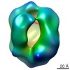
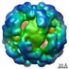
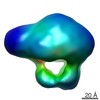
 Map data Map data | cryo-EM structure of full-length C3PO (WT) adopt a closed football-like shape with a hollow interior cavity consisting of four Trax/Translin heterodimers. | |||||||||
 Sample Sample |
| |||||||||
| Biological species |  | |||||||||
| Method | single particle reconstruction / cryo EM / Resolution: 12.0 Å | |||||||||
 Authors Authors | Mo X / Yuan AY | |||||||||
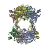
 Citation Citation | Journal: Nucleic Acids Res / Year: 2018 Title: Structural insights into Drosophila-C3PO complex assembly and 'Dynamic Side Port' model in substrate entry and release. Authors: Xiaobing Mo / Xia Yang / Yuren Adam Yuan /   Abstract: In Drosophila and human, component 3 promoter of RISC (C3PO), a heteromeric complex, enhances RISC assembly and promotes RISC activity. Here, we report crystal structure of full-length Drosophila ...In Drosophila and human, component 3 promoter of RISC (C3PO), a heteromeric complex, enhances RISC assembly and promotes RISC activity. Here, we report crystal structure of full-length Drosophila C3PO (E126Q), an inactive C3PO mutant displaying much weaker RNA binding ability, at 2.1 Å resolution. In addition, we also report the cryo-EM structures of full-length Drosophila C3PO (E126Q), C3PO (WT) and SUMO-C3PO (WT, sumo-TRAX + Translin) particles trapped at different conformations at 12, 19.7 and 12.8 Å resolutions, respectively. Crystal structure of C3PO (E126Q) displays a half-barrel architecture consisting of two Trax/Translin heterodimers, whereas cryo-EM structures of C3PO (E126Q), C3PO (WT) and SUMO-C3PO (WT) adopt a closed football-like shape with a hollow interior cavity. Remarkably, both cryo-EM structures of Drosophila C3PO (E126Q) and Drosophila SUMO-C3PO (WT) particles contain a wide side port (∼25 Å × ∼30 Å versus ∼15 Å × ∼20 Å) for RNA substrate entry and release, formed by a pair of anti-parallel packed long α1 helices of TRAX subunits. Notably, cryo-EM structure of SUMO-C3PO showed that four copies of extra densities belonging to N-terminal SUMO tag are located at the outside shell of SUMO-C3PO particle, which demonstrated that the stoichiometry of TRAX/Translin for the in vitro expressed and assembled full-length Drosophila-SUMO-C3PO particle is 4:4, suggesting Drosophila C3PO is composed by TRAX/translin at a ratio of 4:4. Remarkably, the comparison of the cryo-EM structures suggests that the C3PO side ports regulated by α1 helices of TRAX molecules are highly dynamic. Hence, we propose that C3PO particles could adopt a 'Dynamic Side Port' model to capture/digest nucleic acid duplex substrate and release the digested fragments through the dynamic side ports. | |||||||||
| History |
|
- Structure visualization
| Movie |
 Movie viewer Movie viewer |
|---|---|
| Structure viewer | EM map: SurfViewMolmilJmol/JSmol |
| Supplemental images |
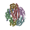
 UCSF Chimera
- Downloads & links
-EMDB archive
| Map data | emd_6722.map.gz | 6 MB | EMDB map data format | |
|---|---|---|---|---|
| Header (meta data) | emd-6722-v30.xmlemd-6722.xml | 13.6 KB 13.6 KB | Display Display | EMDB header |
| FSC (resolution estimation) | emd_6722_fsc.xml | 7.2 KB | Display | FSC data file |
| Images | 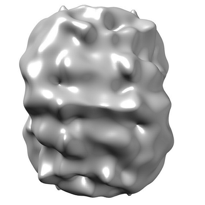 emd_6722.png emd_6722.png | 46.6 KB | ||
| Archive directory |  http://ftp.pdbj.org/pub/emdb/structures/EMD-6722ftp://ftp.pdbj.org/pub/emdb/structures/EMD-6722 http://ftp.pdbj.org/pub/emdb/structures/EMD-6722ftp://ftp.pdbj.org/pub/emdb/structures/EMD-6722 | HTTPS FTP |
-Related structure data


-Links
| EMDB pages | EMDB (EBI/PDBe) / EMDataResource |
|---|
-Map
| File | Download / File: emd_6722.map.gz / Format: CCP4 / Size: 30.5 MB / Type: IMAGE STORED AS FLOATING POINT NUMBER (4 BYTES) | ||||||||||||||||||||||||||||||||||||||||||||||||||||||||||||
|---|---|---|---|---|---|---|---|---|---|---|---|---|---|---|---|---|---|---|---|---|---|---|---|---|---|---|---|---|---|---|---|---|---|---|---|---|---|---|---|---|---|---|---|---|---|---|---|---|---|---|---|---|---|---|---|---|---|---|---|---|---|
| Annotation | cryo-EM structure of full-length C3PO (WT) adopt a closed football-like shape with a hollow interior cavity consisting of four Trax/Translin heterodimers. | ||||||||||||||||||||||||||||||||||||||||||||||||||||||||||||
| Projections & slices | Image control
Images are generated by Spider. | ||||||||||||||||||||||||||||||||||||||||||||||||||||||||||||
| Voxel size | X=Y=Z: 1.14 Å | ||||||||||||||||||||||||||||||||||||||||||||||||||||||||||||
| Density |
| ||||||||||||||||||||||||||||||||||||||||||||||||||||||||||||
| Symmetry | Space group: 1 | ||||||||||||||||||||||||||||||||||||||||||||||||||||||||||||
| Details | EMDB XML:
CCP4 map header:
| ||||||||||||||||||||||||||||||||||||||||||||||||||||||||||||
 Z (Sec.)
 Y (Row.)
 X (Col.)


-Supplemental data
- Sample components
-Entire : C3PO
| Entire | Name: C3PO |
|---|---|
| Components |
|
-Supramolecule #1: C3PO
| Supramolecule | Name: C3PO / type: complex / ID: 1 / Parent: 0 Details: In Drosophila, component 3 promoter of RISC (C3PO)is a complex, consisting of 4 Trax/Translin heterodimers. |
|---|---|
| Source (natural) | Organism: |
| Recombinant expression | Organism: Recombinant strain: DH5a / Recombinant plasmid: pET-Duet |
| Molecular weight | Experimental: 290 KDa |
-Supramolecule #2: TRAX
| Supramolecule | Name: TRAX / type: complex / ID: 2 / Parent: 1 / Details: Translin-associated factor X |
|---|
-Supramolecule #3: Translin
| Supramolecule | Name: Translin / type: complex / ID: 3 / Parent: 1 / Details: A key component in C3PO complex |
|---|
-Experimental details
-Structure determination
| Method | cryo EM |
|---|---|
 Processing Processing | single particle reconstruction |
| Aggregation state | particle |
-Sample preparation
| Concentration | 1 mg/mL | |||||||||||||||
|---|---|---|---|---|---|---|---|---|---|---|---|---|---|---|---|---|
| Buffer | pH: 7.4 Component:
Details: 20mM Tris (pH 7.4), 500mM NaCl, 1mM DTT and 2mM EDTA | |||||||||||||||
| Grid | Model: Quantifoil R2/2 / Material: COPPER / Mesh: 400 / Support film - Material: FORMVAR / Support film - topology: HOLEY / Pretreatment - Type: GLOW DISCHARGE / Pretreatment - Atmosphere: AIR / Pretreatment - Pressure: 101.325 kPa | |||||||||||||||
| Vitrification | Cryogen name: ETHANE / Chamber humidity: 100 % / Chamber temperature: 298 K / Instrument: FEI VITROBOT MARK IV / Details: blot for 5 seconds before pluning. | |||||||||||||||
| Details | This sample was monodisperse. |
- Electron microscopy
| Microscope | FEI TECNAI 12 |
|---|---|
| Temperature | Min: 70.0 K / Max: 70.0 K |
| Details | Preliminary grid screening was performed manually. |
| Image recording | Film or detector model: GATAN ULTRASCAN 4000 (4k x 4k) / Number grids imaged: 20 / Number real images: 500 / Average exposure time: 1.0 sec. / Average electron dose: 20.0 e/Å2 |
| Electron beam | Acceleration voltage: 120 kV / Electron source: LAB6 |
| Electron optics | Calibrated defocus max: 5.0 µm / Calibrated defocus min: 1.2 µm / Calibrated magnification: 67000 / Illumination mode: OTHER / Imaging mode: DARK FIELD / Cs: 2.7 mm / Nominal defocus max: 5.0 µm / Nominal defocus min: 1.2 µm / Nominal magnification: 67000 |
| Sample stage | Specimen holder model: GATAN 626 SINGLE TILT LIQUID NITROGEN CRYO TRANSFER HOLDER Cooling holder cryogen: NITROGEN |
+Image processing

-Atomic model buiding 1
| Refinement | Space: RECIPROCAL / Protocol: AB INITIO MODEL / Overall B value: 300 |
|---|
