 Movie
Movie Controller

[English] 日本語
 Yorodumi
- EMDB-63988: cryo-EM structure of ligand-free active-state M1 muscarinic acety... -
+ Open data

- Basic information
| Entry |  | |||||||||
|---|---|---|---|---|---|---|---|---|---|---|
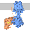
| Title | cryo-EM structure of ligand-free active-state M1 muscarinic acetylcholine receptor with alpha5 helix of G11 protein complex | |||||||||
 Map data Map data | ||||||||||
 Sample Sample |
| |||||||||
 Keywords Keywords | GPCR / active-state / ligand-free / de novo protein / MEMBRANE PROTEIN | |||||||||
| Function / homology |  Function and homology information Function and homology informationphospholipase C-activating G protein-coupled acetylcholine receptor signaling pathway / Fatty Acids bound to GPR40 (FFAR1) regulate insulin secretion / phospholipase C-activating dopamine receptor signaling pathway / Acetylcholine regulates insulin secretion / cranial skeletal system development / endothelin receptor signaling pathway / PLC beta mediated events / entrainment of circadian clock / phototransduction, visible light / action potential ...phospholipase C-activating G protein-coupled acetylcholine receptor signaling pathway / Fatty Acids bound to GPR40 (FFAR1) regulate insulin secretion / phospholipase C-activating dopamine receptor signaling pathway / Acetylcholine regulates insulin secretion / cranial skeletal system development / endothelin receptor signaling pathway / PLC beta mediated events / entrainment of circadian clock / phototransduction, visible light / action potential / photoreceptor outer segment / Turbulent (oscillatory, disturbed) flow shear stress activates signaling by PIEZO1 and integrins in endothelial cells / regulation of blood pressure / G protein-coupled receptor binding / G-protein beta/gamma-subunit complex binding / adenylate cyclase-modulating G protein-coupled receptor signaling pathway / phospholipase C-activating G protein-coupled receptor signaling pathway / G protein-coupled acetylcholine receptor signaling pathway / Thromboxane signalling through TP receptor / G-protein activation / ADP signalling through P2Y purinoceptor 1 / Cooperation of PDCL (PhLP1) and TRiC/CCT in G-protein beta folding / heterotrimeric G-protein complex / Thrombin signalling through proteinase activated receptors (PARs) / G protein activity / High laminar flow shear stress activates signaling by PIEZO1 and PECAM1:CDH5:KDR in endothelial cells / G alpha (q) signalling events / Hydrolases; Acting on acid anhydrides; Acting on GTP to facilitate cellular and subcellular movement / lysosomal membrane / GTPase activity / synapse / GTP binding / signal transduction / extracellular exosome / metal ion binding / plasma membrane / cytoplasm Similarity search - Function | |||||||||
| Biological species |  Homo sapiens (human) Homo sapiens (human) | |||||||||
| Method | single particle reconstruction / cryo EM / Resolution: 3.62 Å | |||||||||
 Authors Authors | Zhang X / Gao K / Liu X | |||||||||
| Funding support |  China, 1 items China, 1 items
| |||||||||
 Citation Citation | Journal: Proc Natl Acad Sci U S A / Year: 2025 Title: Extracellular nanobody screening using conformationally stable GPCR variants. Authors: Xin Zhang / Kaixuan Gao / Jia Nie / Hengyu Meng / Xiaoou Sun / Jiawei Zhao / Xiangyu Liu / Abstract: G protein-coupled receptors (GPCRs) are prominent drug targets that have attracted intensive efforts in drug screening. Binding-based screening methods for GPCR ligands often require conformationally ...G protein-coupled receptors (GPCRs) are prominent drug targets that have attracted intensive efforts in drug screening. Binding-based screening methods for GPCR ligands often require conformationally stable, purified receptors. However, obtaining large quantities of GPCRs in stable states, particularly with unoccupied extracellular ligand-binding pockets and especially in their active conformations, remains challenging due to the inherent dynamic nature of these receptors. To address this challenge, we propose a universal approach for stabilizing GPCRs in specific conformations. Using the M1 muscarinic acetylcholine receptor (M1R) as a model, we successfully stabilized M1R in its active conformation through de novo design of a fusion protein, and further demonstrated the generalizability of this strategy by applying it to other GPCRs. We screened a synthetic yeast display library of nanobodies against both the stabilized active-state and previously reported inactive-state M1R, identifying several nanobodies that specifically recognize each conformation. This method not only facilitates the stabilization of GPCRs in desired states but also provides valuable tools for developing more selective therapeutic agents, enhancing drug discovery efficiency and specificity. | |||||||||
| History |
|
- Structure visualization
| Supplemental images |
|---|

- Downloads & links
-EMDB archive
| Map data | emd_63988.map.gz | 59.7 MB | EMDB map data format | |
|---|---|---|---|---|
| Header (meta data) | emd-63988-v30.xmlemd-63988.xml | 19.8 KB 19.8 KB | Display Display | EMDB header |
| Images |  emd_63988.png emd_63988.png | 49.7 KB | ||
| Masks | emd_63988_msk_1.map | 64 MB | Mask map | |
| Filedesc metadata | emd-63988.cif.gz | 6 KB | ||
| Others | emd_63988_half_map_1.map.gzemd_63988_half_map_2.map.gz | 59.5 MB 59.5 MB | ||
| Archive directory |  http://ftp.pdbj.org/pub/emdb/structures/EMD-63988ftp://ftp.pdbj.org/pub/emdb/structures/EMD-63988 http://ftp.pdbj.org/pub/emdb/structures/EMD-63988ftp://ftp.pdbj.org/pub/emdb/structures/EMD-63988 | HTTPS FTP |
-Related structure data
| Related structure data |  9uapMC  9uazC  9ucpC M: atomic model generated by this map C: citing same article ( |
|---|---|
| Similar structure data |


-Links
| EMDB pages | EMDB (EBI/PDBe) / EMDataResource |
|---|---|
| Related items in Molecule of the Month |







-Map
| File | Download / File: emd_63988.map.gz / Format: CCP4 / Size: 64 MB / Type: IMAGE STORED AS FLOATING POINT NUMBER (4 BYTES) | ||||||||||||||||||||||||||||||||||||
|---|---|---|---|---|---|---|---|---|---|---|---|---|---|---|---|---|---|---|---|---|---|---|---|---|---|---|---|---|---|---|---|---|---|---|---|---|---|
| Projections & slices | Image control
Images are generated by Spider. | ||||||||||||||||||||||||||||||||||||
| Voxel size | X=Y=Z: 1.0979 Å | ||||||||||||||||||||||||||||||||||||
| Density |
| ||||||||||||||||||||||||||||||||||||
| Symmetry | Space group: 1 | ||||||||||||||||||||||||||||||||||||
| Details | EMDB XML:
|
 Z (Sec.)
 Y (Row.)
 X (Col.)


-Supplemental data
-Mask #1
| File | emd_63988_msk_1.map | ||||||||||||
|---|---|---|---|---|---|---|---|---|---|---|---|---|---|
| Projections & Slices |
| ||||||||||||
| Density Histograms |


-Half map: #2
| File | emd_63988_half_map_1.map | ||||||||||||
|---|---|---|---|---|---|---|---|---|---|---|---|---|---|
| Projections & Slices |
| ||||||||||||
| Density Histograms |


-Half map: #1
| File | emd_63988_half_map_2.map | ||||||||||||
|---|---|---|---|---|---|---|---|---|---|---|---|---|---|
| Projections & Slices |
| ||||||||||||
| Density Histograms |


- Sample components
-Entire : Ligand-free active-state M1 muscarinic acetylcholine receptor wit...
| Entire | Name: Ligand-free active-state M1 muscarinic acetylcholine receptor with alpha5 helix of G11 protein complex/de novo design fusion protein |
|---|---|
| Components |
|
-Supramolecule #1: Ligand-free active-state M1 muscarinic acetylcholine receptor wit...
| Supramolecule | Name: Ligand-free active-state M1 muscarinic acetylcholine receptor with alpha5 helix of G11 protein complex/de novo design fusion protein type: complex / ID: 1 / Parent: 0 / Macromolecule list: all |
|---|---|
| Source (natural) | Organism: Homo sapiens (human) |
-Macromolecule #1: M1 muscarinic acetylcholine receptor, de novo design protein
| Macromolecule | Name: M1 muscarinic acetylcholine receptor, de novo design protein type: protein_or_peptide / ID: 1 / Number of copies: 1 / Enantiomer: LEVO |
|---|---|
| Source (natural) | Organism: Homo sapiens (human) |
| Molecular weight | Theoretical: 42.07073 KDa |
| Recombinant expression | Organism:   Spodoptera frugiperda (fall armyworm) Spodoptera frugiperda (fall armyworm) |
| Sequence | String: VAFIGITTGL LSLATVTGNL LVLISFKVNT ELKTVNNYFL LSLACADLII GTFSMNLYTT YLLMGHWALG TLACDLWLAL DYVASNASV MNLLLISFDR YFSVTRPLSY RAKRTPRRAA LMIGLAWLVS FVLWAPAILF WQYLVGERTV LAGQCYIQFL S QPIITFGT ...String: VAFIGITTGL LSLATVTGNL LVLISFKVNT ELKTVNNYFL LSLACADLII GTFSMNLYTT YLLMGHWALG TLACDLWLAL DYVASNASV MNLLLISFDR YFSVTRPLSY RAKRTPRRAA LMIGLAWLVS FVLWAPAILF WQYLVGERTV LAGQCYIQFL S QPIITFGT AMAAFYLPVT VMCTLYWRIY RETKRAGERL AKLLEKFEAL PLEDIVAALK ALLATNRPEI QLAVKTIVEN FP EIKKEAE KLTPEQKAKL AALEAQLADL PEELRKVLLS MYLTGLLYGG SEENRKRAIE KAARTLSAIL LAFILTWTPY NIM VLVSTF CKDCVPETLW ELGYWLCYVN STINPMCYAL CNKAFRDTFR LLLLCR |
-Macromolecule #2: Guanine nucleotide-binding protein subunit alpha-11
| Macromolecule | Name: Guanine nucleotide-binding protein subunit alpha-11 / type: protein_or_peptide / ID: 2 / Number of copies: 1 / Enantiomer: LEVO EC number: Hydrolases; Acting on acid anhydrides; Acting on GTP to facilitate cellular and subcellular movement |
|---|---|
| Source (natural) | Organism: Homo sapiens (human) |
| Molecular weight | Theoretical: 3.15266 KDa |
| Recombinant expression | Organism: Spodoptera frugiperda (fall armyworm) |
| Sequence | String: TPENIRFVFA AVKDTILQLN LKEYNLV UniProtKB: Guanine nucleotide-binding protein subunit alpha-11 |
-Experimental details
-Structure determination
| Method | cryo EM |
|---|---|
 Processing Processing | single particle reconstruction |
| Aggregation state | particle |
-Sample preparation
| Buffer | pH: 7.5 |
|---|---|
| Vitrification | Cryogen name: ETHANE |
- Electron microscopy
| Microscope | TFS KRIOS |
|---|---|
| Image recording | Film or detector model: GATAN K3 (6k x 4k) / Average electron dose: 50.0 e/Å2 |
| Electron beam | Acceleration voltage: 300 kV / Electron source:  FIELD EMISSION GUN FIELD EMISSION GUN |
| Electron optics | Illumination mode: SPOT SCAN / Imaging mode: BRIGHT FIELD / Nominal defocus max: 1.8 µm / Nominal defocus min: 1.1 µm |
| Experimental equipment |  Model: Titan Krios / Image courtesy: FEI Company |
