Ministry of Education, Culture, Sports, Science and Technology (Japan)
JP24ama121005
Japan
Citation
Journal: ACS Chem Neurosci / Year: 2025 Title: Microgravity-Assisted Exploration of the Conformational Space of Amyloid β Affected by Tottori-Type Familial Mutation D7N. Authors: Maho Yagi-Utsumi / Saeko Yanaka / Raymond N Burton-Smith / Chihong Song / Christian Ganser / Chiaki Yamazaki / Haruo Kasahara / Toru Shimazu / Takayuki Uchihashi / Kazuyoshi Murata / Koichi Kato / Abstract: The amyloid β (Aβ) Tottori variant (D7N) exhibits unique aggregation behaviors and altered fibril formation, posing challenges for structural characterization. To overcome this, the microgravity ...The amyloid β (Aβ) Tottori variant (D7N) exhibits unique aggregation behaviors and altered fibril formation, posing challenges for structural characterization. To overcome this, the microgravity environment on the International Space Station was employed to study Tottori-type Aβ40 fibril formation and structure. Under Earth gravity, Tottori-type Aβ40 primarily formed nonfibrillar aggregates, hindering detailed structural analysis. In contrast, microgravity significantly enhanced fibril formation and minimized amorphous aggregates. Cryo-electron microscopy revealed two structurally distinct fibril types, each comprising different protomer conformations. In both types, the N-terminal segment was disordered and nor resolved in the density maps. The D7N mutation disrupts the protection of the core by the N-terminal segment often observed in wild-type Aβ40 fibrils, enhancing the hydrophobicity-mediated aggregation propensity. However, microgravity suppressed kinetic traps and facilitated high-quality fibril formation suitable for structural studies that can explore the free energy landscape of Aβ fibril formation. These findings demonstrate the utility of microgravity for studying familial Aβ variants and potentially accelerate our understanding of Aβ aggregation mechanisms in Alzheimer's disease.
In the structure databanks used in Yorodumi, some data are registered as the other names, "COVID-19 virus" and "2019-nCoV". Here are the details of the virus and the list of structure data.
Jan 31, 2019. EMDB accession codes are about to change! (news from PDBe EMDB page)
EMDB accession codes are about to change! (news from PDBe EMDB page)
The allocation of 4 digits for EMDB accession codes will soon come to an end. Whilst these codes will remain in use, new EMDB accession codes will include an additional digit and will expand incrementally as the available range of codes is exhausted. The current 4-digit format prefixed with “EMD-” (i.e. EMD-XXXX) will advance to a 5-digit format (i.e. EMD-XXXXX), and so on. It is currently estimated that the 4-digit codes will be depleted around Spring 2019, at which point the 5-digit format will come into force.
The EM Navigator/Yorodumi systems omit the EMD- prefix.
Related info.:Q: What is EMD? / ID/Accession-code notation in Yorodumi/EM Navigator
Yorodumi is a browser for structure data from EMDB, PDB, SASBDB, etc.
This page is also the successor to EM Navigator detail page, and also detail information page/front-end page for Omokage search.
The word "yorodu" (or yorozu) is an old Japanese word meaning "ten thousand". "mi" (miru) is to see.
Related info.:EMDB / PDB / SASBDB / Comparison of 3 databanks / Yorodumi Search / Aug 31, 2016. New EM Navigator & Yorodumi / Yorodumi Papers / Jmol/JSmol / Function and homology information / Changes in new EM Navigator and Yorodumi
 Movie
Movie Controller


 Open data
 Basic information
 Map data
 Sample
 Keywords
 Function and homology information
 Homo sapiens (human)
 Authors
 Japan, 1 items
  Citation
 Structure visualization
 Downloads & links
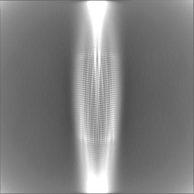
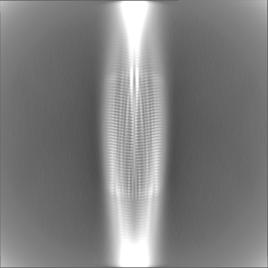
 emd_63647.png
 http://ftp.pdbj.org/pub/emdb/structures/EMD-63647


 Z (Sec.)
 Y (Row.)
 X (Col.)


 Sample components
 Processing
 Electron microscopy
 FIELD EMISSION GUN

