 ムービー
ムービー コントローラー


+ データを開く

- 基本情報
基本情報
| 登録情報 | データベース: EMDB / ID: EMD-6337 | |||||||||
|---|---|---|---|---|---|---|---|---|---|---|
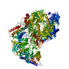
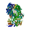
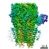
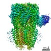
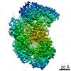
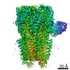
| タイトル | Structure of the L-protein of vesicular stomatitis virus from electron cryomicroscopy | |||||||||
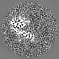
 マップデータ マップデータ | Reconstruction of the L-protein of vesicular stomatitis virus | |||||||||
 試料 試料 |
| |||||||||
 キーワード キーワード | RNA-dependent RNA polymerase / RNA capping / cryoEM single-particle analysis | |||||||||
| 機能・相同性 |  機能・相同性情報 機能・相同性情報NNS virus cap methyltransferase / GDP polyribonucleotidyltransferase / negative stranded viral RNA replication / viral transcription / 加水分解酵素; 酸無水物に作用; リン含有酸無水物に作用 / virion component / mRNA 5'-cap (guanine-N7-)-methyltransferase activity / host cell cytoplasm / RNA-directed RNA polymerase / RNA-dependent RNA polymerase activity ...NNS virus cap methyltransferase / GDP polyribonucleotidyltransferase / negative stranded viral RNA replication / viral transcription / 加水分解酵素; 酸無水物に作用; リン含有酸無水物に作用 / virion component / mRNA 5'-cap (guanine-N7-)-methyltransferase activity / host cell cytoplasm / RNA-directed RNA polymerase / RNA-dependent RNA polymerase activity / GTPase activity / ATP binding / metal ion binding 類似検索 - 分子機能 | |||||||||
| 生物種 |  Vesicular stomatitis virus (ウイルス) Vesicular stomatitis virus (ウイルス) | |||||||||
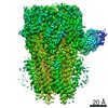
| 手法 | 単粒子再構成法 / クライオ電子顕微鏡法 / 解像度: 3.8 Å | |||||||||
 データ登録者 データ登録者 | Liang B / Li Z / Jenni S / Rameh AA / Morin BM / Grant T / Grigorieff N / Harrison SC / Whelan SPJ | |||||||||
 引用 引用 | ジャーナル: Cell / 年: 2015 タイトル: Structure of the L Protein of Vesicular Stomatitis Virus from Electron Cryomicroscopy. 著者: Bo Liang / Zongli Li / Simon Jenni / Amal A Rahmeh / Benjamin M Morin / Timothy Grant / Nikolaus Grigorieff / Stephen C Harrison / Sean P J Whelan /  要旨: The large (L) proteins of non-segmented, negative-strand RNA viruses, a group that includes Ebola and rabies viruses, catalyze RNA-dependent RNA polymerization with viral ribonucleoprotein as ...The large (L) proteins of non-segmented, negative-strand RNA viruses, a group that includes Ebola and rabies viruses, catalyze RNA-dependent RNA polymerization with viral ribonucleoprotein as template, a non-canonical sequence of capping and methylation reactions, and polyadenylation of viral messages. We have determined by electron cryomicroscopy the structure of the vesicular stomatitis virus (VSV) L protein. The density map, at a resolution of 3.8 Å, has led to an atomic model for nearly all of the 2109-residue polypeptide chain, which comprises three enzymatic domains (RNA-dependent RNA polymerase [RdRp], polyribonucleotidyl transferase [PRNTase], and methyltransferase) and two structural domains. The RdRp resembles the corresponding enzymatic regions of dsRNA virus polymerases and influenza virus polymerase. A loop from the PRNTase (capping) domain projects into the catalytic site of the RdRp, where it appears to have the role of a priming loop and to couple product elongation to large-scale conformational changes in L. | |||||||||
| 履歴 |
|
- 構造の表示
構造の表示
| ムービー |
ムービービューア |
|---|---|
| 構造ビューア | EMマップ: SurfViewMolmilJmol/JSmol |
| 添付画像 |

- ダウンロードとリンク
-EMDBアーカイブ
| マップデータ | emd_6337.map.gz | 6.2 MB | EMDBマップデータ形式 | |
|---|---|---|---|---|
| ヘッダ (付随情報) | emd-6337-v30.xmlemd-6337.xml | 11.3 KB 11.3 KB | 表示 表示 | EMDBヘッダ |
| 画像 | 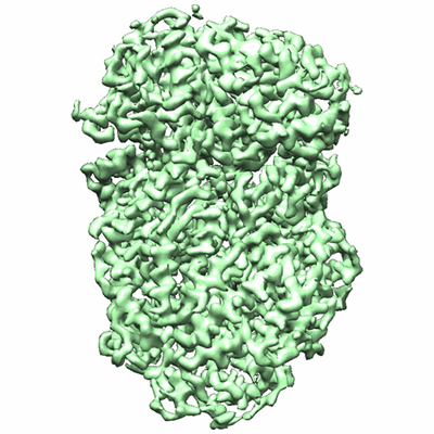 400_6337.gif 400_6337.gif 80_6337.gif 80_6337.gif | 82.4 KB 5 KB | ||
| Filedesc structureFactors | emd_6337_sf.cif.gz | 1.2 MB | ||
| アーカイブディレクトリ |  http://ftp.pdbj.org/pub/emdb/structures/EMD-6337ftp://ftp.pdbj.org/pub/emdb/structures/EMD-6337 http://ftp.pdbj.org/pub/emdb/structures/EMD-6337ftp://ftp.pdbj.org/pub/emdb/structures/EMD-6337 | HTTPS FTP |
-検証レポート
| 文書・要旨 | emd_6337_validation.pdf.gz | 281.1 KB | 表示 | EMDB検証レポート |
|---|---|---|---|---|
| 文書・詳細版 | emd_6337_full_validation.pdf.gz | 280.2 KB | 表示 | |
| XML形式データ | emd_6337_validation.xml.gz | 5.4 KB | 表示 | |
| アーカイブディレクトリ | https://ftp.pdbj.org/pub/emdb/validation_reports/EMD-6337ftp://ftp.pdbj.org/pub/emdb/validation_reports/EMD-6337 | HTTPS FTP |
-関連構造データ
-リンク
| EMDBのページ | EMDB (EBI/PDBe) / EMDataResource |
|---|---|
| 「今月の分子」の関連する項目 |

-マップ
| ファイル | ダウンロード / ファイル: emd_6337.map.gz / 形式: CCP4 / 大きさ: 6.4 MB / タイプ: IMAGE STORED AS FLOATING POINT NUMBER (4 BYTES) | ||||||||||||||||||||||||||||||||||||||||||||||||||||||||||||
|---|---|---|---|---|---|---|---|---|---|---|---|---|---|---|---|---|---|---|---|---|---|---|---|---|---|---|---|---|---|---|---|---|---|---|---|---|---|---|---|---|---|---|---|---|---|---|---|---|---|---|---|---|---|---|---|---|---|---|---|---|---|
| 注釈 | Reconstruction of the L-protein of vesicular stomatitis virus | ||||||||||||||||||||||||||||||||||||||||||||||||||||||||||||
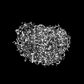
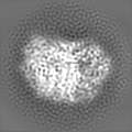
| 投影像・断面図 | 画像のコントロール
画像は Spider により作成 | ||||||||||||||||||||||||||||||||||||||||||||||||||||||||||||
| ボクセルのサイズ | X=Y=Z: 1.237 Å | ||||||||||||||||||||||||||||||||||||||||||||||||||||||||||||
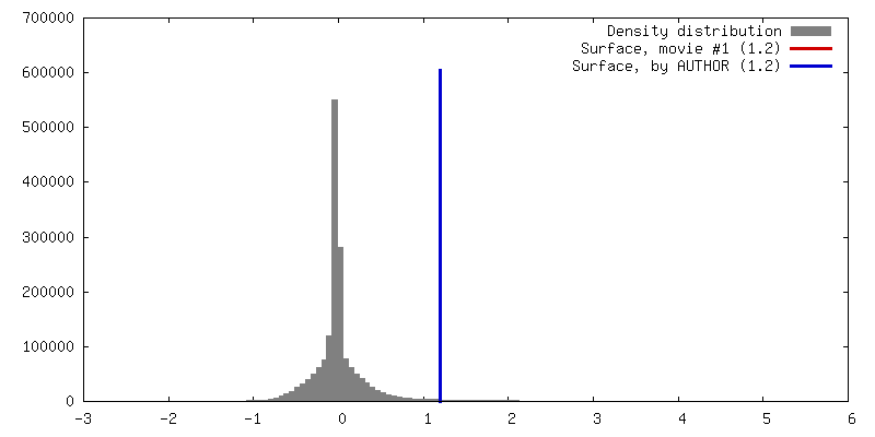
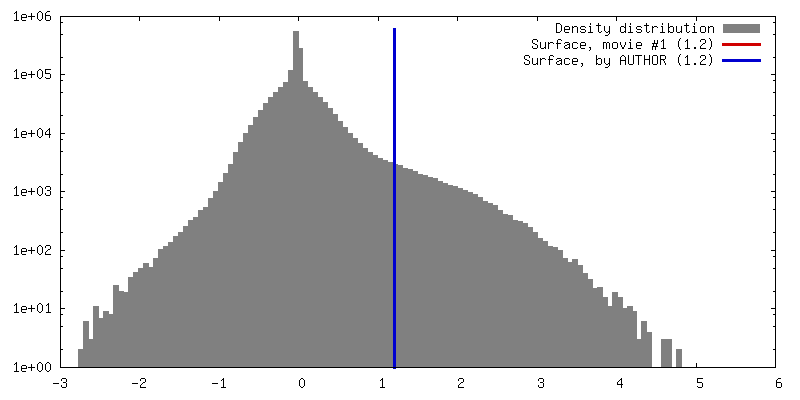
| 密度 |
| ||||||||||||||||||||||||||||||||||||||||||||||||||||||||||||
| 対称性 | 空間群: 1 | ||||||||||||||||||||||||||||||||||||||||||||||||||||||||||||
| 詳細 | EMDB XML:
CCP4マップ ヘッダ情報:
| ||||||||||||||||||||||||||||||||||||||||||||||||||||||||||||
 Z (Sec.)
 Y (Row.)
 X (Col.)


-添付データ
- 試料の構成要素
-全体 : VSV-L
| 全体 | 名称: VSV-L |
|---|---|
| 要素 |
|
-超分子 #1000: VSV-L
| 超分子 | 名称: VSV-L / タイプ: sample / ID: 1000 集合状態: One monomer of VSV-L bound to one monomer of P Number unique components: 2 |
|---|---|
| 分子量 | 理論値: 250 KDa |
-分子 #1: VSV large protein
| 分子 | 名称: VSV large protein / タイプ: protein_or_peptide / ID: 1 / Name.synonym: VSV-L / コピー数: 1 / 集合状態: Monomer / 組換発現: Yes |
|---|---|
| 由来(天然) | 生物種: Vesicular stomatitis virus (ウイルス) |
| 分子量 | 理論値: 240 KDa |
| 組換発現 | 生物種:   Spodoptera frugiperda (ツマジロクサヨトウ) Spodoptera frugiperda (ツマジロクサヨトウ)組換細胞: Sf21 |
-分子 #2: VSV phosphoprotein
| 分子 | 名称: VSV phosphoprotein / タイプ: protein_or_peptide / ID: 2 / Name.synonym: P / コピー数: 1 / 集合状態: Monomer / 組換発現: Yes |
|---|---|
| 由来(天然) | 生物種: Vesicular stomatitis virus (ウイルス) |
| 分子量 | 理論値: 10 KDa |
| 組換発現 | 生物種:  |
-実験情報
-構造解析
| 手法 | クライオ電子顕微鏡法 |
|---|---|
 解析 解析 | 単粒子再構成法 |
| 試料の集合状態 | particle |
-試料調製
| 濃度 | 0.35 mg/mL |
|---|---|
| 緩衝液 | pH: 7.4 / 詳細: 25 mM HEPES, 250 mM NaCl, 6 mM MgSO4, 0.5 mM TCEP |
| グリッド | 詳細: 400 mesh Quantifoil R1.2/1.3 Cu grid |
| 凍結 | 凍結剤: ETHANE / チャンバー内湿度: 65 % / 装置: FEI VITROBOT MARK I 手法: Blot time 2 seconds, drain time 1 second before plunging |
- 電子顕微鏡法
| 顕微鏡 | FEI TECNAI F20 |
|---|---|
| 詳細 | Beam intensity: 8 electrons/pixel/s Movie mode: 30 frames, 5 frames/s |
| 日付 | 2014年8月1日 |
| 撮影 | カテゴリ: CCD / フィルム・検出器のモデル: GATAN K2 (4k x 4k) / デジタル化 - サンプリング間隔: 5 µm / 実像数: 1272 / 平均電子線量: 31 e/Å2 詳細: Images are the sums of all 30 aligned movie frames (high dose) or frames 3 - 12 (low-dose). |
| 電子線 | 加速電圧: 200 kV / 電子線源:  FIELD EMISSION GUN FIELD EMISSION GUN |
| 電子光学系 | 倍率(補正後): 40410 / 照射モード: FLOOD BEAM / 撮影モード: BRIGHT FIELD / Cs: 2.0 mm / 最大 デフォーカス(公称値): 2.3 µm / 最小 デフォーカス(公称値): 0.9 µm / 倍率(公称値): 29000 |
| 試料ステージ | 試料ホルダー: CT3500 / 試料ホルダーモデル: GATAN LIQUID NITROGEN |
| 実験機器 |  モデル: Tecnai F20 / 画像提供: FEI Company |
-画像解析
| 詳細 | An initial map was obtained with EMAN2, IMAGIC, and TIGRIS. CTF was determined using CTFFIND3. Refinement and classification were done using Frealign. |
|---|---|
| CTF補正 | 詳細: Each particle |
| 最終 再構成 | アルゴリズム: OTHER / 解像度のタイプ: BY AUTHOR / 解像度: 3.8 Å / 解像度の算出法: OTHER / ソフトウェア - 名称: EMAN2, IMAGIC, Frealign 詳細: The final map represents the best class out of three classes. 使用した粒子像数: 74940 |
| 最終 2次元分類 | クラス数: 3 |
