Movie
Movie Controller

[English] 日本語
 Yorodumi
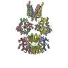
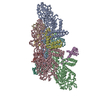
- EMDB-6330: 3D structure of the decameric assembly of the DNA-injection prote... -
+ Open data

- Basic information
| Entry | Database: EMDB / ID: EMD-6330 | |||||||||
|---|---|---|---|---|---|---|---|---|---|---|
| Title | 3D structure of the decameric assembly of the DNA-injection protein gp12 from bacteriophage Sf6 | |||||||||
 Map data Map data | EM reconstruction of DNA injection protein decameric assembly | |||||||||
 Sample Sample |
| |||||||||
 Keywords Keywords | bacteriophage / podovirus / DNA injection / decamer / Gram-negative / cell envelope | |||||||||
| Function / homology | DNA transfer protein / DNA/protein translocase of phage P22 injectosome / Gene 12 protein Function and homology information Function and homology information | |||||||||
| Biological species |  Bacillus phage SF6 (virus) Bacillus phage SF6 (virus) | |||||||||
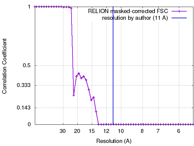
| Method | single particle reconstruction / negative staining / Resolution: 11.0 Å | |||||||||
 Authors Authors | Zhao H / Speir JA / Matsui T / Lin Z / Liang L / Lynn AY / Varnado B / Weiss TM / Tang L | |||||||||
 Citation Citation | Journal: PLoS One / Year: 2016 Title: Structure of a Bacterial Virus DNA-Injection Protein Complex Reveals a Decameric Assembly with a Constricted Molecular Channel. Authors: Haiyan Zhao / Jeffrey A Speir / Tsutomu Matsui / Zihan Lin / Lingfei Liang / Anna Y Lynn / Brittany Varnado / Thomas M Weiss / Liang Tang /  Abstract: The multi-layered cell envelope structure of Gram-negative bacteria represents significant physical and chemical barriers for short-tailed phages to inject phage DNA into the host cytoplasm. Here we ...The multi-layered cell envelope structure of Gram-negative bacteria represents significant physical and chemical barriers for short-tailed phages to inject phage DNA into the host cytoplasm. Here we show that a DNA-injection protein of bacteriophage Sf6, gp12, forms a 465-kDa, decameric assembly in vitro. The electron microscopic structure of the gp12 assembly shows a ~150-Å, mushroom-like architecture consisting of a crown domain and a tube-like domain, which embraces a 25-Å-wide channel that could precisely accommodate dsDNA. The constricted channel suggests that gp12 mediates rapid, uni-directional injection of phage DNA into host cells by providing a molecular conduit for DNA translocation. The assembly exhibits a 10-fold symmetry, which may be a common feature among DNA-injection proteins of P22-like phages and may suggest a symmetry mismatch with respect to the 6-fold symmetric phage tail. The gp12 monomer is highly flexible in solution, supporting a mechanism for translocation of the protein through the conduit of the phage tail toward the host cell envelope, where it assembles into a DNA-injection device. | |||||||||
| History |
|
- Structure visualization
| Movie |
Movie viewer |
|---|---|
| Structure viewer | EM map: SurfViewMolmilJmol/JSmol |
| Supplemental images |

- Downloads & links
-EMDB archive
| Map data | emd_6330.map.gz | 5.5 MB | EMDB map data format | |
|---|---|---|---|---|
| Header (meta data) | emd-6330-v30.xmlemd-6330.xml | 10.9 KB 10.9 KB | Display Display | EMDB header |
| FSC (resolution estimation) | emd_6330_fsc.xml | 4.4 KB | Display | FSC data file |
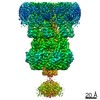
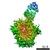
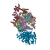
| Images | 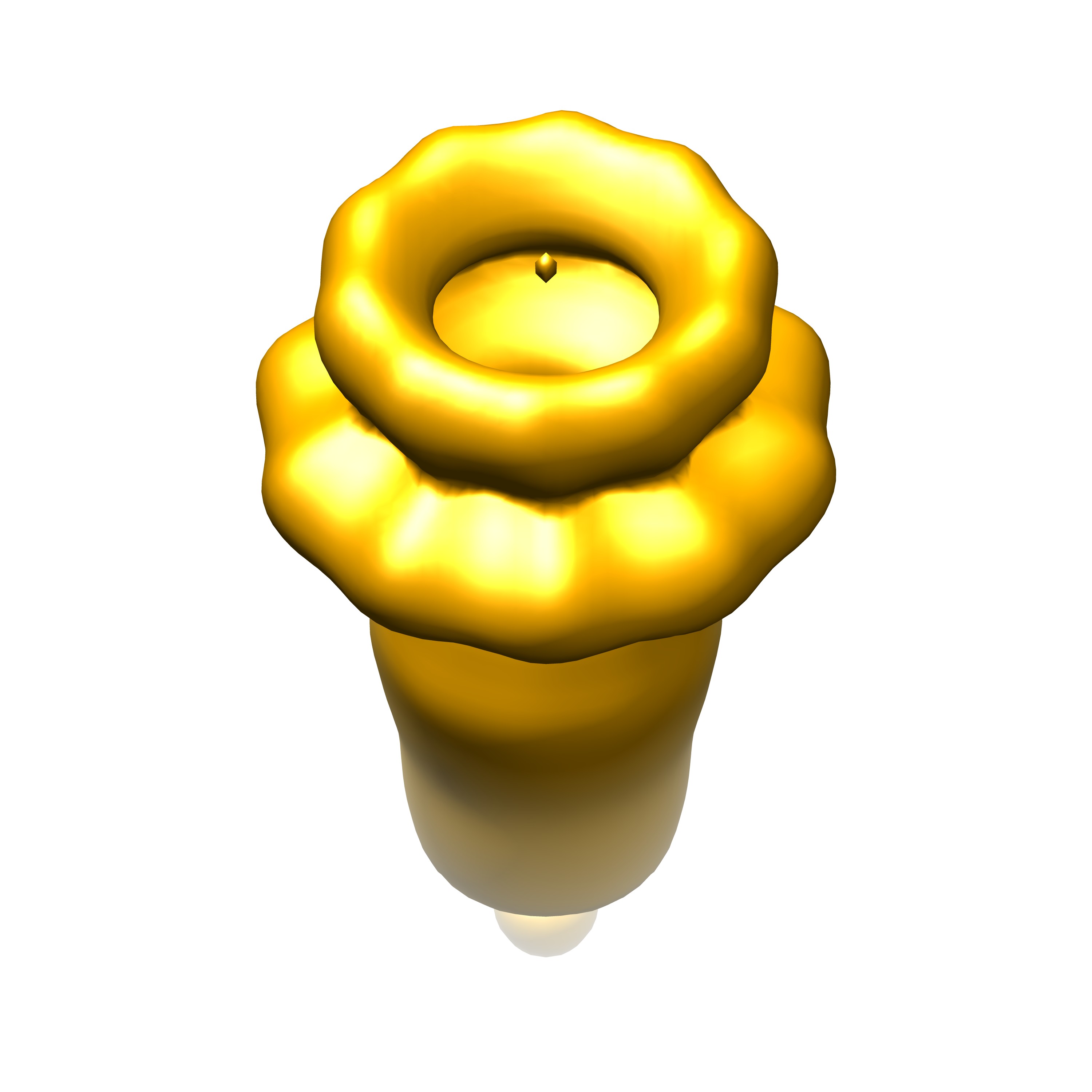 emd_6330.jpg emd_6330.jpg | 298.2 KB | ||
| Archive directory |  http://ftp.pdbj.org/pub/emdb/structures/EMD-6330ftp://ftp.pdbj.org/pub/emdb/structures/EMD-6330 http://ftp.pdbj.org/pub/emdb/structures/EMD-6330ftp://ftp.pdbj.org/pub/emdb/structures/EMD-6330 | HTTPS FTP |
-Related structure data
| Similar structure data |
|---|

-Links
| EMDB pages | EMDB (EBI/PDBe) / EMDataResource |
|---|
-Map
| File | Download / File: emd_6330.map.gz / Format: CCP4 / Size: 7.8 MB / Type: IMAGE STORED AS FLOATING POINT NUMBER (4 BYTES) | ||||||||||||||||||||||||||||||||||||||||||||||||||||||||||||||||||||
|---|---|---|---|---|---|---|---|---|---|---|---|---|---|---|---|---|---|---|---|---|---|---|---|---|---|---|---|---|---|---|---|---|---|---|---|---|---|---|---|---|---|---|---|---|---|---|---|---|---|---|---|---|---|---|---|---|---|---|---|---|---|---|---|---|---|---|---|---|---|
| Annotation | EM reconstruction of DNA injection protein decameric assembly | ||||||||||||||||||||||||||||||||||||||||||||||||||||||||||||||||||||
| Projections & slices | Image control
Images are generated by Spider. | ||||||||||||||||||||||||||||||||||||||||||||||||||||||||||||||||||||
| Voxel size | X=Y=Z: 2.73 Å | ||||||||||||||||||||||||||||||||||||||||||||||||||||||||||||||||||||
| Density |
| ||||||||||||||||||||||||||||||||||||||||||||||||||||||||||||||||||||
| Symmetry | Space group: 1 | ||||||||||||||||||||||||||||||||||||||||||||||||||||||||||||||||||||
| Details | EMDB XML:
CCP4 map header:
| ||||||||||||||||||||||||||||||||||||||||||||||||||||||||||||||||||||
 Z (Sec.)
 Y (Row.)
 X (Col.)


-Supplemental data
- Sample components
-Entire : gp12 of bacteriophage Sf6
| Entire | Name: gp12 of bacteriophage Sf6 |
|---|---|
| Components |
|
-Supramolecule #1000: gp12 of bacteriophage Sf6
| Supramolecule | Name: gp12 of bacteriophage Sf6 / type: sample / ID: 1000 / Details: The sample was mostly monodisperse. / Oligomeric state: decamer / Number unique components: 1 |
|---|---|
| Molecular weight | Experimental: 465 KDa / Theoretical: 465 KDa / Method: SDS PAGE and size exclusion |
-Macromolecule #1: product of gene 12
| Macromolecule | Name: product of gene 12 / type: protein_or_peptide / ID: 1 / Name.synonym: gp12 / Number of copies: 10 / Oligomeric state: decamer / Recombinant expression: Yes |
|---|---|
| Source (natural) | Organism: Bacillus phage SF6 (virus) / synonym: phage Sf6 |
| Molecular weight | Experimental: 46.5 KDa / Theoretical: 46.5 KDa |
| Recombinant expression | Organism:  |
| Sequence | UniProtKB: Gene 12 protein |
-Experimental details
-Structure determination
| Method | negative staining |
|---|---|
 Processing Processing | single particle reconstruction |
| Aggregation state | particle |
-Sample preparation
| Concentration | 0.0415 mg/mL |
|---|---|
| Buffer | pH: 7.4 / Details: 20 mM NaPO4, 100 mM NaCl, 1 mM EDTA |
| Staining | Type: NEGATIVE Details: 3 uL sample was applied onto a grid that was plasma cleaned with Ar/O2 mixture (20 sec, full power) |
| Grid | Details: copper 400 mesh with carbon over vinyl |
| Vitrification | Cryogen name: NONE / Instrument: OTHER |
- Electron microscopy
| Microscope | FEI TECNAI F20 |
|---|---|
| Temperature | Average: 90 K |
| Date | Aug 21, 2014 |
| Image recording | Category: CCD / Film or detector model: TVIPS TEMCAM-F416 (4k x 4k) / Number real images: 494 / Average electron dose: 38.89 e/Å2 / Bits/pixel: 8 |
| Electron beam | Acceleration voltage: 200 kV / Electron source:  FIELD EMISSION GUN FIELD EMISSION GUN |
| Electron optics | Illumination mode: FLOOD BEAM / Imaging mode: BRIGHT FIELD / Nominal defocus max: -2.45 µm / Nominal defocus min: -0.29 µm / Nominal magnification: 62000 |
| Sample stage | Specimen holder model: GATAN LIQUID NITROGEN |
| Experimental equipment |  Model: Tecnai F20 / Image courtesy: FEI Company |