 Movie
Movie Controller

[English] 日本語
 Yorodumi
- EMDB-6292: An EM structure of the helicase-loader complex in G. kaustophilus... -
+ Open data

- Basic information
| Entry | Database: EMDB / ID: EMD-6292 | |||||||||
|---|---|---|---|---|---|---|---|---|---|---|
| Title | An EM structure of the helicase-loader complex in G. kaustophilus suggesting an early stage conformation in Gram-positive bacteria | |||||||||
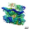
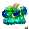
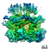
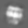
 Map data Map data | Reconstruction of the GkDnaC-GkDnaI helicase-loader complex in a putative early stage conformation | |||||||||
 Sample Sample |
| |||||||||
 Keywords Keywords | DNA replication / primosome assembly / helicase-loader complex | |||||||||
| Biological species |  Geobacillus kaustophilus (bacteria) Geobacillus kaustophilus (bacteria) | |||||||||
| Method | single particle reconstruction / negative staining / Resolution: 19.0 Å | |||||||||
 Authors Authors | Lin Y-C / Vankadari N / Hsiao C-D | |||||||||
 Citation Citation | Journal: To Be Published Title: An EM structure of the helicase-loader complex in G. kaustophilus suggesting an early stage conformation Authors: Lin Y-C / Vankadari N / Hsiao C-D | |||||||||
| History |
|
- Structure visualization
| Movie |
 Movie viewer Movie viewer |
|---|---|
| Structure viewer | EM map: SurfViewMolmilJmol/JSmol |
| Supplemental images |

- Downloads & links
-EMDB archive
| Map data | emd_6292.map.gz | 5 MB | EMDB map data format | |
|---|---|---|---|---|
| Header (meta data) | emd-6292-v30.xmlemd-6292.xml | 9.9 KB 9.9 KB | Display Display | EMDB header |
| Images | 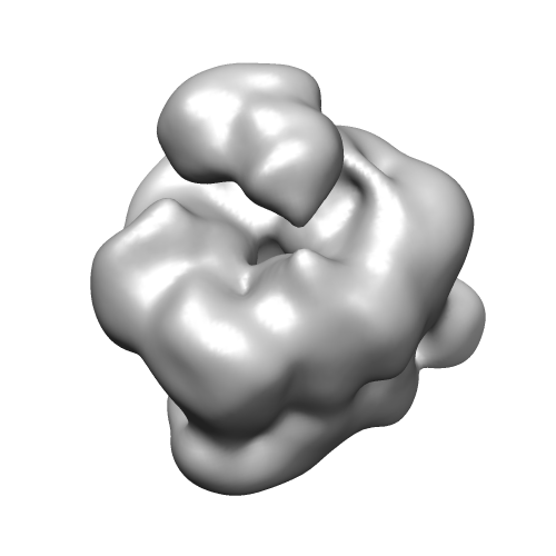 emd_6292.png emd_6292.png | 52.1 KB | ||
| Archive directory |  http://ftp.pdbj.org/pub/emdb/structures/EMD-6292ftp://ftp.pdbj.org/pub/emdb/structures/EMD-6292 http://ftp.pdbj.org/pub/emdb/structures/EMD-6292ftp://ftp.pdbj.org/pub/emdb/structures/EMD-6292 | HTTPS FTP |
-Validation report
| Summary document | emd_6292_validation.pdf.gz | 78.5 KB | Display | EMDB validaton report |
|---|---|---|---|---|
| Full document | emd_6292_full_validation.pdf.gz | 77.6 KB | Display | |
| Data in XML | emd_6292_validation.xml.gz | 493 B | Display | |
| Arichive directory | https://ftp.pdbj.org/pub/emdb/validation_reports/EMD-6292ftp://ftp.pdbj.org/pub/emdb/validation_reports/EMD-6292 | HTTPS FTP |
-Related structure data
| Similar structure data |
|---|

-Links
| EMDB pages | EMDB (EBI/PDBe) / EMDataResource |
|---|
-Map
| File | Download / File: emd_6292.map.gz / Format: CCP4 / Size: 6.4 MB / Type: IMAGE STORED AS FLOATING POINT NUMBER (4 BYTES) | ||||||||||||||||||||||||||||||||||||||||||||||||||||||||||||
|---|---|---|---|---|---|---|---|---|---|---|---|---|---|---|---|---|---|---|---|---|---|---|---|---|---|---|---|---|---|---|---|---|---|---|---|---|---|---|---|---|---|---|---|---|---|---|---|---|---|---|---|---|---|---|---|---|---|---|---|---|---|
| Annotation | Reconstruction of the GkDnaC-GkDnaI helicase-loader complex in a putative early stage conformation | ||||||||||||||||||||||||||||||||||||||||||||||||||||||||||||
| Projections & slices | Image control
Images are generated by Spider. | ||||||||||||||||||||||||||||||||||||||||||||||||||||||||||||
| Voxel size | X=Y=Z: 1.75 Å | ||||||||||||||||||||||||||||||||||||||||||||||||||||||||||||
| Density |
| ||||||||||||||||||||||||||||||||||||||||||||||||||||||||||||
| Symmetry | Space group: 1 | ||||||||||||||||||||||||||||||||||||||||||||||||||||||||||||
| Details | EMDB XML:
CCP4 map header:
| ||||||||||||||||||||||||||||||||||||||||||||||||||||||||||||
 Z (Sec.)
 Y (Row.)
 X (Col.)


-Supplemental data
- Sample components
-Entire : GkDnaC-GkDnaI helicase-loader complex
| Entire | Name: GkDnaC-GkDnaI helicase-loader complex |
|---|---|
| Components |
|
-Supramolecule #1000: GkDnaC-GkDnaI helicase-loader complex
| Supramolecule | Name: GkDnaC-GkDnaI helicase-loader complex / type: sample / ID: 1000 Oligomeric state: One homohexamer of GkDnaC binds to one dimer (or two monomers) of GkDnaI Number unique components: 2 |
|---|---|
| Molecular weight | Theoretical: 378 KDa |
-Macromolecule #1: GkDnaC-GkDnaI complex
| Macromolecule | Name: GkDnaC-GkDnaI complex / type: protein_or_peptide / ID: 1 Oligomeric state: one GkDnaC hexamer + two GkDnaI monomer (or one GkDnaI dimer) Recombinant expression: Yes |
|---|---|
| Source (natural) | Organism: Geobacillus kaustophilus (bacteria) / Strain: HTA426 |
| Molecular weight | Theoretical: 378 KDa |
| Recombinant expression | Organism: |
-Experimental details
-Structure determination
| Method | negative staining |
|---|---|
 Processing Processing | single particle reconstruction |
| Aggregation state | particle |
-Sample preparation
| Concentration | 0.02 mg/mL |
|---|---|
| Buffer | pH: 8.5 Details: 20 mM Tris-HCl pH 8.5, 200 mM NaCl, 5mM MgCl2, 1 mM beta-mercaptoethanol, 1 mM ATP-gamma-S |
| Staining | Type: NEGATIVE Details: Grids with adsorbed protein floated sequentially on three drops of 0.75 % w/v uranyl formate each for 20 seconds and then left for staining for 1 minute. |
| Grid | Details: 300 mesh Cu grid with thin carbon support, glow discharged in atmosphere |
| Vitrification | Cryogen name: NONE / Instrument: OTHER |
- Electron microscopy
| Microscope | FEI TECNAI F20 |
|---|---|
| Temperature | Average: 298 K |
| Date | May 26, 2013 |
| Image recording | Category: CCD / Film or detector model: GATAN ULTRASCAN 4000 (4k x 4k) / Average electron dose: 25 e/Å2 |
| Tilt angle min | 0 |
| Electron beam | Acceleration voltage: 200 kV / Electron source:  FIELD EMISSION GUN FIELD EMISSION GUN |
| Electron optics | Calibrated magnification: 85658 / Illumination mode: SPOT SCAN / Imaging mode: BRIGHT FIELD / Cs: 2 mm / Nominal defocus max: 1.4 µm / Nominal defocus min: 0.6 µm / Nominal magnification: 62000 |
| Sample stage | Specimen holder model: SIDE ENTRY, EUCENTRIC / Tilt angle max: 50 |
| Experimental equipment |  Model: Tecnai F20 / Image courtesy: FEI Company |
-Image processing
| Details | The particles were semi-automatically selected using EMAN2 package and manually checked. |
|---|---|
| CTF correction | Details: CTFFIND3 |
| Final reconstruction | Resolution.type: BY AUTHOR / Resolution: 19.0 Å / Resolution method: OTHER / Software - Name: Spider, Relion / Number images used: 10214 |
-Atomic model buiding 1
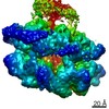
| Initial model | PDB ID: Chain - Chain ID: A |
|---|---|
| Software | Name: Chimera |
| Details | Homologous models of GkDnaC and GkDnaI using 4NMN Chain A and 4M4W Chain O as templates respectively were docked into the reconstructed EM map. |
| Refinement | Space: REAL / Protocol: RIGID BODY FIT |

