 ムービー
ムービー コントローラー


+ データを開く

- 基本情報
基本情報
| 登録情報 | データベース: EMDB / ID: EMD-6290 | |||||||||
|---|---|---|---|---|---|---|---|---|---|---|
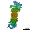
| タイトル | Negative stain reconstruction of bovine dynactin complex | |||||||||
 マップデータ マップデータ | Negative stain reconstruction of bovine dynactin complex | |||||||||
 試料 試料 |
| |||||||||
 キーワード キーワード | dynactin / actin-related proteins / dynein | |||||||||
| 生物種 |  | |||||||||
| 手法 | 単粒子再構成法 / ネガティブ染色法 / 解像度: 24.0 Å | |||||||||
 データ登録者 データ登録者 | Chowdhury S / Ketcham SA / Schroer TA / Lander GC | |||||||||
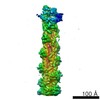
 引用 引用 | ジャーナル: Nat Struct Mol Biol / 年: 2015 タイトル: Structural organization of the dynein-dynactin complex bound to microtubules. 著者: Saikat Chowdhury / Stephanie A Ketcham / Trina A Schroer / Gabriel C Lander /  要旨: Cytoplasmic dynein associates with dynactin to drive cargo movement on microtubules, but the structure of the dynein-dynactin complex is unknown. Using electron microscopy, we determined the ...Cytoplasmic dynein associates with dynactin to drive cargo movement on microtubules, but the structure of the dynein-dynactin complex is unknown. Using electron microscopy, we determined the organization of native bovine dynein, dynactin and the dynein-dynactin-microtubule quaternary complex. In the microtubule-bound complex, the dynein motor domains are positioned for processive unidirectional movement, and the cargo-binding domains of both dynein and dynactin are accessible. | |||||||||
| 履歴 |
|
- 構造の表示
構造の表示
| ムービー |
 ムービービューア ムービービューア |
|---|---|
| 構造ビューア | EMマップ: SurfViewMolmilJmol/JSmol |
| 添付画像 |
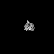
 UCSF Chimera
- ダウンロードとリンク
-EMDBアーカイブ
| マップデータ | emd_6290.map.gz | 576 KB | EMDBマップデータ形式 | |
|---|---|---|---|---|
| ヘッダ (付随情報) | emd-6290-v30.xmlemd-6290.xml | 20 KB 20 KB | 表示 表示 | EMDBヘッダ |
| 画像 | 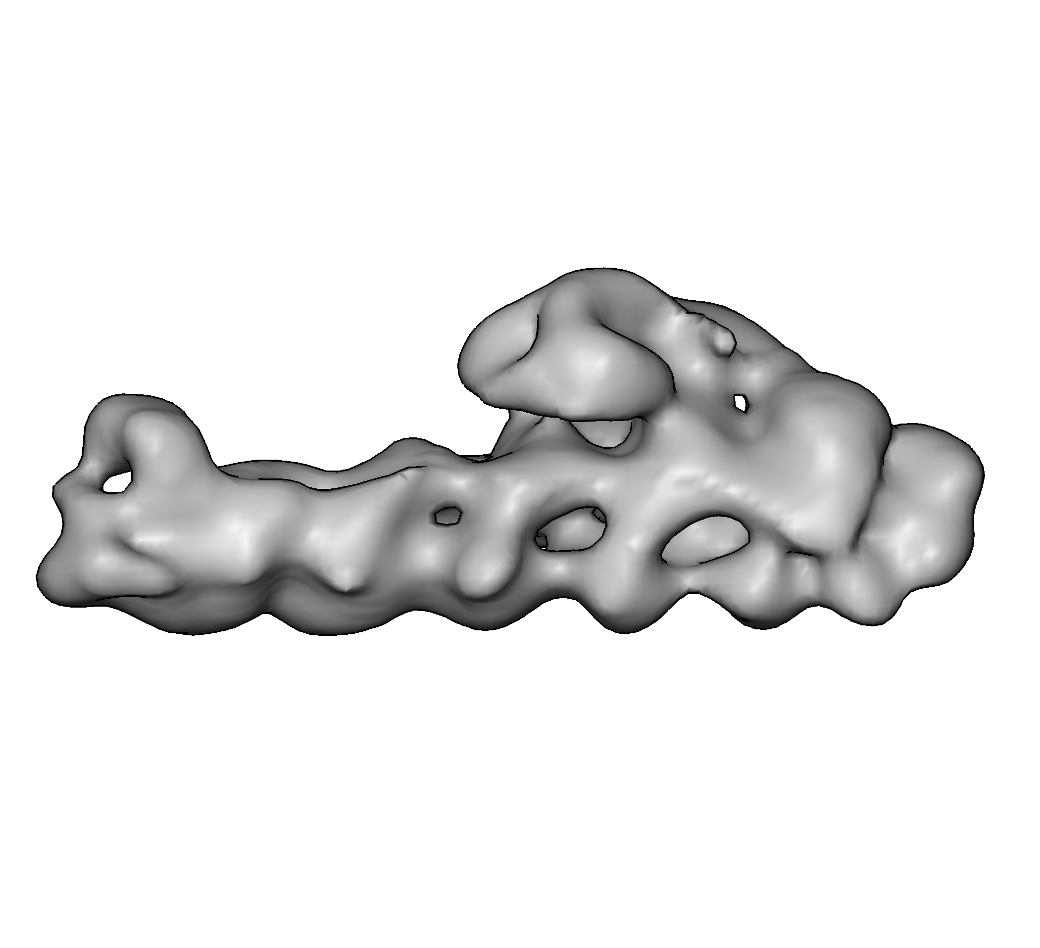 emd_6290.png emd_6290.png | 270.6 KB | ||
| アーカイブディレクトリ |  http://ftp.pdbj.org/pub/emdb/structures/EMD-6290ftp://ftp.pdbj.org/pub/emdb/structures/EMD-6290 http://ftp.pdbj.org/pub/emdb/structures/EMD-6290ftp://ftp.pdbj.org/pub/emdb/structures/EMD-6290 | HTTPS FTP |
-検証レポート
| 文書・要旨 | emd_6290_validation.pdf.gz | 78.1 KB | 表示 | EMDB検証レポート |
|---|---|---|---|---|
| 文書・詳細版 | emd_6290_full_validation.pdf.gz | 77.2 KB | 表示 | |
| XML形式データ | emd_6290_validation.xml.gz | 494 B | 表示 | |
| アーカイブディレクトリ | https://ftp.pdbj.org/pub/emdb/validation_reports/EMD-6290ftp://ftp.pdbj.org/pub/emdb/validation_reports/EMD-6290 | HTTPS FTP |
-関連構造データ
-リンク
| EMDBのページ | EMDB (EBI/PDBe) / EMDataResource |
|---|
-マップ
| ファイル | ダウンロード / ファイル: emd_6290.map.gz / 形式: CCP4 / 大きさ: 22.5 MB / タイプ: IMAGE STORED AS FLOATING POINT NUMBER (4 BYTES) | ||||||||||||||||||||||||||||||||||||||||||||||||||||||||||||||||||||
|---|---|---|---|---|---|---|---|---|---|---|---|---|---|---|---|---|---|---|---|---|---|---|---|---|---|---|---|---|---|---|---|---|---|---|---|---|---|---|---|---|---|---|---|---|---|---|---|---|---|---|---|---|---|---|---|---|---|---|---|---|---|---|---|---|---|---|---|---|---|
| 注釈 | Negative stain reconstruction of bovine dynactin complex | ||||||||||||||||||||||||||||||||||||||||||||||||||||||||||||||||||||
| 投影像・断面図 | 画像のコントロール
画像は Spider により作成 | ||||||||||||||||||||||||||||||||||||||||||||||||||||||||||||||||||||
| ボクセルのサイズ | X=Y=Z: 4.1 Å | ||||||||||||||||||||||||||||||||||||||||||||||||||||||||||||||||||||
| 密度 |
| ||||||||||||||||||||||||||||||||||||||||||||||||||||||||||||||||||||
| 対称性 | 空間群: 1 | ||||||||||||||||||||||||||||||||||||||||||||||||||||||||||||||||||||
| 詳細 | EMDB XML:
CCP4マップ ヘッダ情報:
| ||||||||||||||||||||||||||||||||||||||||||||||||||||||||||||||||||||
 Z (Sec.)
 Y (Row.)
 X (Col.)


-添付データ
- 試料の構成要素
+全体 : Bovine dynactin complex
+超分子 #1000: Bovine dynactin complex
+分子 #1: p150Glued
+分子 #2: p50
+分子 #3: p24
+分子 #4: Arp1
+分子 #5: Actin
+分子 #6: Arp11
+分子 #7: p62
+分子 #8: p25
+分子 #9: p27
+分子 #10: CapZ alpha
+分子 #11: CapZ beta
-実験情報
-構造解析
| 手法 | ネガティブ染色法 |
|---|---|
 解析 解析 | 単粒子再構成法 |
| 試料の集合状態 | particle |
-試料調製
| 濃度 | 0.05 mg/mL |
|---|---|
| 緩衝液 | pH: 7.2 / 詳細: 35 mM Tris, 5 mM MgSO4, 150 mM KCl, 1mM TCEP |
| 染色 | タイプ: NEGATIVE 詳細: 4 uL of sample was applied to a freshly plasma-cleaned thin carbon surface that was pre-treated with 0.1% w/v poly-L-lysine hydrobromide. After removing excess protein, negative staining was ...詳細: 4 uL of sample was applied to a freshly plasma-cleaned thin carbon surface that was pre-treated with 0.1% w/v poly-L-lysine hydrobromide. After removing excess protein, negative staining was performed with 2% w/v uranyl formate solution. |
| グリッド | 詳細: 400 mesh Cu-Rh Maxtaform grid with a thin continuous carbon film on top |
| 凍結 | 凍結剤: NONE / 装置: OTHER |
- 電子顕微鏡法
| 顕微鏡 | FEI TECNAI SPIRIT |
|---|---|
| 温度 | 最低: 294 K / 最高: 297 K / 平均: 295 K |
| アライメント法 | Legacy - 非点収差: Objective astigmatism was corrected using a quadrupole stigmator at 52,000 times magnification. |
| 日付 | 2013年5月1日 |
| 撮影 | カテゴリ: CCD フィルム・検出器のモデル: TVIPS TEMCAM-F416 (4k x 4k) 実像数: 1978 / 平均電子線量: 20 e/Å2 詳細: Data were collected using Leginon automated image acquisition software. |
| 電子線 | 加速電圧: 120 kV / 電子線源: LAB6 |
| 電子光学系 | 倍率(補正後): 52000 / 照射モード: FLOOD BEAM / 撮影モード: BRIGHT FIELD / Cs: 2.20 mm / 最大 デフォーカス(公称値): 1.5 µm / 最小 デフォーカス(公称値): 0.3 µm / 倍率(公称値): 52000 |
| 試料ステージ | 試料ホルダー: Room temperature holder / 試料ホルダーモデル: SIDE ENTRY, EUCENTRIC |
| 実験機器 |  モデル: Tecnai Spirit / 画像提供: FEI Company |
-画像解析
| 詳細 | Processing leading up to 3D reconstruction was performed using the Appion package. Particles were selected from micrographs using an automated template-based particle picker. The stack was subjected to five iterations of iterative 2D alignment and classification using multivariate statistical analysis (MSA) and multi-reference alignment (MRA). The clean particle stack was subsequently subjected to 3D refinement by iterative projection matching using EMAN2 and SPARX libraries. |
|---|---|
| CTF補正 | 詳細: Phase flipping of whole micrograph |
| 最終 再構成 | アルゴリズム: OTHER / 解像度のタイプ: BY AUTHOR / 解像度: 24.0 Å / 解像度の算出法: OTHER / ソフトウェア - 名称: EMAN2, SPARX 詳細: Processing leading up to 3D reconstruction was performed using the Appion package. Particles were selected from micrographs using an automated template-based particle picker. The stack was ...詳細: Processing leading up to 3D reconstruction was performed using the Appion package. Particles were selected from micrographs using an automated template-based particle picker. The stack was subjected to five iterations of iterative 2D alignment and classification using multivariate statistical analysis (MSA) and multi-reference alignment (MRA). The clean particle stack was subsequently subjected to 3D refinement by iterative projection matching using EMAN2 and SPARX libraries. 使用した粒子像数: 46734 |
| 最終 角度割当 | 詳細: EMAN2: az 90 degrees, alt 90 degrees, phi 90 degrees |
