Movie
Movie Controller


+ Open data

- Basic information
| Entry |  | |||||||||
|---|---|---|---|---|---|---|---|---|---|---|
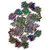
| Title | cryo-EM structure of PSII-ACPII from Rhodomonas sp. NIES-2332 | |||||||||
 Map data Map data | ||||||||||
 Sample Sample |
| |||||||||
 Keywords Keywords | photosynthesis complex / PHOTOSYNTHESIS | |||||||||
| Function / homology | Photosystem II reaction centre protein H / Photosystem II reaction centre protein H superfamily / Photosystem II 10 kDa phosphoprotein / photosystem II / phosphate ion binding / chloroplast thylakoid membrane / photosynthesis / protein stabilization / Photosystem II reaction center protein H Function and homology information Function and homology information | |||||||||
| Biological species |  Rhodomonas sp. NIES-2332 (eukaryote) Rhodomonas sp. NIES-2332 (eukaryote) | |||||||||
| Method | single particle reconstruction / cryo EM / Resolution: 2.17 Å | |||||||||
 Authors Authors | Yonehara N / Akita F / Shen JR | |||||||||
| Funding support |  Japan, 1 items Japan, 1 items
| |||||||||
 Citation Citation | Journal: Front Plant Sci / Year: 2025 Title: Structural analysis of PSI-ACPI and PSII-ACPII supercomplexes from a cryptophyte alga sp. NIES-2332. Authors: Wenyue Zhang / Nozomi Yonehara / Mizuki Ishii / Haowei Jiang / Romain La Rocca / Pi-Cheng Tsai / Hongjie Li / Koji Kato / Fusamichi Akita / Jian-Ren Shen Abstract: Light energy is converted to chemical energy by two photosystems (PSI and PSII) in complex with their light-harvesting complex proteins (LHCI and LHCII) in photosynthesis. is a member of cryptophyte ...Light energy is converted to chemical energy by two photosystems (PSI and PSII) in complex with their light-harvesting complex proteins (LHCI and LHCII) in photosynthesis. is a member of cryptophyte alga whose LHCs contain unique chlorophyll proteins (ACPs) and phycobiliproteins. We purified PSI-ACPI and PSII-ACPII supercomplexes from a cryptophyte sp. NIES-2332 and analyzed their structures at high resolutions of 2.08 Å and 2.17 Å, respectively, using cryo-electron microscopy. These structures are largely similar to those reported previously from two other species of cryptophytes, but exhibited some differences in both the pigment locations and subunit structures. A part of the antenna subunits of both photosystems is shifted compared with the previously reported structures from other species of cryptophytes, suggesting some differences in the energy transfer rates from the antenna to the PSI and PSII cores. Newly identified lipids are found to occupy the interfaces between the antennae and cores, which may be important for assembly and stabilization of the supercomplexes. Water molecules surrounding three iron-sulfur clusters of the PSI core are found in our high-resolution structure, some of which are conserved from cyanobacteria to higher plants but some are different. In addition, our structure of PSII-ACPII lacks the subunits of oxygen-evolving complex as well as the MnCaO cluster, suggesting that the cells are in the S-growth phase, yet the PSI-ACPI structure showed the binding of PsaQ, suggesting that it is in an L-phase. These results suggest that the S-phase and L-phase can co-exist in the cryptophytic cells. The high-resolution structures of both PSI-ACPIs and PSII-ACPIIs solved in this study provide a more solid structural basis for elucidating the energy transfer and quenching mechanisms in this group of the organisms. | |||||||||
| History |
|
- Structure visualization
| Supplemental images |
|---|

- Downloads & links
-EMDB archive
| Map data | emd_62846.map.gz | 413.7 MB | EMDB map data format | |
|---|---|---|---|---|
| Header (meta data) | emd-62846-v30.xmlemd-62846.xml | 45.5 KB 45.5 KB | Display Display | EMDB header |
| FSC (resolution estimation) | emd_62846_fsc.xml | 19.8 KB | Display | FSC data file |
| Images |  emd_62846.png emd_62846.png | 80.6 KB | ||
| Filedesc metadata | emd-62846.cif.gz | 10.2 KB | ||
| Others | emd_62846_half_map_1.map.gzemd_62846_half_map_2.map.gz | 763.2 MB 763.2 MB | ||
| Archive directory |  http://ftp.pdbj.org/pub/emdb/structures/EMD-62846ftp://ftp.pdbj.org/pub/emdb/structures/EMD-62846 http://ftp.pdbj.org/pub/emdb/structures/EMD-62846ftp://ftp.pdbj.org/pub/emdb/structures/EMD-62846 | HTTPS FTP |
-Related structure data
| Related structure data |  9l5vMC  9kz9C  9l0kC M: atomic model generated by this map C: citing same article ( |
|---|---|
| Similar structure data |


-Links
| EMDB pages | EMDB (EBI/PDBe) / EMDataResource |
|---|---|
| Related items in Molecule of the Month |


-Map
| File | Download / File: emd_62846.map.gz / Format: CCP4 / Size: 824 MB / Type: IMAGE STORED AS FLOATING POINT NUMBER (4 BYTES) | ||||||||||||||||||||||||||||||||||||
|---|---|---|---|---|---|---|---|---|---|---|---|---|---|---|---|---|---|---|---|---|---|---|---|---|---|---|---|---|---|---|---|---|---|---|---|---|---|
| Projections & slices | Image control
Images are generated by Spider. | ||||||||||||||||||||||||||||||||||||
| Voxel size | X=Y=Z: 0.727 Å | ||||||||||||||||||||||||||||||||||||
| Density |
| ||||||||||||||||||||||||||||||||||||
| Symmetry | Space group: 1 | ||||||||||||||||||||||||||||||||||||
| Details | EMDB XML:
|
 Z (Sec.)
 Y (Row.)
 X (Col.)


-Supplemental data
-Half map: #2
| File | emd_62846_half_map_1.map | ||||||||||||
|---|---|---|---|---|---|---|---|---|---|---|---|---|---|
| Projections & Slices |
| ||||||||||||
| Density Histograms |


-Half map: #1
| File | emd_62846_half_map_2.map | ||||||||||||
|---|---|---|---|---|---|---|---|---|---|---|---|---|---|
| Projections & Slices |
| ||||||||||||
| Density Histograms |


- Sample components
+Entire : cryo-EM structure of PSII-ACPII from Rhodomonas sp. NIES-2332
+Supramolecule #1: cryo-EM structure of PSII-ACPII from Rhodomonas sp. NIES-2332
+Macromolecule #1: Photosystem II protein D1
+Macromolecule #2: Photosystem II CP47 reaction center protein
+Macromolecule #3: Photosystem II CP43 reaction center protein
+Macromolecule #4: Photosystem II D2 protein
+Macromolecule #5: Cytochrome b559 subunit alpha
+Macromolecule #6: Cytochrome b559 subunit beta
+Macromolecule #7: Photosystem II reaction center protein H
+Macromolecule #8: Photosystem II reaction center protein I
+Macromolecule #9: Photosystem II reaction center protein K
+Macromolecule #10: Photosystem II reaction center protein L
+Macromolecule #11: Photosystem II protein M
+Macromolecule #12: Photosystem II reaction center protein T
+Macromolecule #13: Photosystem II protein W
+Macromolecule #14: Photosystem II reaction center X protein
+Macromolecule #15: Photosystem II reaction center protein Psb30
+Macromolecule #16: Photosystem II reaction center protein Z
+Macromolecule #17: ACPII-1
+Macromolecule #18: ACPII-2
+Macromolecule #19: ACPII-3
+Macromolecule #20: ACPII-4
+Macromolecule #21: ACPII-5
+Macromolecule #22: ACPII-6
+Macromolecule #23: Psb-gama_linker
+Macromolecule #24: Psb-gama_linker
+Macromolecule #25: FE (II) ION
+Macromolecule #26: CHLOROPHYLL A

+Macromolecule #27: PHEOPHYTIN A

+Macromolecule #28: 1,3,3-trimethyl-2-[(1E,3E,5E,7E,9E,11E,13E,15E,17E)-3,7,12,16-tet...
+Macromolecule #29: 2,3-DIMETHYL-5-(3,7,11,15,19,23,27,31,35-NONAMETHYL-2,6,10,14,18,...
+Macromolecule #30: 1,2-DI-O-ACYL-3-O-[6-DEOXY-6-SULFO-ALPHA-D-GLUCOPYRANOSYL]-SN-GLYCEROL

+Macromolecule #31: CHLORIDE ION
+Macromolecule #32: MANGANESE (II) ION
+Macromolecule #33: 1,2-DISTEAROYL-MONOGALACTOSYL-DIGLYCERIDE

+Macromolecule #34: 1,2-DIPALMITOYL-PHOSPHATIDYL-GLYCEROLE

+Macromolecule #35: DIGALACTOSYL DIACYL GLYCEROL (DGDG)

+Macromolecule #36: BICARBONATE ION

+Macromolecule #37: PROTOPORPHYRIN IX CONTAINING FE

+Macromolecule #38: Chlorophyll c2

+Macromolecule #39: (1~{R})-3,5,5-trimethyl-4-[(3~{E},5~{E},7~{E},9~{E},11~{E},13~{E}...

+Macromolecule #40: (1~{R})-3,5,5-trimethyl-4-[(3~{E},5~{E},7~{E},9~{E},11~{E},13~{E}...
+Macromolecule #41: water

-Experimental details
-Structure determination
| Method | cryo EM |
|---|---|
 Processing Processing | single particle reconstruction |
| Aggregation state | particle |
-Sample preparation
| Buffer | pH: 6.5 |
|---|---|
| Vitrification | Cryogen name: ETHANE |
- Electron microscopy
| Microscope | TFS KRIOS |
|---|---|
| Image recording | Film or detector model: FEI FALCON IV (4k x 4k) / Average electron dose: 50.0 e/Å2 |
| Electron beam | Acceleration voltage: 300 kV / Electron source:  FIELD EMISSION GUN FIELD EMISSION GUN |
| Electron optics | Illumination mode: FLOOD BEAM / Imaging mode: BRIGHT FIELD / Nominal defocus max: 1.0 µm / Nominal defocus min: 0.2 µm |
| Experimental equipment |  Model: Titan Krios / Image courtesy: FEI Company |