 Movie
Movie Controller

[English] 日本語
 Yorodumi
- EMDB-62749: Structure of Western equine encephalitis virus 71V1658 strain VLP... -
+ Open data

- Basic information
| Entry |  | ||||||||||||
|---|---|---|---|---|---|---|---|---|---|---|---|---|---|
| Title | Structure of Western equine encephalitis virus 71V1658 strain VLP in complex with human PCDH10 EC1 | ||||||||||||
 Map data Map data | |||||||||||||
 Sample Sample |
| ||||||||||||
 Keywords Keywords | Western Equine Encephalitis virus / WEEV / PCDH10 / EC1 / receptor / complex / glycoprotein / VIRAL PROTEIN | ||||||||||||
| Function / homology |  Function and homology information Function and homology informationT=4 icosahedral viral capsid / homophilic cell-cell adhesion / nervous system development / postsynaptic membrane / host cell cytoplasm / cell adhesion / serine-type endopeptidase activity / fusion of virus membrane with host endosome membrane / calcium ion binding / symbiont entry into host cell ...T=4 icosahedral viral capsid / homophilic cell-cell adhesion / nervous system development / postsynaptic membrane / host cell cytoplasm / cell adhesion / serine-type endopeptidase activity / fusion of virus membrane with host endosome membrane / calcium ion binding / symbiont entry into host cell / virion attachment to host cell / host cell nucleus / host cell plasma membrane / glutamatergic synapse / virion membrane / structural molecule activity / proteolysis / membrane / plasma membrane Similarity search - Function | ||||||||||||
| Biological species |  Western equine encephalitis virus / Western equine encephalitis virus /  Homo sapiens (human) Homo sapiens (human) | ||||||||||||
| Method | single particle reconstruction / cryo EM / Resolution: 3.3 Å | ||||||||||||
 Authors Authors | Cao D / Ma B / Cao Z / Zhang X / Xiang Y | ||||||||||||
| Funding support |  China, 3 items China, 3 items
| ||||||||||||
 Citation Citation | Journal: Cell Rep / Year: 2025 Title: Structural basis for the recognition of two different types of receptors by Western equine encephalitis virus. Authors: Bingting Ma / Ziyi Cao / Weijia Ding / Xinzheng Zhang / Ye Xiang / Duanfang Cao / Abstract: Western equine encephalitis virus (WEEV) enters cells via various receptors. Here, we report the cryoelectron microscopy (cryo-EM) structures of WEEV in complex with its receptors PCDH10 and very-low- ...Western equine encephalitis virus (WEEV) enters cells via various receptors. Here, we report the cryoelectron microscopy (cryo-EM) structures of WEEV in complex with its receptors PCDH10 and very-low-density lipoprotein receptor (VLDLR). Structural analysis shows that PCDH10 binds in the cleft formed by adjacent E2-E1 heterodimers of WEEV through its EC1 ectodomain. Residues of viral envelope proteins involved in the interactions with PCDH10 EC1 are unique to WEEV. The strain-specific receptor VLDLR binds WEEV strain McMillan through two consecutive ecto-LDLR class A (LA) repeats. LA1-2, LA2-3, LA3-4, LA4-5, and LA5-6 of VLDLR all have detectable interactions with WEEV. Detailed structures of WEEV in complex with LA1-2 and LA2-3 show that the N-terminal LA repeat binds in the cleft and that the C-terminal LA repeat is attached to the E2 B domain. The acquisition of a single E2 mutation (V265F) allows WEEV strain 71V-1658, originally unable to bind VLDLR, to gain this receptor-binding ability. The binding of VLDLR to WEEV is in a mode different from those of other alphaviruses. | ||||||||||||
| History |
|
- Structure visualization
| Supplemental images |
|---|

- Downloads & links
-EMDB archive
| Map data | emd_62749.map.gz | 4.9 MB | EMDB map data format | |
|---|---|---|---|---|
| Header (meta data) | emd-62749-v30.xmlemd-62749.xml | 21.1 KB 21.1 KB | Display Display | EMDB header |
| Images |  emd_62749.png emd_62749.png | 133.2 KB | ||
| Masks | emd_62749_msk_1.map | 30.5 MB | Mask map | |
| Filedesc metadata | emd-62749.cif.gz | 6.6 KB | ||
| Others | emd_62749_half_map_1.map.gzemd_62749_half_map_2.map.gz | 23.1 MB 23.1 MB | ||
| Archive directory |  http://ftp.pdbj.org/pub/emdb/structures/EMD-62749ftp://ftp.pdbj.org/pub/emdb/structures/EMD-62749 http://ftp.pdbj.org/pub/emdb/structures/EMD-62749ftp://ftp.pdbj.org/pub/emdb/structures/EMD-62749 | HTTPS FTP |
-Validation report
| Summary document | emd_62749_validation.pdf.gz | 825.7 KB | Display | EMDB validaton report |
|---|---|---|---|---|
| Full document | emd_62749_full_validation.pdf.gz | 825.2 KB | Display | |
| Data in XML | emd_62749_validation.xml.gz | 10.6 KB | Display | |
| Data in CIF | emd_62749_validation.cif.gz | 12.5 KB | Display | |
| Arichive directory | https://ftp.pdbj.org/pub/emdb/validation_reports/EMD-62749ftp://ftp.pdbj.org/pub/emdb/validation_reports/EMD-62749 | HTTPS FTP |
-Related structure data
| Related structure data |  9l1nMC  9l99C 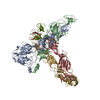 9l9aC M: atomic model generated by this map C: citing same article ( |
|---|---|
| Similar structure data |


-Links
| EMDB pages | EMDB (EBI/PDBe) / EMDataResource |
|---|---|
| Related items in Molecule of the Month |


-Map
| File | Download / File: emd_62749.map.gz / Format: CCP4 / Size: 30.5 MB / Type: IMAGE STORED AS FLOATING POINT NUMBER (4 BYTES) | ||||||||||||||||||||||||||||||||||||
|---|---|---|---|---|---|---|---|---|---|---|---|---|---|---|---|---|---|---|---|---|---|---|---|---|---|---|---|---|---|---|---|---|---|---|---|---|---|
| Projections & slices | Image control
Images are generated by Spider. | ||||||||||||||||||||||||||||||||||||
| Voxel size | X=Y=Z: 1.34 Å | ||||||||||||||||||||||||||||||||||||
| Density |
| ||||||||||||||||||||||||||||||||||||
| Symmetry | Space group: 1 | ||||||||||||||||||||||||||||||||||||
| Details | EMDB XML:
|
 Z (Sec.)
 Y (Row.)
 X (Col.)


-Supplemental data
-Mask #1
| File | emd_62749_msk_1.map | ||||||||||||
|---|---|---|---|---|---|---|---|---|---|---|---|---|---|
| Projections & Slices |
| ||||||||||||
| Density Histograms |


-Half map: #1
| File | emd_62749_half_map_1.map | ||||||||||||
|---|---|---|---|---|---|---|---|---|---|---|---|---|---|
| Projections & Slices |
| ||||||||||||
| Density Histograms |


-Half map: #2
| File | emd_62749_half_map_2.map | ||||||||||||
|---|---|---|---|---|---|---|---|---|---|---|---|---|---|
| Projections & Slices |
| ||||||||||||
| Density Histograms |


- Sample components
-Entire : Western equine encephalitis virus strain 71V1658 VLP in complex w...
| Entire | Name: Western equine encephalitis virus strain 71V1658 VLP in complex with its receptor PCDH10 EC1 at the asymmetric unit |
|---|---|
| Components |
|
-Supramolecule #1: Western equine encephalitis virus strain 71V1658 VLP in complex w...
| Supramolecule | Name: Western equine encephalitis virus strain 71V1658 VLP in complex with its receptor PCDH10 EC1 at the asymmetric unit type: complex / ID: 1 / Parent: 0 / Macromolecule list: #1-#4 |
|---|---|
| Source (natural) | Organism: Western equine encephalitis virus / Strain: strian 71V1658 |
-Macromolecule #1: E1 glycoprotein
| Macromolecule | Name: E1 glycoprotein / type: protein_or_peptide / ID: 1 / Number of copies: 4 / Enantiomer: LEVO |
|---|---|
| Source (natural) | Organism: Western equine encephalitis virus / Strain: 71V1658 |
| Molecular weight | Theoretical: 47.326781 KDa |
| Recombinant expression | Organism: Homo sapiens (human) |
| Sequence | String: FEHATTVPNV PGIPYKALVE RAGYAPLNLE ITVVSSELTP STNKEYVTCK FHTVIPSPQV KCCGSLECKA SSKADYTCRV FGGVYPFMW GGAQCFCDSE NTQLSEAYVE FAPDCTIDHA VALKVHTAAL KVGLRIVYGN TTAHLDTFVN GVTPGSSRDL K VIAGPISA ...String: FEHATTVPNV PGIPYKALVE RAGYAPLNLE ITVVSSELTP STNKEYVTCK FHTVIPSPQV KCCGSLECKA SSKADYTCRV FGGVYPFMW GGAQCFCDSE NTQLSEAYVE FAPDCTIDHA VALKVHTAAL KVGLRIVYGN TTAHLDTFVN GVTPGSSRDL K VIAGPISA AFSPFDHKVV IRKGLVYNYD FPEYGAMKPG AFGDIQASSL DATDIVARTD IRLLKPSVKN IHVPYTQAVS GY EMWKNNS GRPLQETAPF GCKIEVEPLR ASNCAYGHIP ISIDIPDAAF VRSSESPTIL EVSCTVADCI YSADFGGSLT LQY KADREG HCPVHSHSTT AVLKEATTHV TAVGSITLHF STSSPQANFI VSLCGKKSTC NAECKPPADH IIGEPHKVDQ EFQA AVSKT SWNWLLALFG GASSLIVVGL IVLVCSSMLI NTRR UniProtKB: Structural polyprotein |
-Macromolecule #2: E2 glycoprotein
| Macromolecule | Name: E2 glycoprotein / type: protein_or_peptide / ID: 2 / Number of copies: 4 / Enantiomer: LEVO |
|---|---|
| Source (natural) | Organism: Western equine encephalitis virus / Strain: 71V1658 |
| Molecular weight | Theoretical: 46.218676 KDa |
| Recombinant expression | Organism: Homo sapiens (human) |
| Sequence | String: SITDDFTLTS PYLGFCPYCR HSTPCFSPIK IENVWDESDD GSIRIQVSAQ FGYNQAGTAD VTKFRYMSFD HDHDIKEDSM EKIAISTSG PCRRLGHKGY FLLAQCPPGD SVTVSITSGA SENSCTVEKK IRRKFVGREE YLFPPVHGKL VKCHVYDHLK E TSAGYITM ...String: SITDDFTLTS PYLGFCPYCR HSTPCFSPIK IENVWDESDD GSIRIQVSAQ FGYNQAGTAD VTKFRYMSFD HDHDIKEDSM EKIAISTSG PCRRLGHKGY FLLAQCPPGD SVTVSITSGA SENSCTVEKK IRRKFVGREE YLFPPVHGKL VKCHVYDHLK E TSAGYITM HRPGPHAYKS YLEEASGEVY IKPPSGKNVT YECKCGDYST GIVSTRTKMN GCTKAKQCIA YKSDQTKWVF NS PDLIRHT DHSVQGKLHI PFRLTPTVCP VPLAHTPTVT KWFKGITLHL TAMRPTLLTT RKLGLRADAT AEWITGSTSR NFS VGREGL EYVWGNHEPV RVWAQESAPG DPHGWPHEII IHYYHRHPVY TVIVLCGVAL AILVGTASSA ACIAKARRDC LTPY ALAPN ATVPTALAVL C UniProtKB: Structural polyprotein |
-Macromolecule #3: Capsid glycoprotein
| Macromolecule | Name: Capsid glycoprotein / type: protein_or_peptide / ID: 3 / Number of copies: 4 / Enantiomer: LEVO |
|---|---|
| Source (natural) | Organism: Western equine encephalitis virus / Strain: 71V1658 |
| Molecular weight | Theoretical: 16.409549 KDa |
| Recombinant expression | Organism: Homo sapiens (human) |
| Sequence | String: KTFPIMLNGQ VNGYACVVGG RLMKPLHVEG KIDNEQLAAV KLKKASMYDL EYGDVPQNMK SDTLQYTSDK PPGFYNWHHG AVQYENGRF TVPRGVGGKG DSGRPILDNR GRVVAIVLGG ANEGTRTALS VVTWNQKGVT IRDTPEGSEP W UniProtKB: Structural polyprotein |
-Macromolecule #4: Protocadherin-10
| Macromolecule | Name: Protocadherin-10 / type: protein_or_peptide / ID: 4 / Number of copies: 1 / Enantiomer: LEVO |
|---|---|
| Source (natural) | Organism: Homo sapiens (human) |
| Molecular weight | Theoretical: 10.866193 KDa |
| Recombinant expression | Organism: Homo sapiens (human) |
| Sequence | String: QLHYTVQEEQ EHGTFVGNIA EDLGLDITKL SARGFQTVPN SRTPYLDLNL ETGVLYVNEK IDREQICKQS PSCVLHLEVF LENPLELFQ VEIEVL UniProtKB: Protocadherin-10 |
-Macromolecule #5: 2-acetamido-2-deoxy-beta-D-glucopyranose
| Macromolecule | Name: 2-acetamido-2-deoxy-beta-D-glucopyranose / type: ligand / ID: 5 / Number of copies: 8 / Formula: NAG |
|---|---|
| Molecular weight | Theoretical: 221.208 Da |
| Chemical component information |  ChemComp-NAG: |
-Experimental details
-Structure determination
| Method | cryo EM |
|---|---|
 Processing Processing | single particle reconstruction |
| Aggregation state | particle |
-Sample preparation
| Buffer | pH: 8 |
|---|---|
| Vitrification | Cryogen name: ETHANE |
- Electron microscopy
| Microscope | TFS KRIOS |
|---|---|
| Image recording | Film or detector model: GATAN K2 SUMMIT (4k x 4k) / Average electron dose: 50.0 e/Å2 |
| Electron beam | Acceleration voltage: 300 kV / Electron source:  FIELD EMISSION GUN FIELD EMISSION GUN |
| Electron optics | Illumination mode: FLOOD BEAM / Imaging mode: BRIGHT FIELD / Nominal defocus max: 1.7 µm / Nominal defocus min: 1.2 µm |
| Experimental equipment |  Model: Titan Krios / Image courtesy: FEI Company |
