 Movie
Movie Controller

[English] 日本語
 Yorodumi
- EMDB-62298: Cryo-EM structure of the compound 4-bound human relaxin family pe... -
+ Open data

- Basic information
| Entry |  | ||||||||||||
|---|---|---|---|---|---|---|---|---|---|---|---|---|---|
| Title | Cryo-EM structure of the compound 4-bound human relaxin family peptide receptor 3 (RXFP3)-Gi complex | ||||||||||||
 Map data Map data | |||||||||||||
 Sample Sample |
| ||||||||||||
 Keywords Keywords | human relaxin family peptide receptor 3 / G protein-coupled receptor / ligand recognition / STRUCTURAL PROTEIN / STRUCTURAL PROTEIN-IMMUNE SYSTEM complex | ||||||||||||
| Function / homology |  Function and homology information Function and homology informationRelaxin receptors / galanin receptor activity / negative regulation of adenylate cyclase-activating adrenergic receptor signaling pathway / negative regulation of calcium ion-dependent exocytosis / negative regulation of adenylate cyclase activity / G protein-coupled adenosine receptor signaling pathway / positive regulation of neural precursor cell proliferation / negative regulation of synaptic transmission / positive regulation of urine volume / gamma-aminobutyric acid signaling pathway ...Relaxin receptors / galanin receptor activity / negative regulation of adenylate cyclase-activating adrenergic receptor signaling pathway / negative regulation of calcium ion-dependent exocytosis / negative regulation of adenylate cyclase activity / G protein-coupled adenosine receptor signaling pathway / positive regulation of neural precursor cell proliferation / negative regulation of synaptic transmission / positive regulation of urine volume / gamma-aminobutyric acid signaling pathway / positive regulation of cytokinesis / regulation of calcium ion transport / negative regulation of apoptotic signaling pathway / neuronal dense core vesicle / positive regulation of vascular associated smooth muscle cell proliferation / positive regulation of superoxide anion generation / response to nutrient / Adenylate cyclase inhibitory pathway / hippocampal mossy fiber to CA3 synapse / Regulation of insulin secretion / neuropeptide signaling pathway / electron transport chain / G protein-coupled receptor binding / G protein-coupled receptor activity / G-protein beta/gamma-subunit complex binding / adenylate cyclase-inhibiting G protein-coupled receptor signaling pathway / Olfactory Signaling Pathway / Activation of the phototransduction cascade / G protein-coupled acetylcholine receptor signaling pathway / G beta:gamma signalling through PLC beta / Presynaptic function of Kainate receptors / Thromboxane signalling through TP receptor / Activation of G protein gated Potassium channels / Inhibition of voltage gated Ca2+ channels via Gbeta/gamma subunits / G-protein activation / Glucagon signaling in metabolic regulation / G beta:gamma signalling through CDC42 / Prostacyclin signalling through prostacyclin receptor / Synthesis, secretion, and inactivation of Glucagon-like Peptide-1 (GLP-1) / G beta:gamma signalling through BTK / photoreceptor disc membrane / ADP signalling through P2Y purinoceptor 12 / Glucagon-type ligand receptors / Sensory perception of sweet, bitter, and umami (glutamate) taste / Adrenaline,noradrenaline inhibits insulin secretion / Vasopressin regulates renal water homeostasis via Aquaporins / Glucagon-like Peptide-1 (GLP1) regulates insulin secretion / G alpha (z) signalling events / cellular response to catecholamine stimulus / ADP signalling through P2Y purinoceptor 1 / G beta:gamma signalling through PI3Kgamma / ADORA2B mediated anti-inflammatory cytokines production / adenylate cyclase-activating dopamine receptor signaling pathway / Cooperation of PDCL (PhLP1) and TRiC/CCT in G-protein beta folding / GPER1 signaling / cellular response to prostaglandin E stimulus / heterotrimeric G-protein complex / Inactivation, recovery and regulation of the phototransduction cascade / G alpha (12/13) signalling events / G-protein beta-subunit binding / extracellular vesicle / sensory perception of taste / Thrombin signalling through proteinase activated receptors (PARs) / signaling receptor complex adaptor activity / adenylate cyclase-activating G protein-coupled receptor signaling pathway / retina development in camera-type eye / fibroblast proliferation / cell body / GTPase binding / midbody / Ca2+ pathway / High laminar flow shear stress activates signaling by PIEZO1 and PECAM1:CDH5:KDR in endothelial cells / G alpha (i) signalling events / G alpha (s) signalling events / G alpha (q) signalling events / phospholipase C-activating G protein-coupled receptor signaling pathway / Hydrolases; Acting on acid anhydrides; Acting on GTP to facilitate cellular and subcellular movement / Ras protein signal transduction / positive regulation of ERK1 and ERK2 cascade / periplasmic space / electron transfer activity / cell population proliferation / Extra-nuclear estrogen signaling / ciliary basal body / positive regulation of cell migration / iron ion binding / G protein-coupled receptor signaling pathway / cell division / lysosomal membrane / GTPase activity / heme binding / centrosome / positive regulation of cell population proliferation / dendrite / synapse / GTP binding / protein-containing complex binding / signal transduction / extracellular exosome / nucleoplasm Similarity search - Function | ||||||||||||
| Biological species |  Homo sapiens (human) / synthetic construct (others) Homo sapiens (human) / synthetic construct (others) | ||||||||||||
| Method | single particle reconstruction / cryo EM / Resolution: 3.1 Å | ||||||||||||
 Authors Authors | Chen Y / Zhou QT / Yan SY / Yan JH / Yang DH / Chen J / Wang MW / Rao QD / Dai AT / Yin WC ...Chen Y / Zhou QT / Yan SY / Yan JH / Yang DH / Chen J / Wang MW / Rao QD / Dai AT / Yin WC / Shen DD / Zhang Y / Xia T / Stevens RC / Xu HE / Zhao LH | ||||||||||||
| Funding support |  China, 3 items China, 3 items
| ||||||||||||
 Citation Citation | Journal: Commun Biol / Year: 2025 Title: Molecular mechanism underlying non-discriminatory recognition of relaxin-3 by RXFP3 and RXFP4. Authors: Yan Chen / Qingtong Zhou / Shiyu Yan / Jiahui Yan / Dehua Yang / Jian Chen / Ming-Wei Wang / Abstract: The human relaxin family peptide receptors RXFP3 and RXFP4 play important physiological roles through interactions with endogenous hormones, relaxin-3 and insulin-like peptide 5 (INSL5). They are ...The human relaxin family peptide receptors RXFP3 and RXFP4 play important physiological roles through interactions with endogenous hormones, relaxin-3 and insulin-like peptide 5 (INSL5). They are implicated in certain neurological and metabolic disorders. While INSL5 only activates RXFP4, relaxin-3 is recognized by both receptors. Here, we report the cryo-electron microscopy structures of RXFP3-G complexes bound by relaxin-3 or a small-molecule dual agonist (compound 4), and relaxin-3 in complex with RXFP4-G, with global resolutions of 2.91 Å, 2.95 Å, and 3.10 Å, respectively. It is found that relaxin-3 adopts a conserved binding conformation within the transmembrane domain (TMD) bundle of RXFP3 and RXFP4, where the C-terminal tip residues of its B chain, R26 and W27, make extensive contacts with conserved receptor residues, thereby activating RXFP3 and RXFP4. Compound 4 mimics these key interactions by binding to both receptors. In contrast, the C-terminal residues composition and TMD-binding angle of INSL5 in RXFP4 differ significantly from that of relaxin-3, ensuring its selectivity for RXFP4. These findings deepen our understanding about the structural basis of ligand recognition and selectivity in this G protein-coupled receptor subfamily. | ||||||||||||
| History |
|
- Structure visualization
| Supplemental images |
|---|

- Downloads & links
-EMDB archive
| Map data | emd_62298.map.gz | 57.2 MB | EMDB map data format | |
|---|---|---|---|---|
| Header (meta data) | emd-62298-v30.xmlemd-62298.xml | 23.2 KB 23.2 KB | Display Display | EMDB header |
| FSC (resolution estimation) | emd_62298_fsc.xml | 9.6 KB | Display | FSC data file |
| Images |  emd_62298.png emd_62298.png | 86.9 KB | ||
| Filedesc metadata | emd-62298.cif.gz | 7.2 KB | ||
| Others | emd_62298_half_map_1.map.gzemd_62298_half_map_2.map.gz | 59.4 MB 59.4 MB | ||
| Archive directory |  http://ftp.pdbj.org/pub/emdb/structures/EMD-62298ftp://ftp.pdbj.org/pub/emdb/structures/EMD-62298 http://ftp.pdbj.org/pub/emdb/structures/EMD-62298ftp://ftp.pdbj.org/pub/emdb/structures/EMD-62298 | HTTPS FTP |
-Related structure data
| Related structure data |  9kfjMC  9kfiC  9kfkC M: atomic model generated by this map C: citing same article ( |
|---|---|
| Similar structure data |


-Links
| EMDB pages | EMDB (EBI/PDBe) / EMDataResource |
|---|---|
| Related items in Molecule of the Month |


-Map
| File | Download / File: emd_62298.map.gz / Format: CCP4 / Size: 64 MB / Type: IMAGE STORED AS FLOATING POINT NUMBER (4 BYTES) | ||||||||||||||||||||||||||||||||||||
|---|---|---|---|---|---|---|---|---|---|---|---|---|---|---|---|---|---|---|---|---|---|---|---|---|---|---|---|---|---|---|---|---|---|---|---|---|---|
| Projections & slices | Image control
Images are generated by Spider. | ||||||||||||||||||||||||||||||||||||
| Voxel size | X=Y=Z: 0.824 Å | ||||||||||||||||||||||||||||||||||||
| Density |
| ||||||||||||||||||||||||||||||||||||
| Symmetry | Space group: 1 | ||||||||||||||||||||||||||||||||||||
| Details | EMDB XML:
|
 Z (Sec.)
 Y (Row.)
 X (Col.)


-Supplemental data
-Half map: #2
| File | emd_62298_half_map_1.map | ||||||||||||
|---|---|---|---|---|---|---|---|---|---|---|---|---|---|
| Projections & Slices |
| ||||||||||||
| Density Histograms |


-Half map: #1
| File | emd_62298_half_map_2.map | ||||||||||||
|---|---|---|---|---|---|---|---|---|---|---|---|---|---|
| Projections & Slices |
| ||||||||||||
| Density Histograms |


- Sample components
+Entire : Cryo-EM structure of the compound 4-bound human relaxin family pe...
+Supramolecule #1: Cryo-EM structure of the compound 4-bound human relaxin family pe...
+Supramolecule #2: human relaxin family peptide receptor 3
+Supramolecule #3: scFv16
+Supramolecule #4: G protein
+Macromolecule #1: Soluble cytochrome b562,Relaxin-3 receptor 1,LgBiT

 Spodoptera frugiperda (fall armyworm)
+Macromolecule #2: Guanine nucleotide-binding protein G(I)/G(S)/G(O) subunit gamma-2
+Macromolecule #3: Guanine nucleotide-binding protein G(i) subunit alpha-2
+Macromolecule #4: scFv16
+Macromolecule #5: Guanine nucleotide-binding protein G(I)/G(S)/G(T) subunit beta-1
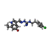
+Macromolecule #6: 1-[2-(4-chlorophenyl)ethyl]-3-[(7-ethyl-5-oxidanyl-1H-indol-3-yl)...
-Experimental details
-Structure determination
| Method | cryo EM |
|---|---|
 Processing Processing | single particle reconstruction |
| Aggregation state | particle |
-Sample preparation
| Buffer | pH: 7.4 |
|---|---|
| Vitrification | Cryogen name: ETHANE |
- Electron microscopy
| Microscope | FEI TALOS ARCTICA |
|---|---|
| Image recording | Film or detector model: GATAN K3 (6k x 4k) / Average electron dose: 80.0 e/Å2 |
| Electron beam | Acceleration voltage: 300 kV / Electron source: OTHER |
| Electron optics | Illumination mode: OTHER / Imaging mode: BRIGHT FIELD / Nominal defocus max: 2.0 µm / Nominal defocus min: 1.0 µm |
| Experimental equipment |  Model: Talos Arctica / Image courtesy: FEI Company |


