 Movie
Movie Controller
 Structure viewers
 About Yorodumi Papers


+Search query
-Structure paper
| Title | Molecular mechanism underlying non-discriminatory recognition of relaxin-3 by RXFP3 and RXFP4. |
|---|---|
| Journal, issue, pages | Commun Biol, Vol. 8, Issue 1, Page 794, Year 2025 |
| Publish date | May 23, 2025 |
 Authors Authors | Yan Chen / Qingtong Zhou / Shiyu Yan / Jiahui Yan / Dehua Yang / Jian Chen / Ming-Wei Wang /  |
| PubMed Abstract | The human relaxin family peptide receptors RXFP3 and RXFP4 play important physiological roles through interactions with endogenous hormones, relaxin-3 and insulin-like peptide 5 (INSL5). They are ...The human relaxin family peptide receptors RXFP3 and RXFP4 play important physiological roles through interactions with endogenous hormones, relaxin-3 and insulin-like peptide 5 (INSL5). They are implicated in certain neurological and metabolic disorders. While INSL5 only activates RXFP4, relaxin-3 is recognized by both receptors. Here, we report the cryo-electron microscopy structures of RXFP3-G complexes bound by relaxin-3 or a small-molecule dual agonist (compound 4), and relaxin-3 in complex with RXFP4-G, with global resolutions of 2.91 Å, 2.95 Å, and 3.10 Å, respectively. It is found that relaxin-3 adopts a conserved binding conformation within the transmembrane domain (TMD) bundle of RXFP3 and RXFP4, where the C-terminal tip residues of its B chain, R26 and W27, make extensive contacts with conserved receptor residues, thereby activating RXFP3 and RXFP4. Compound 4 mimics these key interactions by binding to both receptors. In contrast, the C-terminal residues composition and TMD-binding angle of INSL5 in RXFP4 differ significantly from that of relaxin-3, ensuring its selectivity for RXFP4. These findings deepen our understanding about the structural basis of ligand recognition and selectivity in this G protein-coupled receptor subfamily. |
 External links External links | Commun Biol / PubMed:40410443 / PubMed Central |
| Methods | EM (single particle) |
| Resolution | 2.91 - 3.1 Å |
| Structure data | EMDB-62297, PDB-9kfi: EMDB-62298, PDB-9kfj: EMDB-62299, PDB-9kfk: |
| Chemicals | 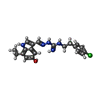 ChemComp-IYF: |
| Source |
|
 Keywords Keywords | STRUCTURAL PROTEN/IMMUNE SYSTEM / human relaxin family peptide receptor 3 / G protein-coupled receptor / ligand recognition / STRUCTURAL PROTEIN / STRUCTURAL PROTEN-IMMUNE SYSTEM complex / STRUCTURAL PROTEIN/IMMUNE SYSTEM / STRUCTURAL PROTEIN-IMMUNE SYSTEM complex / human relaxin family peptide receptor 4 |


 homo sapiens (human)

