 Movie
Movie Controller

[English] 日本語
 Yorodumi
- EMDB-6185: Electron cryo-microscopy of Melanoides tuberculata mega-hemocyanin -
+ Open data

- Basic information
| Entry | Database: EMDB / ID: EMD-6185 | |||||||||
|---|---|---|---|---|---|---|---|---|---|---|
| Title | Electron cryo-microscopy of Melanoides tuberculata mega-hemocyanin | |||||||||
 Map data Map data | Reconstruction of Melanoides tuberculata mega-hemocyanin | |||||||||
 Sample Sample |
| |||||||||
 Keywords Keywords | oxygen transport / origami | |||||||||
| Function / homology |  Function and homology information Function and homology information | |||||||||
| Biological species |  Melanoides tuberculata (invertebrata) Melanoides tuberculata (invertebrata) | |||||||||
| Method | single particle reconstruction / cryo EM / Resolution: 12.2 Å | |||||||||
 Authors Authors | Gatsogiannis C / Hofnagel O / Markl J / Raunser S | |||||||||
 Citation Citation | Journal: Structure / Year: 2015 Title: Structure of mega-hemocyanin reveals protein origami in snails. Authors: Christos Gatsogiannis / Oliver Hofnagel / Jürgen Markl / Stefan Raunser /  Abstract: Mega-hemocyanin is a 13.5 MDa oxygen transporter found in the hemolymph of some snails. Similar to typical gastropod hemocyanins, it is composed of 400 kDa building blocks but has additional ...Mega-hemocyanin is a 13.5 MDa oxygen transporter found in the hemolymph of some snails. Similar to typical gastropod hemocyanins, it is composed of 400 kDa building blocks but has additional 550 kDa subunits. Together, they form a large, completely filled cylinder. The structural basis for this highly complex protein packing is not known so far. Here, we report the electron cryomicroscopy (cryo-EM) structure of mega-hemocyanin complexes from two different snail species. The structures reveal that mega-hemocyanin is composed of flexible building blocks that differ in their conformation, but not in their primary structure. Like a protein origami, these flexible blocks are optimally packed, implementing different local symmetries and pseudosymmetries. A comparison between the two structures suggests a surprisingly simple evolutionary mechanism leading to these large oxygen transporters. | |||||||||
| History |
|
- Structure visualization
| Movie |
Movie viewer |
|---|---|
| Structure viewer | EM map: SurfViewMolmilJmol/JSmol |
| Supplemental images |

- Downloads & links
-EMDB archive
| Map data | emd_6185.map.gz | 26 MB | EMDB map data format | |
|---|---|---|---|---|
| Header (meta data) | emd-6185-v30.xmlemd-6185.xml | 10.9 KB 10.9 KB | Display Display | EMDB header |
| Images | 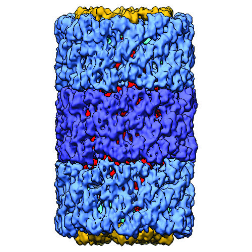 emd_6185.jpg emd_6185.jpg | 737.9 KB | ||
| Archive directory |  http://ftp.pdbj.org/pub/emdb/structures/EMD-6185ftp://ftp.pdbj.org/pub/emdb/structures/EMD-6185 http://ftp.pdbj.org/pub/emdb/structures/EMD-6185ftp://ftp.pdbj.org/pub/emdb/structures/EMD-6185 | HTTPS FTP |
-Related structure data


-Links
| EMDB pages | EMDB (EBI/PDBe) / EMDataResource |
|---|
-Map
| File | Download / File: emd_6185.map.gz / Format: CCP4 / Size: 28.1 MB / Type: IMAGE STORED AS FLOATING POINT NUMBER (4 BYTES) | ||||||||||||||||||||||||||||||||||||||||||||||||||||||||||||||||||||
|---|---|---|---|---|---|---|---|---|---|---|---|---|---|---|---|---|---|---|---|---|---|---|---|---|---|---|---|---|---|---|---|---|---|---|---|---|---|---|---|---|---|---|---|---|---|---|---|---|---|---|---|---|---|---|---|---|---|---|---|---|---|---|---|---|---|---|---|---|---|
| Annotation | Reconstruction of Melanoides tuberculata mega-hemocyanin | ||||||||||||||||||||||||||||||||||||||||||||||||||||||||||||||||||||
| Projections & slices | Image control
Images are generated by Spider. | ||||||||||||||||||||||||||||||||||||||||||||||||||||||||||||||||||||
| Voxel size | X=Y=Z: 3.64 Å | ||||||||||||||||||||||||||||||||||||||||||||||||||||||||||||||||||||
| Density |
| ||||||||||||||||||||||||||||||||||||||||||||||||||||||||||||||||||||
| Symmetry | Space group: 1 | ||||||||||||||||||||||||||||||||||||||||||||||||||||||||||||||||||||
| Details | EMDB XML:
CCP4 map header:
| ||||||||||||||||||||||||||||||||||||||||||||||||||||||||||||||||||||
 Z (Sec.)
 Y (Row.)
 X (Col.)


-Supplemental data
- Sample components
-Entire : Melanoides tuberculata Mega-Hemocyanin (MtH)
| Entire | Name: Melanoides tuberculata Mega-Hemocyanin (MtH) |
|---|---|
| Components |
|
-Supramolecule #1000: Melanoides tuberculata Mega-Hemocyanin (MtH)
| Supramolecule | Name: Melanoides tuberculata Mega-Hemocyanin (MtH) / type: sample / ID: 1000 Oligomeric state: Two peripheral decamers of the MtH400 subunit bind to a central decamer of the MtH550 subunit. Number unique components: 2 |
|---|---|
| Molecular weight | Experimental: 13.5 MDa / Theoretical: 13.5 MDa |
-Macromolecule #1: mega-hemocyanin, strain MtH400
| Macromolecule | Name: mega-hemocyanin, strain MtH400 / type: protein_or_peptide / ID: 1 / Name.synonym: MtH400 / Number of copies: 2 / Oligomeric state: Decamer / Recombinant expression: No / Database: NCBI |
|---|---|
| Source (natural) | Organism: Melanoides tuberculata (invertebrata) / synonym: red-rimmed melania |
| Molecular weight | Experimental: 4 MDa / Theoretical: 4 MDa |
| Sequence | UniProtKB: Mega-hemocyanin |
-Macromolecule #2: mega-hemocyanin, strain MtH550
| Macromolecule | Name: mega-hemocyanin, strain MtH550 / type: protein_or_peptide / ID: 2 / Name.synonym: MtH550 / Number of copies: 1 / Oligomeric state: Decamer / Recombinant expression: No / Database: NCBI |
|---|---|
| Source (natural) | Organism: Melanoides tuberculata (invertebrata) / synonym: red-rimmed melania |
| Molecular weight | Experimental: 5.5 MDa / Theoretical: 5.5 MDa |
| Sequence | UniProtKB: Mega-hemocyanin |
-Experimental details
-Structure determination
| Method | cryo EM |
|---|---|
 Processing Processing | single particle reconstruction |
| Aggregation state | particle |
-Sample preparation
| Concentration | 0.2 mg/mL |
|---|---|
| Buffer | pH: 7.4 Details: 50 mM Tris-HCl, 150 mM NaCl, 5 mM CaCl2, 5 mM MgCl2 |
| Grid | Details: C-Flat 2/1-4C copper 400 mesh, with additional thin carbon support |
| Vitrification | Cryogen name: ETHANE / Chamber humidity: 90 % / Chamber temperature: 100 K / Instrument: GATAN CRYOPLUNGE 3 / Method: Blot for 3 seconds before plunging. |
- Electron microscopy
| Microscope | JEOL 3200FSC |
|---|---|
| Alignment procedure | Legacy - Astigmatism: Objective lens astigmatism was corrected at 120,000 times magnification. |
| Specialist optics | Energy filter - Name: in-column Omega / Energy filter - Lower energy threshold: 0.0 eV / Energy filter - Upper energy threshold: 15.0 eV |
| Date | Jun 7, 2011 |
| Image recording | Category: CCD / Film or detector model: TVIPS TEMCAM-F816 (8k x 8k) / Number real images: 652 / Average electron dose: 15 e/Å2 |
| Electron beam | Acceleration voltage: 200 kV / Electron source:  FIELD EMISSION GUN FIELD EMISSION GUN |
| Electron optics | Calibrated magnification: 170978 / Illumination mode: FLOOD BEAM / Imaging mode: BRIGHT FIELD / Cs: 4.1 mm / Nominal defocus max: 2.4 µm / Nominal defocus min: 0.4 µm / Nominal magnification: 80000 |
| Sample stage | Specimen holder model: JEOL 3200FSC CRYOHOLDER |
-Image processing
| Details | C5 symmetry was imposed locally after each iteration during the refinement. |
|---|---|
| CTF correction | Details: each particle |
| Final reconstruction | Algorithm: OTHER / Resolution.type: BY AUTHOR / Resolution: 12.2 Å / Resolution method: OTHER / Software - Name: SPARX / Number images used: 6764 |
