 Movie
Movie Controller


+ Open data

- Basic information
| Entry | 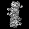 | |||||||||
|---|---|---|---|---|---|---|---|---|---|---|
| Title | Helical structure of EfAvs5(SIR2-STAND) | |||||||||
 Map data Map data | ||||||||||
 Sample Sample |
| |||||||||
 Keywords Keywords | anti-phage / sir2 / STAND / IMMUNE SYSTEM | |||||||||
| Function / homology | : / Novel STAND NTPase 3 / SIR2-like domain / P-loop containing nucleoside triphosphate hydrolase / SIR2 family protein Function and homology information Function and homology information | |||||||||
| Biological species |  | |||||||||
| Method | single particle reconstruction / cryo EM / Resolution: 3.4 Å | |||||||||
 Authors Authors | Wang Y / Zheng J | |||||||||
| Funding support |  China, 1 items China, 1 items
| |||||||||
 Citation Citation | Journal: Nat Commun / Year: 2025 Title: Filamentation activates bacterial Avs5 antiviral protein. Authors: Yiqun Wang / Yuqing Tian / Xu Yang / Feng Yu / Jianting Zheng / Abstract: Bacterial antiviral STANDs (Avs) are evolutionarily related to the nucleotide-binding oligomerization domain (NOD)-like receptors widely distributed in immune systems across animals and plants. ...Bacterial antiviral STANDs (Avs) are evolutionarily related to the nucleotide-binding oligomerization domain (NOD)-like receptors widely distributed in immune systems across animals and plants. EfAvs5, a type 5 Avs from Escherichia fergusonii, contains an N-terminal SIR2 effector domain, a NOD, and a C-terminal sensor domain, conferring protection against diverse phage invasions. Despite the established roles of SIR2 and STAND in prokaryotic and eukaryotic immunity, the mechanism underlying their collaboration remains unclear. Here we present cryo-EM structures of EfAvs5 filaments, elucidating the mechanisms of dimerization, filamentation, filament bundling, ATP binding, and NAD hydrolysis, all of which are crucial for anti-phage defense. The SIR2 and NOD domains engage in intra- and inter-dimer interaction to form an individual filament, while the outward C-terminal sensor domains contribute to bundle formation. Filamentation potentially stabilizes the dimeric SIR2 configuration, thereby activating the NADase activity of EfAvs5. Furthermore, we identify the nucleotide kinase gp1.7 of phage T7 as an activator of EfAvs5, demonstrating its ability to induce filamentation and NADase activity. Together, we uncover the filament assembly of Avs5 as a unique mechanism to switch enzyme activities and perform anti-phage defenses. | |||||||||
| History |
|
- Structure visualization
| Supplemental images |
|---|

- Downloads & links
-EMDB archive
| Map data | emd_61299.map.gz | 19.1 MB | EMDB map data format | |
|---|---|---|---|---|
| Header (meta data) | emd-61299-v30.xmlemd-61299.xml | 18.2 KB 18.2 KB | Display Display | EMDB header |
| FSC (resolution estimation) | emd_61299_fsc.xml | 11.9 KB | Display | FSC data file |
| Images |  emd_61299.png emd_61299.png | 54.9 KB | ||
| Filedesc metadata | emd-61299.cif.gz | 6.7 KB | ||
| Others | emd_61299_half_map_1.map.gzemd_61299_half_map_2.map.gz | 165.4 MB 165.4 MB | ||
| Archive directory |  http://ftp.pdbj.org/pub/emdb/structures/EMD-61299ftp://ftp.pdbj.org/pub/emdb/structures/EMD-61299 http://ftp.pdbj.org/pub/emdb/structures/EMD-61299ftp://ftp.pdbj.org/pub/emdb/structures/EMD-61299 | HTTPS FTP |
-Related structure data
| Related structure data | 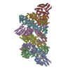 9japMC M: atomic model generated by this map C: citing same article ( |
|---|---|
| Similar structure data |


-Links
| EMDB pages | EMDB (EBI/PDBe) / EMDataResource |
|---|
-Map
| File | Download / File: emd_61299.map.gz / Format: CCP4 / Size: 178 MB / Type: IMAGE STORED AS FLOATING POINT NUMBER (4 BYTES) | ||||||||||||||||||||||||||||||||||||
|---|---|---|---|---|---|---|---|---|---|---|---|---|---|---|---|---|---|---|---|---|---|---|---|---|---|---|---|---|---|---|---|---|---|---|---|---|---|
| Projections & slices | Image control
Images are generated by Spider. | ||||||||||||||||||||||||||||||||||||
| Voxel size | X=Y=Z: 0.89 Å | ||||||||||||||||||||||||||||||||||||
| Density |
| ||||||||||||||||||||||||||||||||||||
| Symmetry | Space group: 1 | ||||||||||||||||||||||||||||||||||||
| Details | EMDB XML:
|
 Z (Sec.)
 Y (Row.)
 X (Col.)


-Supplemental data
-Half map: #2
| File | emd_61299_half_map_1.map | ||||||||||||
|---|---|---|---|---|---|---|---|---|---|---|---|---|---|
| Projections & Slices |
| ||||||||||||
| Density Histograms |


-Half map: #1
| File | emd_61299_half_map_2.map | ||||||||||||
|---|---|---|---|---|---|---|---|---|---|---|---|---|---|
| Projections & Slices |
| ||||||||||||
| Density Histograms |


- Sample components
-Entire : EfAvs5(SIR2-STAND)
| Entire | Name: EfAvs5(SIR2-STAND) |
|---|---|
| Components |
|
-Supramolecule #1: EfAvs5(SIR2-STAND)
| Supramolecule | Name: EfAvs5(SIR2-STAND) / type: complex / ID: 1 / Parent: 0 / Macromolecule list: #1 |
|---|---|
| Source (natural) | Organism: |
| Molecular weight | Theoretical: 800 KDa |
-Macromolecule #1: SIR2-like domain-containing protein
| Macromolecule | Name: SIR2-like domain-containing protein / type: protein_or_peptide / ID: 1 Details: The protein belongs to Escherichia fergusonii, the NCBI Taxonomy ID is QML19490.1. Sequence reference for Escherichia fergusonii (564) is not available in UniProt at the time of biocuration. ...Details: The protein belongs to Escherichia fergusonii, the NCBI Taxonomy ID is QML19490.1. Sequence reference for Escherichia fergusonii (564) is not available in UniProt at the time of biocuration. Current sequence reference is from UniProt ID A0AA94GZI5. Number of copies: 8 / Enantiomer: LEVO |
|---|---|
| Source (natural) | Organism: |
| Molecular weight | Theoretical: 100.285055 KDa |
| Recombinant expression | Organism: |
| Sequence | String: MGSSHHHHHH SSGLVPRGSH MDAMLNSLKL ANSANESISD ETYERLKRLI STGNAIVFVG AGFSKESINI IGSTPPLAKD LALQISNKS ANYLKEVGAD SHYIEEIKQC DDLMVASDFF LNNIPQKDEL LQLLKDNYTI KDVTQEQIDI FSMKWRRIYT T NYDNAIEL ...String: MGSSHHHHHH SSGLVPRGSH MDAMLNSLKL ANSANESISD ETYERLKRLI STGNAIVFVG AGFSKESINI IGSTPPLAKD LALQISNKS ANYLKEVGAD SHYIEEIKQC DDLMVASDFF LNNIPQKDEL LQLLKDNYTI KDVTQEQIDI FSMKWRRIYT T NYDNAIEL SLIKSGKSVT PLTLEDAPNQ YKSAEDICLH INGRIERSKE SDLDSAIKLT TSSYLSPEQF LTSSWYRQFK AD IDNASAI VFLGYSMYDI DIQKIFFNDS SIKSKTFFIT REGTTKFQNY KLAMFGEVIN IGVNAFSHIA AKCIEESHQD KEV GLINSL ELYTPGEEHD EIRDNDIANF MIFGKVSDRY IDEVTLNDNM HDKIILREEV SKIIEHIETD NDILIASDLG NGKS IMTRM LMSKLSRKGY LCFYYLYNEF SFSKDIERLS KLGQKIVIFI DDYSNCIDDT RYAIENRKDN IQLVLTTRHF GYENT KQHL LTMDMSSFKT HSVDYLSDSE VDNFVHIVDH LGAWGEKAGL SRHEKLSELD ENARNQLSFL LLSILKSEAI QSRIRE ISN LALNDKEYKE TVFAILLLDV IGLPLVRSLI SDVAVNEKIY SAEFTENEGV KNLFIISNGM VKTKSSTLSR FLIANIF EH KYVVNQLLKV IEHLYVINKD AKDHRLQTLI TSLLRFSIIE KLLPQRRVEI NYFYEKVKHI IPNLINDPHF WVQYAMSM I PFKDYPSADR YLATAYSLAA RKDNYHTKNI DTQRARLHLL VSLTKTGNEA YLEFEAGDNL IRIIPNDIYK YRQVLRYRD IYEKVYPTFN AKQKVFYEHA IKRIIKESES PELVEDLTYK IGVNWLDKLR GNLKLIVENI QENRPKGKK UniProtKB: SIR2 family protein |
-Macromolecule #2: ADENOSINE-5'-TRIPHOSPHATE
| Macromolecule | Name: ADENOSINE-5'-TRIPHOSPHATE / type: ligand / ID: 2 / Number of copies: 8 / Formula: ATP |
|---|---|
| Molecular weight | Theoretical: 507.181 Da |
| Chemical component information |  ChemComp-ATP: |
-Macromolecule #3: MAGNESIUM ION
| Macromolecule | Name: MAGNESIUM ION / type: ligand / ID: 3 / Number of copies: 8 / Formula: MG |
|---|---|
| Molecular weight | Theoretical: 24.305 Da |
-Experimental details
-Structure determination
| Method | cryo EM |
|---|---|
 Processing Processing | single particle reconstruction |
| Aggregation state | particle |
-Sample preparation
| Concentration | 2 mg/mL | |||||||||||||||
|---|---|---|---|---|---|---|---|---|---|---|---|---|---|---|---|---|
| Buffer | pH: 8 Component:
| |||||||||||||||
| Vitrification | Cryogen name: ETHANE / Chamber humidity: 100 % / Chamber temperature: 289 K / Instrument: FEI VITROBOT MARK IV |
- Electron microscopy
| Microscope | FEI TITAN KRIOS |
|---|---|
| Temperature | Min: 77.0 K / Max: 77.0 K |
| Specialist optics | Energy filter - Name: GIF Bioquantum |
| Image recording | Film or detector model: GATAN K2 QUANTUM (4k x 4k) / Detector mode: COUNTING / Average electron dose: 40.0 e/Å2 |
| Electron beam | Acceleration voltage: 300 kV / Electron source:  FIELD EMISSION GUN FIELD EMISSION GUN |
| Electron optics | C2 aperture diameter: 70.0 µm / Illumination mode: FLOOD BEAM / Imaging mode: BRIGHT FIELD / Cs: 0.1 mm / Nominal defocus max: 2.0 µm / Nominal defocus min: 1.0 µm / Nominal magnification: 130000 |
| Sample stage | Specimen holder model: FEI TITAN KRIOS AUTOGRID HOLDER / Cooling holder cryogen: NITROGEN |
| Experimental equipment |  Model: Titan Krios / Image courtesy: FEI Company |
+Image processing

-Atomic model buiding 1
| Refinement | Space: REAL |
|---|---|
| Output model | PDB-9jap: |
