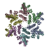
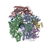
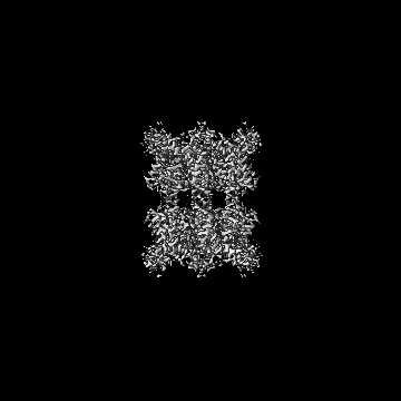
ジャーナル: Nat Commun / 年: 2024 タイトル: Open architecture of archaea MCM and dsDNA complexes resolved using monodispersed streptavidin affinity CryoEM. 著者: Jianbing Ma / Gangshun Yi / Mingda Ye / Craig MacGregor-Chatwin / Yuewen Sheng / Ying Lu / Ming Li / Qingrong Li / Dong Wang / Robert J C Gilbert / Peijun Zhang / 要旨: The cryo-electron microscopy (cryoEM) method has enabled high-resolution structure determination of numerous biomolecules and complexes. Nevertheless, cryoEM sample preparation of challenging ...The cryo-electron microscopy (cryoEM) method has enabled high-resolution structure determination of numerous biomolecules and complexes. Nevertheless, cryoEM sample preparation of challenging proteins and complexes, especially those with low abundance or with preferential orientation, remains a major hurdle. We developed an affinity-grid method employing monodispersed single particle streptavidin on a lipid monolayer to enhance particle absorption on the grid surface and alleviate sample exposure to the air-water interface. Using this approach, we successfully enriched the Thermococcus kodakarensis mini-chromosome maintenance complex 3 (MCM3) on cryoEM grids through biotinylation and resolved its structure. We further utilized this affinity method to tether the biotin-tagged dsDNA to selectively enrich a stable MCM3-ATP-dsDNA complex for cryoEM structure determination. Intriguingly, both MCM3 apo and dsDNA bound structures exhibit left-handed open spiral conformations, distinct from other reported MCM structures. The large open gate is sufficient to accommodate a dsDNA which could potentially be melted. The value of mspSA affinity method was further demonstrated by mitigating the issue of preferential angular distribution of HIV-1 capsid protein hexamer and RNA polymerase II elongation complex from Saccharomyces cerevisiae.
 ムービー
ムービー コントローラー


 データを開く
 基本情報
基本情報
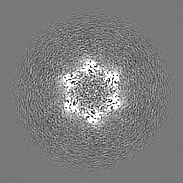
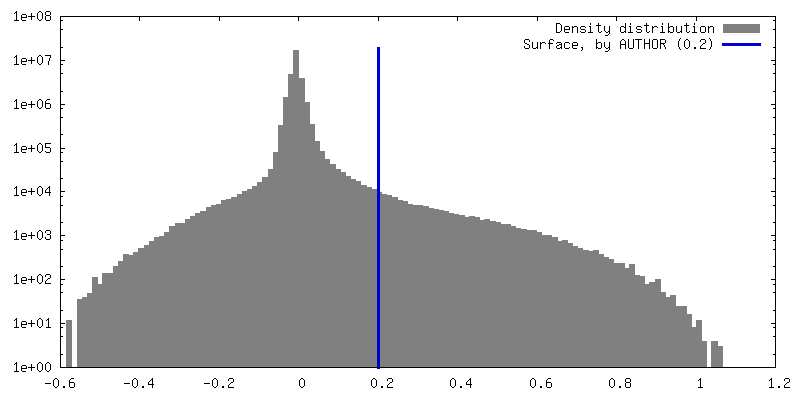
 マップデータ
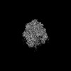
 試料
試料 キーワード
キーワード 機能・相同性情報
 Human immunodeficiency virus 1 (ヒト免疫不全ウイルス)
 データ登録者
 英国, 1件
  引用
引用

 構造の表示
構造の表示
 ダウンロードとリンク
 emd_61286.png
 http://ftp.pdbj.org/pub/emdb/structures/EMD-61286


 Z (Sec.)
 Y (Row.)
 X (Col.)


 試料の構成要素
 解析
解析 電子顕微鏡法
 FIELD EMISSION GUN