Movie
Movie Controller

[English] 日本語
 Yorodumi
- EMDB-61131: Cryo-EM structure of aPlexinA1-19-43 Fab in complex with PlexinA1... -
+ Open data

- Basic information
| Entry |  | |||||||||
|---|---|---|---|---|---|---|---|---|---|---|
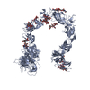
| Title | Cryo-EM structure of aPlexinA1-19-43 Fab in complex with PlexinA1 dimer | |||||||||
 Map data Map data | Cryo-EM structure of aPlexinA1-19-43 Fab in complex with PlexinA1 dimer. Sharpened main map. | |||||||||
 Sample Sample |
| |||||||||
 Keywords Keywords | Signalling / Fab / Complex / PROTEIN BINDING / PROTEIN BINDING-IMMUNE SYSTEM complex | |||||||||
| Function / homology |  Function and homology information Function and homology informationolfactory nerve formation / neuron projection guidance / dichotomous subdivision of terminal units involved in salivary gland branching / gonadotrophin-releasing hormone neuronal migration to the hypothalamus / Other semaphorin interactions / T cell activation via T cell receptor contact with antigen bound to MHC molecule on antigen presenting cell / semaphorin receptor complex / SEMA3A-Plexin repulsion signaling by inhibiting Integrin adhesion / CRMPs in Sema3A signaling / semaphorin receptor activity ...olfactory nerve formation / neuron projection guidance / dichotomous subdivision of terminal units involved in salivary gland branching / gonadotrophin-releasing hormone neuronal migration to the hypothalamus / Other semaphorin interactions / T cell activation via T cell receptor contact with antigen bound to MHC molecule on antigen presenting cell / semaphorin receptor complex / SEMA3A-Plexin repulsion signaling by inhibiting Integrin adhesion / CRMPs in Sema3A signaling / semaphorin receptor activity / regulation of smooth muscle cell migration / RHOD GTPase cycle / RND1 GTPase cycle / neuron projection extension / semaphorin-plexin signaling pathway / Sema3A PAK dependent Axon repulsion / synapse assembly / regulation of cell migration / glutamatergic synapse / extracellular exosome / nucleoplasm / plasma membrane / cytosol Similarity search - Function | |||||||||
| Biological species |  Homo sapiens (human) / Homo sapiens (human) /  | |||||||||
| Method | single particle reconstruction / cryo EM / Resolution: 3.33 Å | |||||||||
 Authors Authors | Tian H / Fung CP | |||||||||
| Funding support |  Hong Kong, 1 items Hong Kong, 1 items
| |||||||||
 Citation Citation | Journal: J Biol Chem / Year: 2026 Title: A bispecific antibody designed to act as a NRP2/PLXNA1 agonist mimics anticancer activity of SEMA3F. Authors: Honglei Tian / Chun Po Fung / Luke Burman / Yeeting E Chong / Changdong Liu / Yanyan Geng / Lam Yang / Man Wai Chow / Yingyi Zhang / Kwok Wa Hugo Ho / Guang Zhu / Zhenguo Wu / Xiang-Lei Yang ...Authors: Honglei Tian / Chun Po Fung / Luke Burman / Yeeting E Chong / Changdong Liu / Yanyan Geng / Lam Yang / Man Wai Chow / Yingyi Zhang / Kwok Wa Hugo Ho / Guang Zhu / Zhenguo Wu / Xiang-Lei Yang / Zhiwen Xu / Leslie A Nangle /  Abstract: Neuropilin-2 (NRP2) is a pleiotropic receptor with diverse roles across biological systems. Recent work detailed its role as an immunomodulatory receptor target that is currently being explored in ...Neuropilin-2 (NRP2) is a pleiotropic receptor with diverse roles across biological systems. Recent work detailed its role as an immunomodulatory receptor target that is currently being explored in clinical development for interstitial lung diseases, establishing it as a viable therapeutic target. To mediate its diverse effects, NRP2 interacts with endogenous ligands, including semaphorins (SEMAs) and vascular endothelial growth factors, signaling via ligand-induced heterodimerization with various receptor families. One of these ligands, SEMA3F exhibits well-documented tumor-suppressive activities mediated through NRP2 and plexinA1 (PLXNA1). Despite its observed benefits, SEMA3F is not therapeutically viable due to the multifaceted nature of its functions through non-NRP2-mediated interactions, leading to concerns around potential toxicity. Here, we describe development of bispecific antibodies (bsAbs) that dimerize PLXNA1 and NRP2, selectively mimicking the beneficial aspects of SEMA3F signaling as a basis for a novel anticancer therapy. Using a single B cell-based mAb discovery platform, anti-PLXNA1 mAbs with diverse lineages were generated and combined with anti-NRP2 mAbs to produce over 200 PLXNA1-NRP2 bsAbs. Antibodies were screened in cell-based assays (receptor dimerization, phospho-AKT, oncogene expression, and cell proliferation), yielding one bsAb capable of mimicking NRP2-mediated SEMA3F activities in all assays. Structural studies revealed that this bsAb binds to PLXNA1/NRP2 at sites distinct from the SEMA3F-binding site, but in a manner that allows proper spacing for receptor complex formation and flexibility of conformational changes for signaling. This study demonstrates the potential of these receptors as targets for agonistic bsAbs development and provides the groundwork for further exploration in tumor models. | |||||||||
| History |
|
- Structure visualization
| Supplemental images |
|---|

- Downloads & links
-EMDB archive
| Map data | emd_61131.map.gz | 79.1 MB | EMDB map data format | |
|---|---|---|---|---|
| Header (meta data) | emd-61131-v30.xmlemd-61131.xml | 28.1 KB 28.1 KB | Display Display | EMDB header |
| FSC (resolution estimation) | emd_61131_fsc.xml | 9.3 KB | Display | FSC data file |
| Images |  emd_61131.png emd_61131.png | 58.4 KB | ||
| Filedesc metadata | emd-61131.cif.gz | 8 KB | ||
| Others | emd_61131_additional_1.map.gzemd_61131_half_map_1.map.gzemd_61131_half_map_2.map.gz | 41.7 MB 77.9 MB 77.9 MB | ||
| Archive directory |  http://ftp.pdbj.org/pub/emdb/structures/EMD-61131ftp://ftp.pdbj.org/pub/emdb/structures/EMD-61131 http://ftp.pdbj.org/pub/emdb/structures/EMD-61131ftp://ftp.pdbj.org/pub/emdb/structures/EMD-61131 | HTTPS FTP |
-Related structure data
| Related structure data |  9j4cMC M: atomic model generated by this map C: citing same article ( |
|---|---|
| Similar structure data |
-Links
| EMDB pages | EMDB (EBI/PDBe) / EMDataResource |
|---|---|
| Related items in Molecule of the Month |


-Map
| File | Download / File: emd_61131.map.gz / Format: CCP4 / Size: 83.7 MB / Type: IMAGE STORED AS FLOATING POINT NUMBER (4 BYTES) | ||||||||||||||||||||||||||||||||||||
|---|---|---|---|---|---|---|---|---|---|---|---|---|---|---|---|---|---|---|---|---|---|---|---|---|---|---|---|---|---|---|---|---|---|---|---|---|---|
| Annotation | Cryo-EM structure of aPlexinA1-19-43 Fab in complex with PlexinA1 dimer. Sharpened main map. | ||||||||||||||||||||||||||||||||||||
| Projections & slices | Image control
Images are generated by Spider. | ||||||||||||||||||||||||||||||||||||
| Voxel size | X=Y=Z: 1.026 Å | ||||||||||||||||||||||||||||||||||||
| Density |
| ||||||||||||||||||||||||||||||||||||
| Symmetry | Space group: 1 | ||||||||||||||||||||||||||||||||||||
| Details | EMDB XML:
|
 Z (Sec.)
 Y (Row.)
 X (Col.)


-Supplemental data
-Additional map: Cryo-EM structure of aPlexinA1-19-43 Fab in complex with...
| File | emd_61131_additional_1.map | ||||||||||||
|---|---|---|---|---|---|---|---|---|---|---|---|---|---|
| Annotation | Cryo-EM structure of aPlexinA1-19-43 Fab in complex with PlexinA1 dimer. Raw Map | ||||||||||||
| Projections & Slices |
| ||||||||||||
| Density Histograms |


-Half map: Cryo-EM structure of aPlexinA1-19-43 Fab in complex with...
| File | emd_61131_half_map_1.map | ||||||||||||
|---|---|---|---|---|---|---|---|---|---|---|---|---|---|
| Annotation | Cryo-EM structure of aPlexinA1-19-43 Fab in complex with PlexinA1 dimer. Half Map B | ||||||||||||
| Projections & Slices |
| ||||||||||||
| Density Histograms |


-Half map: Cryo-EM structure of aPlexinA1-19-43 Fab in complex with...
| File | emd_61131_half_map_2.map | ||||||||||||
|---|---|---|---|---|---|---|---|---|---|---|---|---|---|
| Annotation | Cryo-EM structure of aPlexinA1-19-43 Fab in complex with PlexinA1 dimer. Half Map A | ||||||||||||
| Projections & Slices |
| ||||||||||||
| Density Histograms |


- Sample components
-Entire : Dimer complex of Human Plexin-A1 27-710 with aPlexinA1-19-43 Fab ...
| Entire | Name: Dimer complex of Human Plexin-A1 27-710 with aPlexinA1-19-43 Fab binding |
|---|---|
| Components |
|
-Supramolecule #1: Dimer complex of Human Plexin-A1 27-710 with aPlexinA1-19-43 Fab ...
| Supramolecule | Name: Dimer complex of Human Plexin-A1 27-710 with aPlexinA1-19-43 Fab binding type: complex / ID: 1 / Parent: 0 / Macromolecule list: all Details: Fab fragments generated from cloning the variable region of PlexinA1 antibody generated from mouse, into Fab vector. Fab fragments and PlexinA1 27-710 are expressed and purified from CHO cells. |
|---|
-Supramolecule #2: Dimer of Human Plexin-A1 27-710
| Supramolecule | Name: Dimer of Human Plexin-A1 27-710 / type: complex / ID: 2 / Parent: 1 / Macromolecule list: #1 |
|---|---|
| Source (natural) | Organism: Homo sapiens (human) |
-Supramolecule #3: aPlexinA1-19-43 Fab
| Supramolecule | Name: aPlexinA1-19-43 Fab / type: complex / ID: 3 / Parent: 1 / Macromolecule list: #2-#3 Details: Heavy chain and light chain of aPlexinA1-19-43 Fab expressed and purified from CHO cells |
|---|---|
| Source (natural) | Organism: |
-Macromolecule #1: Plexin-A1
| Macromolecule | Name: Plexin-A1 / type: protein_or_peptide / ID: 1 / Details: Human Plexin A1 27 to 710 with His Tag / Number of copies: 1 / Enantiomer: LEVO |
|---|---|
| Source (natural) | Organism: Homo sapiens (human) |
| Molecular weight | Theoretical: 76.710734 KDa |
| Recombinant expression | Organism:  Cricetulus griseus (Chinese hamster) Cricetulus griseus (Chinese hamster) |
| Sequence | String: EAGLPRAGGG SQPPFRTFSA SDWGLTHLVV HEQTGEVYVG AVNRIYKLSG NLTLLRAHVT GPVEDNEKCY PPPSVQSCPH GLGSTDNVN KLLLLDYAAN RLLACGSASQ GICQFLRLDD LFKLGEPHHR KEHYLSSVQE AGSMAGVLIA GPPGQGQAKL F VGTPIDGK ...String: EAGLPRAGGG SQPPFRTFSA SDWGLTHLVV HEQTGEVYVG AVNRIYKLSG NLTLLRAHVT GPVEDNEKCY PPPSVQSCPH GLGSTDNVN KLLLLDYAAN RLLACGSASQ GICQFLRLDD LFKLGEPHHR KEHYLSSVQE AGSMAGVLIA GPPGQGQAKL F VGTPIDGK SEYFPTLSSR RLMANEEDAD MFGFVYQDEF VSSQLKIPSD TLSKFPAFDI YYVYSFRSEQ FVYYLTLQLD TQ LTSPDAA GEHFFTSKIV RLCVDDPKFY SYVEFPIGCE QAGVEYRLVQ DAYLSRPGRA LAHQLGLAED EDVLFTVFAQ GQK NRVKPP KESALCLFTL RAIKEKIKER IQSCYRGEGK LSLPWLLNKE LGCINSPLQI DDDFCGQDFN QPLGGTVTIE GTPL FVDKD DGLTAVAAYD YRGRTVVFAG TRSGRIRKIL VDLSNPGGRP ALAYESVVAQ EGSPILRDLV LSPNHQYLYA MTEKQ VTRV PVESCVQYTS CELCLGSRDP HCGWCVLHSI CSRRDACERA DEPQRFAADL LQCVQLTVQP RNVSVTMSQV PLVLQA WNV PDLSAGVNCS FEDFTESESV LEDGRIHCRS PSAREVAPIT RGQGDQRVVK LYLKSKETGK KFASVDFVFY NCSVHQS CL SCVNGSFPCH WCKYRHVCTH NVADCAFLEG RVNVSEDCPQ ILHHHHHH UniProtKB: Plexin-A1 |
-Macromolecule #2: Light chain of aPlexinA1-19-43 Fab
| Macromolecule | Name: Light chain of aPlexinA1-19-43 Fab / type: protein_or_peptide / ID: 2 / Number of copies: 1 / Enantiomer: LEVO |
|---|---|
| Source (natural) | Organism: |
| Molecular weight | Theoretical: 23.413873 KDa |
| Recombinant expression | Organism: Cricetulus griseus (Chinese hamster) |
| Sequence | String: QIVLTQSPAI LSASPGEKVT MSCSVSSSIT YMHWYQQKPG TSPKRWIYDT SKLASGVPAR FSGSGSGTSF SLTISNMEAE DAATYYCHQ RSSYPYSFGG GTKLEIKRAD AAPTVSIFPP SSEQLTSGGA SVVCFLNNFY PKDINVKWKI DGSERQNGVL N SWTDQDSK ...String: QIVLTQSPAI LSASPGEKVT MSCSVSSSIT YMHWYQQKPG TSPKRWIYDT SKLASGVPAR FSGSGSGTSF SLTISNMEAE DAATYYCHQ RSSYPYSFGG GTKLEIKRAD AAPTVSIFPP SSEQLTSGGA SVVCFLNNFY PKDINVKWKI DGSERQNGVL N SWTDQDSK DSTYSMSSTL TLTKDEYERH NSYTCEATHK TSTSPIVKSF NRNEC |
-Macromolecule #3: Heavy chain of aPlexinA1-19-43 Fab
| Macromolecule | Name: Heavy chain of aPlexinA1-19-43 Fab / type: protein_or_peptide / ID: 3 / Number of copies: 1 / Enantiomer: LEVO |
|---|---|
| Source (natural) | Organism: |
| Molecular weight | Theoretical: 25.302014 KDa |
| Recombinant expression | Organism: Cricetulus griseus (Chinese hamster) |
| Sequence | String: EVQLQQSGPE LVKPGASVKI SCKASGYKFT ENYMDWVKQS HGESLEWIGD ISPDNGDTSY NQKFRDKATL TVDKSSSTAY MELRSLTSE DSAVYYCAQI IYYDYVGYAL DYWGQGTSVT VSSAKTTPPS VYPLAPGSAA QTNSMVTLGC LVKGYFPEPV T VTWNSGSL ...String: EVQLQQSGPE LVKPGASVKI SCKASGYKFT ENYMDWVKQS HGESLEWIGD ISPDNGDTSY NQKFRDKATL TVDKSSSTAY MELRSLTSE DSAVYYCAQI IYYDYVGYAL DYWGQGTSVT VSSAKTTPPS VYPLAPGSAA QTNSMVTLGC LVKGYFPEPV T VTWNSGSL SSGVHTFPAV LQSDLYTLSS SVTVPSSTWP SETVTCNVAH PASSTKVDKK IVPRDCGGSH HHHHH |
-Experimental details
-Structure determination
| Method | cryo EM |
|---|---|
 Processing Processing | single particle reconstruction |
| Aggregation state | particle |
-Sample preparation
| Concentration | 0.1 mg/mL |
|---|---|
| Buffer | pH: 7.4 / Details: 1x PBS 7.4 |
| Grid | Model: Quantifoil R2/2 / Material: GOLD / Mesh: 300 / Support film - Material: CARBON / Support film - topology: HOLEY / Pretreatment - Type: GLOW DISCHARGE / Pretreatment - Time: 30 sec. |
| Vitrification | Cryogen name: ETHANE / Chamber humidity: 100 % / Chamber temperature: 291.15 K / Instrument: FEI VITROBOT MARK IV Details: To prepare cryo-grids, 3 ul samples were applied to glow discharged Quantifoil Au grids (R2/2, 300 mesh), which were subsequently blotted with filter paper (Ted Pella) for 3 seconds at 18oC ...Details: To prepare cryo-grids, 3 ul samples were applied to glow discharged Quantifoil Au grids (R2/2, 300 mesh), which were subsequently blotted with filter paper (Ted Pella) for 3 seconds at 18oC and 100% humidity. The grids were immediately plunge frozen in liquid ethane using a FEI Vitrobot IV (ThermoFisher). |
| Details | This sample is a 1:1 mixture of aPlexinA1-19-43 Fab in complex with PlexinA1 dimer. The sample was mono disperse. |
- Electron microscopy
| Microscope | FEI TITAN KRIOS |
|---|---|
| Image recording | Film or detector model: FEI FALCON III (4k x 4k) / Number real images: 1989 / Average electron dose: 50.0 e/Å2 Details: A total of 2,084 movies were acquired and imported into Cryo-SPARC using the following parameters: Raw pixel size 1.026 A, Accelerating Voltage 300 kV, Spherical Aberration 2.7 mm, and Total ...Details: A total of 2,084 movies were acquired and imported into Cryo-SPARC using the following parameters: Raw pixel size 1.026 A, Accelerating Voltage 300 kV, Spherical Aberration 2.7 mm, and Total exposure dose 50 e/A^2. Motion correction and CTF estimation were performed using Full-frame Motion Correction and Patch CTF. After that, 1,989 micrographs were selected from the 2,084 micrographs based on average intensity (-10055.05 to 2041.77) |
| Electron beam | Acceleration voltage: 300 kV / Electron source: OTHER |
| Electron optics | Illumination mode: OTHER / Imaging mode: OTHER / Nominal defocus max: 2.5 µm / Nominal defocus min: 1.2 µm |
| Experimental equipment |  Model: Titan Krios / Image courtesy: FEI Company |
+Image processing

-Atomic model buiding 1
| Initial model |
| ||||||||||||
|---|---|---|---|---|---|---|---|---|---|---|---|---|---|
| Details | Initial local fitting was done using Chimera X, followed by manual fitting using Coot. Finally it was refined using Phenix real space refinement | ||||||||||||
| Refinement | Space: REAL / Protocol: AB INITIO MODEL | ||||||||||||
| Output model | PDB-9j4c: |