 Movie
Movie Controller

[English] 日本語
 Yorodumi
- EMDB-60931: 24-mer DARPin-apoferritin scaffold in complex with the maltose bi... -
+ Open data

- Basic information
| Entry |  | |||||||||
|---|---|---|---|---|---|---|---|---|---|---|
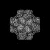
| Title | 24-mer DARPin-apoferritin scaffold in complex with the maltose binding protein | |||||||||
 Map data Map data | ||||||||||
 Sample Sample |
| |||||||||
 Keywords Keywords | DARPin / apoferritin / scaffold / maltose binding protein / BIOSYNTHETIC PROTEIN | |||||||||
| Function / homology |  Function and homology information Function and homology informationiron ion sequestering activity / ferritin complex / Scavenging by Class A Receptors / negative regulation of ferroptosis / Golgi Associated Vesicle Biogenesis / ferroxidase / autolysosome / ferroxidase activity / carbohydrate transmembrane transporter activity / maltose binding ...iron ion sequestering activity / ferritin complex / Scavenging by Class A Receptors / negative regulation of ferroptosis / Golgi Associated Vesicle Biogenesis / ferroxidase / autolysosome / ferroxidase activity / carbohydrate transmembrane transporter activity / maltose binding / maltose transport / maltodextrin transmembrane transport / negative regulation of fibroblast proliferation / ATP-binding cassette (ABC) transporter complex, substrate-binding subunit-containing / ferric iron binding / autophagosome / Iron uptake and transport / iron ion transport / ferrous iron binding / tertiary granule lumen / outer membrane-bounded periplasmic space / ficolin-1-rich granule lumen / intracellular iron ion homeostasis / immune response / iron ion binding / negative regulation of cell population proliferation / Neutrophil degranulation / extracellular exosome / extracellular region / identical protein binding / nucleus / cytoplasm / cytosol Similarity search - Function | |||||||||
| Biological species |  Homo sapiens (human) / Homo sapiens (human) /  | |||||||||
| Method | single particle reconstruction / cryo EM / Resolution: 3.0 Å | |||||||||
 Authors Authors | Lu X / Yan M / Zhang HM / Hao Q | |||||||||
| Funding support |  China, 1 items China, 1 items
| |||||||||
 Citation Citation | Journal: IUCrJ / Year: 2025 Title: A large, general and modular DARPin-apoferritin scaffold enables the visualization of small proteins by cryo-EM. Authors: Xin Lu / Ming Yan / Yang Cai / Xi Song / Huan Chen / Mengtan Du / Zhenyi Wang / Jia'an Li / Liwen Niu / Fuxing Zeng / Quan Hao / Hongmin Zhang / Abstract: Single-particle cryo-electron microscopy (cryo-EM) has emerged as an indispensable technique in structural biology that is pivotal for deciphering protein architectures. However, the medium-sized ...Single-particle cryo-electron microscopy (cryo-EM) has emerged as an indispensable technique in structural biology that is pivotal for deciphering protein architectures. However, the medium-sized proteins (30-40 kDa) that are prevalent in both eukaryotic and prokaryotic organisms often elude the resolving capabilities of contemporary cryo-EM methods. To address this challenge, we engineered a scaffold strategy that securely anchors proteins of interest to a robust, symmetric base via a selective adapter. Our most efficacious constructs, namely models 4 and 6c, feature a designed ankyrin-repeat protein (DARPin) rigidly linked to an octahedral human apoferritin via a helical linker. By utilizing these large, highly symmetric scaffolds (∼1 MDa), we achieved near-atomic-resolution cryo-EM structures of green fluorescent protein (GFP) and maltose-binding protein (MBP), revealing nearly all side-chain densities of GFP and the distinct structural features of MBP. The modular design of our scaffold allows the adaptation of new DARPins through minor amino-acid-sequence modifications, enabling the binding and visualization of a diverse array of proteins. The high symmetry and near-spherical shape of the scaffold not only mitigates the prevalent challenge of preferred particle orientation in cryo-EM but also significantly reduces the demands of image collection and data processing. This approach presents a versatile solution, breaking through the size constraints that have traditionally limited single-particle cryo-EM. | |||||||||
| History |
|
- Structure visualization
| Supplemental images |
|---|

- Downloads & links
-EMDB archive
| Map data | emd_60931.map.gz | 62.7 MB | EMDB map data format | |
|---|---|---|---|---|
| Header (meta data) | emd-60931-v30.xmlemd-60931.xml | 15.6 KB 15.6 KB | Display Display | EMDB header |
| FSC (resolution estimation) | emd_60931_fsc.xml | 14.6 KB | Display | FSC data file |
| Images |  emd_60931.png emd_60931.png | 93.7 KB | ||
| Filedesc metadata | emd-60931.cif.gz | 6.6 KB | ||
| Others | emd_60931_half_map_1.map.gzemd_60931_half_map_2.map.gz | 115.9 MB 115.9 MB | ||
| Archive directory |  http://ftp.pdbj.org/pub/emdb/structures/EMD-60931ftp://ftp.pdbj.org/pub/emdb/structures/EMD-60931 http://ftp.pdbj.org/pub/emdb/structures/EMD-60931ftp://ftp.pdbj.org/pub/emdb/structures/EMD-60931 | HTTPS FTP |
-Validation report
| Summary document | emd_60931_validation.pdf.gz | 832.2 KB | Display | EMDB validaton report |
|---|---|---|---|---|
| Full document | emd_60931_full_validation.pdf.gz | 831.9 KB | Display | |
| Data in XML | emd_60931_validation.xml.gz | 18.8 KB | Display | |
| Data in CIF | emd_60931_validation.cif.gz | 24.8 KB | Display | |
| Arichive directory | https://ftp.pdbj.org/pub/emdb/validation_reports/EMD-60931ftp://ftp.pdbj.org/pub/emdb/validation_reports/EMD-60931 | HTTPS FTP |
-Related structure data
| Related structure data |  9ivpMC  9irvC  9j48C M: atomic model generated by this map C: citing same article ( |
|---|---|
| Similar structure data |

-Links
| EMDB pages | EMDB (EBI/PDBe) / EMDataResource |
|---|---|
| Related items in Molecule of the Month |


-Map
| File | Download / File: emd_60931.map.gz / Format: CCP4 / Size: 125 MB / Type: IMAGE STORED AS FLOATING POINT NUMBER (4 BYTES) | ||||||||||||||||||||||||||||||||||||
|---|---|---|---|---|---|---|---|---|---|---|---|---|---|---|---|---|---|---|---|---|---|---|---|---|---|---|---|---|---|---|---|---|---|---|---|---|---|
| Projections & slices | Image control
Images are generated by Spider. | ||||||||||||||||||||||||||||||||||||
| Voxel size | X=Y=Z: 1.076 Å | ||||||||||||||||||||||||||||||||||||
| Density |
| ||||||||||||||||||||||||||||||||||||
| Symmetry | Space group: 1 | ||||||||||||||||||||||||||||||||||||
| Details | EMDB XML:
|
 Z (Sec.)
 Y (Row.)
 X (Col.)


-Supplemental data
-Half map: #2
| File | emd_60931_half_map_1.map | ||||||||||||
|---|---|---|---|---|---|---|---|---|---|---|---|---|---|
| Projections & Slices |
| ||||||||||||
| Density Histograms |


-Half map: #1
| File | emd_60931_half_map_2.map | ||||||||||||
|---|---|---|---|---|---|---|---|---|---|---|---|---|---|
| Projections & Slices |
| ||||||||||||
| Density Histograms |


- Sample components
-Entire : 24-mer DARPin-apoferritin in complex with the maltose binding protein
| Entire | Name: 24-mer DARPin-apoferritin in complex with the maltose binding protein |
|---|---|
| Components |
|
-Supramolecule #1: 24-mer DARPin-apoferritin in complex with the maltose binding protein
| Supramolecule | Name: 24-mer DARPin-apoferritin in complex with the maltose binding protein type: complex / ID: 1 / Parent: 0 / Macromolecule list: all |
|---|---|
| Source (natural) | Organism: Homo sapiens (human) |
-Macromolecule #1: DARPin,Ferritin heavy chain, N-terminally processed
| Macromolecule | Name: DARPin,Ferritin heavy chain, N-terminally processed / type: protein_or_peptide / ID: 1 / Number of copies: 24 / Enantiomer: LEVO |
|---|---|
| Source (natural) | Organism: Homo sapiens (human) |
| Molecular weight | Theoretical: 41.047145 KDa |
| Recombinant expression | Organism: |
| Sequence | String: MGHHHHHHGP GSAKEEILEE IKKAKQEIAG GGGGSELGKE LLEAARAGQD DEVRILMARG AEVNAADNTG TTPLHLAAYS GHLEIVEVL LKYGAEVNAA DVFGYTPLHL AAYWGHLEIV EVLLKNGADV NARDSDGMTP LHLAAKWGHL EIVEVLLRYG A DVEAQDKF ...String: MGHHHHHHGP GSAKEEILEE IKKAKQEIAG GGGGSELGKE LLEAARAGQD DEVRILMARG AEVNAADNTG TTPLHLAAYS GHLEIVEVL LKYGAEVNAA DVFGYTPLHL AAYWGHLEIV EVLLKNGADV NARDSDGMTP LHLAAKWGHL EIVEVLLRYG A DVEAQDKF GKTPFDLAID NGNEDIAEVL QALLAINRQI NLELYASYVY LSMSYYFDRD DVALKNFAKY FLHQSHEERE HA EKLMKLQ NQRGGAISLQ DIKKPDCDDW ESGLNAMECA LHLEKNVNQS LLELHKLATD CNDPHLCDFI ETHYLNEQVK AIK ELGDHV TNLRKMGAPE SGLAEYLFDK HTLGSGSGAE IEQAKKEIAY LIKK UniProtKB: Ferritin heavy chain |
-Macromolecule #2: Maltodextrin-binding protein
| Macromolecule | Name: Maltodextrin-binding protein / type: protein_or_peptide / ID: 2 / Number of copies: 24 / Enantiomer: LEVO |
|---|---|
| Source (natural) | Organism: |
| Molecular weight | Theoretical: 45.694176 KDa |
| Recombinant expression | Organism: |
| Sequence | String: MGSSHHHHHH SSGLVPRGSH MKIEEGKLVI WINGDKGYNG LAEVGKKFEK DTGIKVTVEH PDKLEEKFPQ VAATGDGPDI IFWAHDRFG GYAQSGLLAE ITPDKAFQDK LYPFTWDAVR YNGKLIAYPI AVEALSLIYN KDLLPNPPKT WEEIPALDKE L KAKGKSAL ...String: MGSSHHHHHH SSGLVPRGSH MKIEEGKLVI WINGDKGYNG LAEVGKKFEK DTGIKVTVEH PDKLEEKFPQ VAATGDGPDI IFWAHDRFG GYAQSGLLAE ITPDKAFQDK LYPFTWDAVR YNGKLIAYPI AVEALSLIYN KDLLPNPPKT WEEIPALDKE L KAKGKSAL MFNLQEPYFT WPLIAADGGY AFKYENGKYD IKDVGVDNAG AKAGLTFLVD LIKNKHMNAD TDYSIAEAAF NK GETAMTI NGPWAWSNID TSKVNYGVTV LPTFKGQPSK PFVGVLSAGI NAASPNKELA KEFLENYLLT DEGLEAVNKD KPL GAVALK SYEEELAKDP RIAATMENAQ KGEIMPNIPQ MSAFWYAVRT AVINAASGRQ TVDEALKDAQ TNSSSNNNNN NNNN NLGIE GRENLYFQGG S UniProtKB: Maltodextrin-binding protein |
-Experimental details
-Structure determination
| Method | cryo EM |
|---|---|
 Processing Processing | single particle reconstruction |
| Aggregation state | particle |
-Sample preparation
| Buffer | pH: 8 |
|---|---|
| Vitrification | Cryogen name: ETHANE |
- Electron microscopy
| Microscope | FEI TITAN KRIOS |
|---|---|
| Image recording | Film or detector model: GATAN K2 SUMMIT (4k x 4k) / Average electron dose: 1.28 e/Å2 |
| Electron beam | Acceleration voltage: 300 kV / Electron source:  FIELD EMISSION GUN FIELD EMISSION GUN |
| Electron optics | Illumination mode: SPOT SCAN / Imaging mode: BRIGHT FIELD / Nominal defocus max: 2.0 µm / Nominal defocus min: 0.8 µm |
| Experimental equipment |  Model: Titan Krios / Image courtesy: FEI Company |


