 Movie
Movie Controller


+ Open data

- Basic information
| Entry | Database: PDB / ID: 9j48 | ||||||
|---|---|---|---|---|---|---|---|
| Title | GFP bound to 24-mer DARPin-apoferritin model 6c | ||||||
 Components Components |
| ||||||
 Keywords Keywords | METAL BINDING PROTEIN/LUMINESCENT PROTEIN / GFP / DARPin / apoferritin / scaffold / METAL BINDING PROTEIN-LUMINESCENT PROTEIN complex | ||||||
| Function / homology |  Function and homology information Function and homology informationiron ion sequestering activity / ferritin complex / Scavenging by Class A Receptors / negative regulation of ferroptosis / Golgi Associated Vesicle Biogenesis / ferroxidase / autolysosome / ferroxidase activity / negative regulation of fibroblast proliferation / ferric iron binding ...iron ion sequestering activity / ferritin complex / Scavenging by Class A Receptors / negative regulation of ferroptosis / Golgi Associated Vesicle Biogenesis / ferroxidase / autolysosome / ferroxidase activity / negative regulation of fibroblast proliferation / ferric iron binding / autophagosome / bioluminescence / generation of precursor metabolites and energy / iron ion transport / Iron uptake and transport / ferrous iron binding / tertiary granule lumen / ficolin-1-rich granule lumen / intracellular iron ion homeostasis / immune response / iron ion binding / negative regulation of cell population proliferation / Neutrophil degranulation / extracellular exosome / extracellular region / identical protein binding / nucleus / cytoplasm / cytosol Similarity search - Function | ||||||
| Biological species |  Homo sapiens (human) Homo sapiens (human)  Aequorea victoria (jellyfish) Aequorea victoria (jellyfish) | ||||||
| Method | ELECTRON MICROSCOPY / single particle reconstruction / cryo EM / Resolution: 3.04 Å | ||||||
 Authors Authors | Lu, X. / Yan, M. / Zhang, H.M. / Hao, Q. | ||||||
| Funding support |  China, 1items China, 1items
| ||||||
 Citation Citation | Journal: IUCrJ / Year: 2025 Title: A large, general and modular DARPin-apoferritin scaffold enables the visualization of small proteins by cryo-EM. Authors: Xin Lu / Ming Yan / Yang Cai / Xi Song / Huan Chen / Mengtan Du / Zhenyi Wang / Jia'an Li / Liwen Niu / Fuxing Zeng / Quan Hao / Hongmin Zhang / Abstract: Single-particle cryo-electron microscopy (cryo-EM) has emerged as an indispensable technique in structural biology that is pivotal for deciphering protein architectures. However, the medium-sized ...Single-particle cryo-electron microscopy (cryo-EM) has emerged as an indispensable technique in structural biology that is pivotal for deciphering protein architectures. However, the medium-sized proteins (30-40 kDa) that are prevalent in both eukaryotic and prokaryotic organisms often elude the resolving capabilities of contemporary cryo-EM methods. To address this challenge, we engineered a scaffold strategy that securely anchors proteins of interest to a robust, symmetric base via a selective adapter. Our most efficacious constructs, namely models 4 and 6c, feature a designed ankyrin-repeat protein (DARPin) rigidly linked to an octahedral human apoferritin via a helical linker. By utilizing these large, highly symmetric scaffolds (∼1 MDa), we achieved near-atomic-resolution cryo-EM structures of green fluorescent protein (GFP) and maltose-binding protein (MBP), revealing nearly all side-chain densities of GFP and the distinct structural features of MBP. The modular design of our scaffold allows the adaptation of new DARPins through minor amino-acid-sequence modifications, enabling the binding and visualization of a diverse array of proteins. The high symmetry and near-spherical shape of the scaffold not only mitigates the prevalent challenge of preferred particle orientation in cryo-EM but also significantly reduces the demands of image collection and data processing. This approach presents a versatile solution, breaking through the size constraints that have traditionally limited single-particle cryo-EM. | ||||||
| History |
|
- Structure visualization
| Structure viewer | Molecule: MolmilJmol/JSmol |
|---|
- Downloads & links
-Download
| PDBx/mmCIF format | 9j48.cif.gz | 2.1 MB | Display | PDBx/mmCIF format |
|---|---|---|---|---|
| PDB format | pdb9j48.ent.gz | Display | PDB format | |
| PDBx/mmJSON format | 9j48.json.gz | Tree view | PDBx/mmJSON format | |
| Others |  Other downloads Other downloads |
-Validation report
| Summary document | 9j48_validation.pdf.gz | 1.6 MB | Display | wwPDB validaton report |
|---|---|---|---|---|
| Full document | 9j48_full_validation.pdf.gz | 1.8 MB | Display | |
| Data in XML | 9j48_validation.xml.gz | 340.4 KB | Display | |
| Data in CIF | 9j48_validation.cif.gz | 519.2 KB | Display | |
| Arichive directory | https://data.pdbj.org/pub/pdb/validation_reports/j4/9j48ftp://data.pdbj.org/pub/pdb/validation_reports/j4/9j48 | HTTPS FTP |
-Related structure data
| Related structure data | 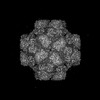 61130MC  9irvC  9ivpC M: map data used to model this data C: citing same article ( |
|---|---|
| Similar structure data |


-Links
 PDBj
PDBj


- Assembly
| Deposited unit | 
|
|---|---|
| 1 |
|
-Components
| #1: Protein | Mass: 43268.602 Da / Num. of mol.: 24 Source method: isolated from a genetically manipulated source Source: (gene. exp.) Homo sapiens (human) / Gene: FTH1, FTH, FTHL6, OK/SW-cl.84, PIG15 / Production host:  #2: Protein | Mass: 26623.918 Da / Num. of mol.: 24 Source method: isolated from a genetically manipulated source Source: (gene. exp.) Aequorea victoria (jellyfish) / Gene: gfp / Production host: Has ligand of interest | Y | Has protein modification | Y | |
|---|
-Experimental details
-Experiment
| Experiment | Method: ELECTRON MICROSCOPY |
|---|---|
| EM experiment | Aggregation state: PARTICLE / 3D reconstruction method: single particle reconstruction |
- Sample preparation
| Component | Name: GFP bound with the 24-mer DARPin-apoferritin scaffold / Type: COMPLEX / Entity ID: all / Source: MULTIPLE SOURCES | |||||||||||||||
|---|---|---|---|---|---|---|---|---|---|---|---|---|---|---|---|---|
| Source (natural) | Organism: Homo sapiens (human) | |||||||||||||||
| Source (recombinant) | Organism: | |||||||||||||||
| Buffer solution | pH: 8 | |||||||||||||||
| Buffer component |
| |||||||||||||||
| Specimen | Embedding applied: NO / Shadowing applied: NO / Staining applied: NO / Vitrification applied: YES | |||||||||||||||
| Specimen support | Grid material: GOLD / Grid mesh size: 300 divisions/in. / Grid type: Homemade | |||||||||||||||
| Vitrification | Instrument: FEI VITROBOT MARK IV / Cryogen name: ETHANE / Humidity: 100 % / Chamber temperature: 281 K |
- Electron microscopy imaging
| Experimental equipment |  Model: Titan Krios / Image courtesy: FEI Company |
|---|---|
| Microscopy | Model: FEI TITAN KRIOS |
| Electron gun | Electron source:  FIELD EMISSION GUN / Accelerating voltage: 300 kV / Illumination mode: SPOT SCAN FIELD EMISSION GUN / Accelerating voltage: 300 kV / Illumination mode: SPOT SCAN |
| Electron lens | Mode: BRIGHT FIELD / Nominal magnification: 130000 X / Nominal defocus max: 2000 nm / Nominal defocus min: 800 nm / Cs: 2.7 mm / C2 aperture diameter: 70 µm |
| Specimen holder | Cryogen: NITROGEN / Specimen holder model: FEI TITAN KRIOS AUTOGRID HOLDER |
| Image recording | Electron dose: 1.28 e/Å2 / Detector mode: SUPER-RESOLUTION / Film or detector model: GATAN K2 SUMMIT (4k x 4k) |
| Image scans | Movie frames/image: 39 |
- Processing
| EM software |
| ||||||||||||||||||||||||||||||||||||||||||||
|---|---|---|---|---|---|---|---|---|---|---|---|---|---|---|---|---|---|---|---|---|---|---|---|---|---|---|---|---|---|---|---|---|---|---|---|---|---|---|---|---|---|---|---|---|---|
| CTF correction | Type: PHASE FLIPPING AND AMPLITUDE CORRECTION | ||||||||||||||||||||||||||||||||||||||||||||
| Particle selection | Num. of particles selected: 750123 | ||||||||||||||||||||||||||||||||||||||||||||
| Symmetry | Point symmetry: O (octahedral) | ||||||||||||||||||||||||||||||||||||||||||||
| 3D reconstruction | Resolution: 3.04 Å / Resolution method: FSC 0.143 CUT-OFF / Num. of particles: 17397 / Algorithm: FOURIER SPACE / Num. of class averages: 1 / Symmetry type: POINT | ||||||||||||||||||||||||||||||||||||||||||||
| Atomic model building | Protocol: RIGID BODY FIT / Space: REAL | ||||||||||||||||||||||||||||||||||||||||||||
| Atomic model building | PDB-ID: 3AJO Accession code: 3AJO / Source name: PDB / Type: experimental model |

