[English] 日本語
Yorodumi
- EMDB-60837: Cryo-EM structure of KpFtsZ-ZapA complex -

+
Open data


ID or keywords:

Loading...

-
Basic information

Entry
Database: EMDB / ID: EMD-60837
TitleCryo-EM structure of KpFtsZ-ZapA complex
Map data
Sample
  • Complex: KpFtsZ-ZapA complex
    • Protein or peptide: Cell division protein FtsZ
    • Protein or peptide: Cell division protein ZapA
  • Ligand: PHOSPHOMETHYLPHOSPHONIC ACID GUANYLATE ESTER
  • Ligand: MAGNESIUM ION
  • Ligand: POTASSIUM ION
Keywordsbacterial cell division / divisome / FtsZ / ZapA / CELL CYCLE
Function / homology
Function and homology information


septin ring assembly / cell septum / division septum assembly / FtsZ-dependent cytokinesis / cell division site / protein polymerization / GTPase activity / GTP binding / plasma membrane / cytosol / cytoplasm
Similarity search - Function
Cell division protein ZapA, eubacteria / Cell division protein ZapA, N-terminal / Cell division protein ZapA-like / Cell division protein ZapA-like superfamily / Cell division protein ZapA / Cell division protein FtsZ / Cell division protein FtsZ, conserved site / Cell division protein FtsZ, C-terminal / FtsZ family, C-terminal domain / FtsZ protein signature 1. ...Cell division protein ZapA, eubacteria / Cell division protein ZapA, N-terminal / Cell division protein ZapA-like / Cell division protein ZapA-like superfamily / Cell division protein ZapA / Cell division protein FtsZ / Cell division protein FtsZ, conserved site / Cell division protein FtsZ, C-terminal / FtsZ family, C-terminal domain / FtsZ protein signature 1. / FtsZ protein signature 2. / Tubulin-like protein FtsZ/CetZ / Tubulin/FtsZ family, C-terminal domain / Tubulin/FtsZ-like, C-terminal domain / Tubulin/FtsZ, C-terminal / Tubulin/FtsZ, 2-layer sandwich domain / Tubulin/FtsZ family, GTPase domain / Tubulin/FtsZ family, GTPase domain / Tubulin/FtsZ, GTPase domain / Tubulin/FtsZ, GTPase domain superfamily
Similarity search - Domain/homology
Cell division protein FtsZ / Cell division protein ZapA
Similarity search - Component
Biological speciesKlebsiella pneumoniae (bacteria) / Klebsiella pneumoniae subsp. pneumoniae MGH 78578 (bacteria) / Klebsiella pneumoniae 342 (bacteria)
Methodhelical reconstruction / cryo EM / Resolution: 2.73 Å
AuthorsFujita J / Hibino K / Kagoshima G / Kamimura N / Kato Y / Uehara R / Namba K / Uchihashi T / Matsumura H
Funding support Japan, 11 items
OrganizationGrant numberCountry
Japan Society for the Promotion of Science (JSPS)JP20K22630 Japan
Japan Society for the Promotion of Science (JSPS)JP23K06418 Japan
Japan Society for the Promotion of Science (JSPS)JP24K01994 Japan
Japan Society for the Promotion of Science (JSPS)JP24H02277 Japan
Japan Society for the Promotion of Science (JSPS)JP24H02270 Japan
Japan Society for the Promotion of Science (JSPS)JP23K18033 Japan
Japan Science and TechnologyJPMJOP1861 Japan
Japan Agency for Medical Research and Development (AMED)JP21am0101117 Japan
Japan Agency for Medical Research and Development (AMED)JP22ama121003 Japan
Japan Agency for Medical Research and Development (AMED)JP23ama121001 Japan
Japan Agency for Medical Research and Development (AMED)JP17pc0101020 Japan
Citation
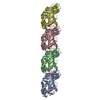
Journal: Nat Commun / Year: 2025
Title: Structural basis for the interaction between the bacterial cell division proteins FtsZ and ZapA.
Authors: Junso Fujita / Kazuki Kasai / Kota Hibino / Gota Kagoshima / Natsuki Kamimura / Shungo Tobita / Yuki Kato / Ryo Uehara / Keiichi Namba / Takayuki Uchihashi / Hiroyoshi Matsumura /
Abstract: Cell division in most bacteria is regulated by the tubulin homolog FtsZ as well as ZapA, a FtsZ-associated protein. However, how FtsZ and ZapA function coordinately has remained elusive. Here we ...Cell division in most bacteria is regulated by the tubulin homolog FtsZ as well as ZapA, a FtsZ-associated protein. However, how FtsZ and ZapA function coordinately has remained elusive. Here we report the cryo-electron microscopy structure of the ZapA-FtsZ complex at 2.73 Å resolution. The complex forms an asymmetric ladder-like structure, in which the double antiparallel FtsZ protofilament on one side and a single protofilament on the other side are tethered by ZapA tetramers. In the complex, the extensive interactions of FtsZ with ZapA cause a structural change of the FtsZ protofilament, and the formation of the double FtsZ protofilament increases electrostatic repulsion. High-speed atomic force microscopy analysis revealed cooperative interactions of ZapA with FtsZ at a molecular level. Our findings not only provide a structural basis for the interaction between FtsZ and ZapA but also shed light on how ZapA binds to FtsZ protofilaments without disturbing FtsZ dynamics to promote cell division.
History
DepositionJul 18, 2024-
Header (metadata) releaseJul 23, 2025-
Map releaseJul 23, 2025-
UpdateJul 23, 2025-
Current statusJul 23, 2025Processing site: PDBj / Status: Released

-
Structure visualization

Supplemental images

Downloads & links

-
Map

FileDownload / File: emd_60837.map.gz / Format: CCP4 / Size: 125 MB / Type: IMAGE STORED AS FLOATING POINT NUMBER (4 BYTES)
Projections & slices

Image control

Size
Brightness
Contrast
Others
AxesZ (Sec.)Y (Row.)X (Col.)
0.88 Å/pix.
x 320 pix.
= 280.96 Å
0.88 Å/pix.
x 320 pix.
= 280.96 Å
0.88 Å/pix.
x 320 pix.
= 280.96 Å

Surface

Projections

Slices (1/3)

Slices (1/2)

Slices (2/3)

Images are generated by Spider.

Voxel sizeX=Y=Z: 0.878 Å
Density
Contour LevelBy AUTHOR: 0.13
Minimum - Maximum-0.49943247 - 0.8740063
Average (Standard dev.)0.0030433696 (±0.031334404)
SymmetrySpace group: 1
Details

EMDB XML:

Map geometry
Axis orderXYZ
Origin000
Dimensions320320320
Spacing320320320
CellA=B=C: 280.96002 Å
α=β=γ: 90.0 °

-
Supplemental data

-
Mask #1

Fileemd_60837_msk_1.map
Projections & Slices
AxesZYX

Projections

Slices (1/2)
Density Histograms

-
Half map: #2

Fileemd_60837_half_map_1.map
Projections & Slices
AxesZYX

Projections

Slices (1/2)
Density Histograms

-
Half map: #1

Fileemd_60837_half_map_2.map
Projections & Slices
AxesZYX

Projections

Slices (1/2)
Density Histograms

-
Sample components

-
Entire : KpFtsZ-ZapA complex

EntireName: KpFtsZ-ZapA complex
Components
  • Complex: KpFtsZ-ZapA complex
    • Protein or peptide: Cell division protein FtsZ
    • Protein or peptide: Cell division protein ZapA
  • Ligand: PHOSPHOMETHYLPHOSPHONIC ACID GUANYLATE ESTER
  • Ligand: MAGNESIUM ION
  • Ligand: POTASSIUM ION

-
Supramolecule #1: KpFtsZ-ZapA complex

SupramoleculeName: KpFtsZ-ZapA complex / type: complex / ID: 1 / Parent: 0 / Macromolecule list: #1-#2
Source (natural)Organism: Klebsiella pneumoniae (bacteria)

-
Macromolecule #1: Cell division protein FtsZ

MacromoleculeName: Cell division protein FtsZ / type: protein_or_peptide / ID: 1 / Number of copies: 6 / Enantiomer: LEVO
Source (natural)Organism: Klebsiella pneumoniae subsp. pneumoniae MGH 78578 (bacteria)
Strain: ATCC 700721 / MGH 78578
Molecular weightTheoretical: 40.37973 KDa
Recombinant expressionOrganism: Escherichia coli (E. coli)
SequenceString: MFEPMELTND AVIKVIGVGG GGGNAVEHMV RERIEGVEFF AVNTDAQALR KTAVGQTIQI GSGITKGLGA GANPEVGRNA ADEDREALR AALDGADMVF IAAGMGGGTG TGAAPVVAEV AKDLGILTVA VVTKPFNFEG KKRMAFAEQG ITELSKHVDS L ITIPNDKL ...String:
MFEPMELTND AVIKVIGVGG GGGNAVEHMV RERIEGVEFF AVNTDAQALR KTAVGQTIQI GSGITKGLGA GANPEVGRNA ADEDREALR AALDGADMVF IAAGMGGGTG TGAAPVVAEV AKDLGILTVA VVTKPFNFEG KKRMAFAEQG ITELSKHVDS L ITIPNDKL LKVLGRGISL LDAFGAANDV LKGAVQGIAE LITRPGLMNV DFADVRTVMS EMGYAMMGSG VASGEDRAEE AA EMAISSP LLEDIDLSGA RGVLVNITAG FDLRLDEFET VGNTIRAFAS DNATVVIGTS LDPDMNDELR VTVVATGIGM DKR PEITLV TNKQVQQPVM DRYQQHGMSP LTQEQKPAAK VVNDNTPQTA KEPDYLDIPA FLRKQAD

UniProtKB: Cell division protein FtsZ

-
Macromolecule #2: Cell division protein ZapA

MacromoleculeName: Cell division protein ZapA / type: protein_or_peptide / ID: 2 / Number of copies: 8 / Enantiomer: LEVO
Source (natural)Organism: Klebsiella pneumoniae 342 (bacteria) / Strain: 342
Molecular weightTheoretical: 12.609186 KDa
Recombinant expressionOrganism: Escherichia coli (E. coli)
SequenceString:
MSAQPVDLQI FGRSLRVNCP PEQRDALNQA AEDLNQRLQD LKERTRVTNT EQLVFIAALN ISYELTQEKA KTRDYASSME QRIRMLQQT IEQALLEQGR ISERPGSKFE

UniProtKB: Cell division protein ZapA

-
Macromolecule #3: PHOSPHOMETHYLPHOSPHONIC ACID GUANYLATE ESTER

MacromoleculeName: PHOSPHOMETHYLPHOSPHONIC ACID GUANYLATE ESTER / type: ligand / ID: 3 / Number of copies: 6 / Formula: G2P
Molecular weightTheoretical: 521.208 Da
Chemical component information

ChemComp-G2P:
PHOSPHOMETHYLPHOSPHONIC ACID GUANYLATE ESTER / GMP-CPP, energy-carrying molecule analogue*YM

-
Macromolecule #4: MAGNESIUM ION

MacromoleculeName: MAGNESIUM ION / type: ligand / ID: 4 / Number of copies: 6 / Formula: MG
Molecular weightTheoretical: 24.305 Da

-
Macromolecule #5: POTASSIUM ION

MacromoleculeName: POTASSIUM ION / type: ligand / ID: 5 / Number of copies: 6 / Formula: K
Molecular weightTheoretical: 39.098 Da

-
Experimental details

-
Structure determination

Methodcryo EM
Processinghelical reconstruction
Aggregation statefilament

-
Sample preparation

BufferpH: 7.5
Component:
ConcentrationFormulaName
17.0 mMHEPES2-[4-(2-Hydroxyethyl)-1-piperazinyl]ethanesulfonic acid
8.6 mMNaClsodium chloride
60.0 mMKClpotassium chloride
3.0 mMMgCl2magnesium chloride
0.6 mMGMPCPPguanosine-5'-[(alpha,beta)-methyleno]triphosphate
GridModel: Quantifoil R1.2/1.3 / Material: COPPER / Mesh: 200 / Pretreatment - Type: GLOW DISCHARGE / Pretreatment - Time: 20 sec. / Pretreatment - Atmosphere: AIR / Details: 20 mA
VitrificationCryogen name: ETHANE / Chamber humidity: 100 % / Chamber temperature: 277 K / Instrument: FEI VITROBOT MARK IV
Details0.3 mg/ml KpFtsZ and 0.19 mg/ml KpZapA

-
Electron microscopy

MicroscopeJEOL CRYO ARM 300
Specialist opticsEnergy filter - Name: In-column Omega Filter / Energy filter - Slit width: 20 eV
Image recordingFilm or detector model: GATAN K3 (6k x 4k) / Number grids imaged: 1 / Average exposure time: 2.3 sec. / Average electron dose: 60.0 e/Å2
Electron beamAcceleration voltage: 300 kV / Electron source: FIELD EMISSION GUN
Electron opticsIllumination mode: FLOOD BEAM / Imaging mode: BRIGHT FIELD / Cs: 2.7 mm / Nominal defocus max: 2.0 µm / Nominal defocus min: 0.5 µm / Nominal magnification: 60000
Sample stageSpecimen holder model: JEOL CRYOSPECPORTER / Cooling holder cryogen: NITROGEN

+
Image processing

Final reconstructionApplied symmetry - Helical parameters - Δz: 44.58 Å
Applied symmetry - Helical parameters - Δ&Phi: -3.11 °
Applied symmetry - Helical parameters - Axial symmetry: D1 (2x1 fold dihedral)
Algorithm: FOURIER SPACE / Resolution.type: BY AUTHOR / Resolution: 2.73 Å / Resolution method: FSC 0.143 CUT-OFF / Software - Name: cryoSPARC (ver. 4.2.1) / Number images used: 54670
CTF correctionSoftware - Name: cryoSPARC (ver. 4.2.1) / Type: PHASE FLIPPING AND AMPLITUDE CORRECTION
Segment selectionNumber selected: 4374348 / Software - Name: cryoSPARC (ver. 4.2.1)
Startup modelType of model: NONE
Final angle assignmentType: MAXIMUM LIKELIHOOD / Software - Name: cryoSPARC (ver. 4.2.1)
FSC plot (resolution estimation)

-
Atomic model buiding 1

Initial model
PDB IDChain

source_name: PDB, initial_model_type: experimental model

source_name: PDB, initial_model_type: experimental model
RefinementSpace: REAL
Output model

PDB-9isk:
Cryo-EM structure of KpFtsZ-ZapA complex

+
About Yorodumi

-
News

-
Feb 9, 2022. New format data for meta-information of EMDB entries

New format data for meta-information of EMDB entries

  • Version 3 of the EMDB header file is now the official format.
  • The previous official version 1.9 will be removed from the archive.

Related info.:EMDB header

External links:wwPDB to switch to version 3 of the EMDB data model

-
Aug 12, 2020. Covid-19 info

Covid-19 info

URL: https://pdbj.org/emnavi/covid19.php

New page: Covid-19 featured information page in EM Navigator.

Related info.:Covid-19 info / Mar 5, 2020. Novel coronavirus structure data

+
Mar 5, 2020. Novel coronavirus structure data

Novel coronavirus structure data

Related info.:Yorodumi Speices / Aug 12, 2020. Covid-19 info

External links:COVID-19 featured content - PDBj / Molecule of the Month (242):Coronavirus Proteases

+
Jan 31, 2019. EMDB accession codes are about to change! (news from PDBe EMDB page)

EMDB accession codes are about to change! (news from PDBe EMDB page)

  • The allocation of 4 digits for EMDB accession codes will soon come to an end. Whilst these codes will remain in use, new EMDB accession codes will include an additional digit and will expand incrementally as the available range of codes is exhausted. The current 4-digit format prefixed with “EMD-” (i.e. EMD-XXXX) will advance to a 5-digit format (i.e. EMD-XXXXX), and so on. It is currently estimated that the 4-digit codes will be depleted around Spring 2019, at which point the 5-digit format will come into force.
  • The EM Navigator/Yorodumi systems omit the EMD- prefix.

Related info.:Q: What is EMD? / ID/Accession-code notation in Yorodumi/EM Navigator

External links:EMDB Accession Codes are Changing Soon! / Contact to PDBj

+
Jul 12, 2017. Major update of PDB

Major update of PDB

  • wwPDB released updated PDB data conforming to the new PDBx/mmCIF dictionary.
  • This is a major update changing the version number from 4 to 5, and with Remediation, in which all the entries are updated.
  • In this update, many items about electron microscopy experimental information are reorganized (e.g. em_software).
  • Now, EM Navigator and Yorodumi are based on the updated data.

External links:wwPDB Remediation / Enriched Model Files Conforming to OneDep Data Standards Now Available in the PDB FTP Archive

-
Yorodumi

Thousand views of thousand structures

  • Yorodumi is a browser for structure data from EMDB, PDB, SASBDB, etc.
  • This page is also the successor to EM Navigator detail page, and also detail information page/front-end page for Omokage search.
  • The word "yorodu" (or yorozu) is an old Japanese word meaning "ten thousand". "mi" (miru) is to see.

Related info.:EMDB / PDB / SASBDB / Comparison of 3 databanks / Yorodumi Search / Aug 31, 2016. New EM Navigator & Yorodumi / Yorodumi Papers / Jmol/JSmol / Function and homology information / Changes in new EM Navigator and Yorodumi

Read more