Movie
Movie Controller


+ Open data

- Basic information
| Entry | Database: PDB / ID: 9isk | ||||||||||||||||||||||||||||||||||||
|---|---|---|---|---|---|---|---|---|---|---|---|---|---|---|---|---|---|---|---|---|---|---|---|---|---|---|---|---|---|---|---|---|---|---|---|---|---|
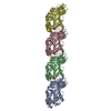
| Title | Cryo-EM structure of KpFtsZ-ZapA complex | ||||||||||||||||||||||||||||||||||||
 Components Components |
| ||||||||||||||||||||||||||||||||||||
 Keywords Keywords | CELL CYCLE / bacterial cell division / divisome / FtsZ / ZapA | ||||||||||||||||||||||||||||||||||||
| Function / homology |  Function and homology information Function and homology informationseptin ring assembly / cell septum / division septum assembly / FtsZ-dependent cytokinesis / cell division site / protein polymerization / GTPase activity / GTP binding / plasma membrane / cytosol / cytoplasm Similarity search - Function | ||||||||||||||||||||||||||||||||||||
| Biological species |  Klebsiella pneumoniae subsp. pneumoniae MGH 78578 (bacteria)Klebsiella pneumoniae 342 (bacteria) Klebsiella pneumoniae subsp. pneumoniae MGH 78578 (bacteria)Klebsiella pneumoniae 342 (bacteria) | ||||||||||||||||||||||||||||||||||||
| Method | ELECTRON MICROSCOPY / helical reconstruction / cryo EM / Resolution: 2.73 Å | ||||||||||||||||||||||||||||||||||||
 Authors Authors | Fujita, J. / Hibino, K. / Kagoshima, G. / Kamimura, N. / Kato, Y. / Uehara, R. / Namba, K. / Uchihashi, T. / Matsumura, H. | ||||||||||||||||||||||||||||||||||||
| Funding support |  Japan, 11items Japan, 11items
| ||||||||||||||||||||||||||||||||||||
 Citation Citation | Journal: Nat Commun / Year: 2025 Title: Structural basis for the interaction between the bacterial cell division proteins FtsZ and ZapA. Authors: Junso Fujita / Kazuki Kasai / Kota Hibino / Gota Kagoshima / Natsuki Kamimura / Shungo Tobita / Yuki Kato / Ryo Uehara / Keiichi Namba / Takayuki Uchihashi / Hiroyoshi Matsumura / Abstract: Cell division in most bacteria is regulated by the tubulin homolog FtsZ as well as ZapA, a FtsZ-associated protein. However, how FtsZ and ZapA function coordinately has remained elusive. Here we ...Cell division in most bacteria is regulated by the tubulin homolog FtsZ as well as ZapA, a FtsZ-associated protein. However, how FtsZ and ZapA function coordinately has remained elusive. Here we report the cryo-electron microscopy structure of the ZapA-FtsZ complex at 2.73 Å resolution. The complex forms an asymmetric ladder-like structure, in which the double antiparallel FtsZ protofilament on one side and a single protofilament on the other side are tethered by ZapA tetramers. In the complex, the extensive interactions of FtsZ with ZapA cause a structural change of the FtsZ protofilament, and the formation of the double FtsZ protofilament increases electrostatic repulsion. High-speed atomic force microscopy analysis revealed cooperative interactions of ZapA with FtsZ at a molecular level. Our findings not only provide a structural basis for the interaction between FtsZ and ZapA but also shed light on how ZapA binds to FtsZ protofilaments without disturbing FtsZ dynamics to promote cell division. | ||||||||||||||||||||||||||||||||||||
| History |
|
- Structure visualization
| Structure viewer | Molecule: MolmilJmol/JSmol |
|---|
- Downloads & links
-Download
| PDBx/mmCIF format | 9isk.cif.gz | 437.5 KB | Display | PDBx/mmCIF format |
|---|---|---|---|---|
| PDB format | pdb9isk.ent.gz | 352 KB | Display | PDB format |
| PDBx/mmJSON format | 9isk.json.gz | Tree view | PDBx/mmJSON format | |
| Others |  Other downloads Other downloads |
-Validation report
| Arichive directory | https://data.pdbj.org/pub/pdb/validation_reports/is/9iskftp://data.pdbj.org/pub/pdb/validation_reports/is/9isk | HTTPS FTP |
|---|
-Related structure data
| Related structure data |  60837MC  9isjC M: map data used to model this data C: citing same article ( |
|---|---|
| Similar structure data |
-Links
 PDBj
PDBj


- Assembly
| Deposited unit | 
|
|---|---|
| 1 |
|
-Components
| #1: Protein | Mass: 40379.730 Da / Num. of mol.: 6 Source method: isolated from a genetically manipulated source Source: (gene. exp.) Klebsiella pneumoniae subsp. pneumoniae MGH 78578 (bacteria)Strain: ATCC 700721 / MGH 78578 / Gene: ftsZ, KPN_00099 / Production host: #2: Protein | Mass: 12609.186 Da / Num. of mol.: 8 Source method: isolated from a genetically manipulated source Source: (gene. exp.) Klebsiella pneumoniae 342 (bacteria) / Strain: 342 / Gene: zapA, KPK_0754 / Production host: #3: Chemical | ChemComp-G2P /   Mass: 521.208 Da / Num. of mol.: 6 / Source method: obtained synthetically / Formula: C11H18N5O13P3 / Feature type: SUBJECT OF INVESTIGATION / Comment: GMP-CPP, energy-carrying molecule analogue*YM Mass: 521.208 Da / Num. of mol.: 6 / Source method: obtained synthetically / Formula: C11H18N5O13P3 / Feature type: SUBJECT OF INVESTIGATION / Comment: GMP-CPP, energy-carrying molecule analogue*YM#4: Chemical | ChemComp-MG /   Mass: 24.305 Da / Num. of mol.: 6 / Source method: obtained synthetically / Formula: Mg / Feature type: SUBJECT OF INVESTIGATION Mass: 24.305 Da / Num. of mol.: 6 / Source method: obtained synthetically / Formula: Mg / Feature type: SUBJECT OF INVESTIGATION#5: Chemical | ChemComp-K /   Mass: 39.098 Da / Num. of mol.: 6 / Source method: obtained synthetically / Formula: K / Feature type: SUBJECT OF INVESTIGATION Mass: 39.098 Da / Num. of mol.: 6 / Source method: obtained synthetically / Formula: K / Feature type: SUBJECT OF INVESTIGATIONHas ligand of interest | Y | Has protein modification | N | |
|---|
-Experimental details
-Experiment
| Experiment | Method: ELECTRON MICROSCOPY |
|---|---|
| EM experiment | Aggregation state: FILAMENT / 3D reconstruction method: helical reconstruction |
- Sample preparation
| Component | Name: KpFtsZ-ZapA complex / Type: COMPLEX / Entity ID: #1-#2 / Source: RECOMBINANT | ||||||||||||||||||||||||||||||
|---|---|---|---|---|---|---|---|---|---|---|---|---|---|---|---|---|---|---|---|---|---|---|---|---|---|---|---|---|---|---|---|
| Molecular weight | Experimental value: NO | ||||||||||||||||||||||||||||||
| Source (natural) | Organism: Klebsiella pneumoniae (bacteria) | ||||||||||||||||||||||||||||||
| Source (recombinant) | Organism: | ||||||||||||||||||||||||||||||
| Buffer solution | pH: 7.5 | ||||||||||||||||||||||||||||||
| Buffer component |
| ||||||||||||||||||||||||||||||
| Specimen | Embedding applied: NO / Shadowing applied: NO / Staining applied: NO / Vitrification applied: YES / Details: 0.3 mg/ml KpFtsZ and 0.19 mg/ml KpZapA | ||||||||||||||||||||||||||||||
| Specimen support | Details: 20 mA / Grid material: COPPER / Grid mesh size: 200 divisions/in. / Grid type: Quantifoil R1.2/1.3 | ||||||||||||||||||||||||||||||
| Vitrification | Instrument: FEI VITROBOT MARK IV / Cryogen name: ETHANE / Humidity: 100 % / Chamber temperature: 277 K |
- Electron microscopy imaging
| Microscopy | Model: JEOL CRYO ARM 300 |
|---|---|
| Electron gun | Electron source:  FIELD EMISSION GUN / Accelerating voltage: 300 kV / Illumination mode: FLOOD BEAM FIELD EMISSION GUN / Accelerating voltage: 300 kV / Illumination mode: FLOOD BEAM |
| Electron lens | Mode: BRIGHT FIELD / Nominal magnification: 60000 X / Nominal defocus max: 2000 nm / Nominal defocus min: 500 nm / Cs: 2.7 mm |
| Specimen holder | Cryogen: NITROGEN / Specimen holder model: JEOL CRYOSPECPORTER |
| Image recording | Average exposure time: 2.3 sec. / Electron dose: 60 e/Å2 / Film or detector model: GATAN K3 (6k x 4k) / Num. of grids imaged: 1 |
| EM imaging optics | Energyfilter name: In-column Omega Filter / Energyfilter slit width: 20 eV |
- Processing
| EM software |
| ||||||||||||||||||||||||||||||||||||
|---|---|---|---|---|---|---|---|---|---|---|---|---|---|---|---|---|---|---|---|---|---|---|---|---|---|---|---|---|---|---|---|---|---|---|---|---|---|
| CTF correction | Type: PHASE FLIPPING AND AMPLITUDE CORRECTION | ||||||||||||||||||||||||||||||||||||
| Helical symmerty | Angular rotation/subunit: -3.11 ° / Axial rise/subunit: 44.58 Å / Axial symmetry: D1 | ||||||||||||||||||||||||||||||||||||
| Particle selection | Num. of particles selected: 4374348 | ||||||||||||||||||||||||||||||||||||
| 3D reconstruction | Resolution: 2.73 Å / Resolution method: FSC 0.143 CUT-OFF / Num. of particles: 54670 / Algorithm: FOURIER SPACE / Symmetry type: HELICAL | ||||||||||||||||||||||||||||||||||||
| Atomic model building | Space: REAL | ||||||||||||||||||||||||||||||||||||
| Atomic model building |
| ||||||||||||||||||||||||||||||||||||
| Refinement | Highest resolution: 2.73 Å Stereochemistry target values: REAL-SPACE (WEIGHTED MAP SUM AT ATOM CENTERS) | ||||||||||||||||||||||||||||||||||||
| Refine LS restraints |
|